Story
Mr. M is a recreational athlete who sought physiotherapy in the fall of 2019 for musculoskeletal issues related to his running. Mr. M noticed that if he started to run more than three times per week, or increased the intensity of his running, he would experience inner thigh tightness on the right leg. He noted this issue had been going on for about 10 years on and off. At the time of the assessment, Mr. M was consistently running three times per week, two 5km interval paced runs, and one 10-15 km run per week of a low intensity pace.
Previously, Mr. M had sought physiotherapy and massage therapy treatment for this complaint and had some relief but still felt he couldn’t progress his running distance or pace for fear of aggravating this issue.
Diane’s question:
Can you describe what type, or where, the physio and massage therapy was applied? i.e. was it only local to the right inner thigh?
Kjersti’s response:
Yes, he mentioned there was release work done locally on the right inner thigh and right glutes. He was also given a core stabilization program.
Diane’s question:
Do you think his lack of distance progression was secondary to this cognitive belief (fear of aggravation of his symptoms) or to reality and therefore not a barrier to progression within the context of your treatment?
Kjersti’s response:
Mr. M’s cognitive belief around fear of aggravation of his symptoms with an increase in distance or pace was based upon the fact if he did try to increase these parameters he would experience a flare of his symptoms. I did not see this cognitive belief as a barrier to progression in his treatment.
About 8 years ago, Mr. M trained for a marathon and had severe symphysis pubis pain which lasted for 8-9 months afterwards. At the time the physiotherapist he was seeing diagnosed him with osteitis pubis. An X-ray was done on the right hip which revealed an os trigonum on the femoral head.
Diane’s question:
An os trigonum is more commonly found in the foot. What is your hypothesis as to the source of this ‘accessory bone’ and its relationship to the severe symphysis pubis pain? Could it have been an incidental finding? Is there anything in his story that supports your answer?
Kjersti’s response:
My hypothesis regarding the os trigonum on the femoral head is that it was an incidental finding and not related to his symphysis pubis pain. There was no report of pain or noted dysfunction in the right hip that bothered him in the past prior to the substantial increase in load that occurred when running a marathon. After which the severe pubis pain started. Mr. M also mentioned during training for his marathon, he had a change in footwear to a ‘free runner’ type shoe with less support than he was used to. It was soon after this change his symptoms in the pubic bone increased.
Mr. M felt he fully recovered from this injury. In terms of previous and concurrent musculoskeletal issues, Mr. M reported an ongoing history of middle back pain when sitting at work and a left acromioclavicular joint separation he sustained during a bench press maneuver as a teenager. Mr. M wears a neutral shoe with a custom orthotic while running, which he has done for 10 years.
Diane’s question:
Given the length of time Mr. M has experienced right inner thigh tightness (on and off for 10 years), using the current neuroscience of pain and findings from this story, can you comment on whether this story supports classifying his symptoms as nociceptive, neuropathic, or nociplastic, or a combination?
Kjersti’s response:
The International Association for the Study of Pain (IASP) defines nociceptive pain as pain that arises from actual or threatened damage to non-neural tissue and is due to the activation of nociceptors. Neuropathic pain is defined as pain which is caused by a lesion or disease of the somatosensory nervous system. Nociplastic pain is defined as pain that arises from altered nociception despite no clear evidence of actual or threatened tissue damage causing the activation of peripheral nociceptors or evidence for disease or lesion of the somatosensory system causing the pain.
Pain science research suggests that with injury chronicity, the nervous system can become hypersensitive and can show an increased response to normal stimulus. However in this case, Mr. M was experiencing symptoms more directly related to increasing load on his tissue, suggesting his pain was mechanical and peripherally mediated. His subjective reports were mechanically based on specific aggravating factors (i.e. increasing distance and pace of running) and would subside with a few days rest from running. He also experienced long stretches of time with no pain or discomfort at all, thus supporting more a nociceptive type pain.
Meaningful Complaint
Mr. M’s meaningful complaint was adductor tightness (right greater than left), progressing to pain after running longer distances (more than 15km’s at a time).
Cognitive Beliefs
Mr. M believed that his adductors may be tight due to instability in his core or that there could be an issue related to his feet.
Diane’s question:
How does Mr. M’s cognitive belief guide, or influence, the tests/regions you choose to use/examine in your initial assessment?
Kjersti’s response:
Listening to Mr. M’s beliefs around where his issues were coming from in his body influenced the regions I chose to investigate but more so, influenced my explanation of his assessment.
Because of the long history of adductor tightness and something being ‘wrong’ with his feet and core muscles, I knew it would be important for him understand how these body regions were influencing his symptoms. During the assessment, whether or not I was led to the body region he was curious about or not, I would explain what was happening with the adductors, core and feet in terms of their interaction with one another and in relation to the primary driver. We also discussed that the area where he felt the obvious symptoms, may not be the area of the primary driver, it may just be the ‘reactor’ or the tissue being affected by more load or strain.
Meaningful Task
Based on Mr. M’s subjective history the meaningful task of running was chosen and specifically a distance of greater than 15 km as this was noted to be when his symptoms typically would begin.
Screening Tasks
Right one leg standing
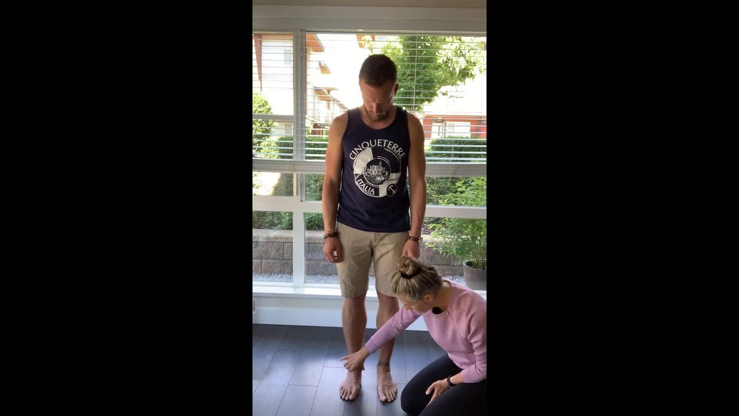
The screening task of right one leg standing was chosen for the initial assessment. One leg standing is required for gait and running.
Standing postural screen
As a starting point of the assessment, it is important to note the position of the body at the starting position of the screening task that relates to the meaningful task. For Mr. M, the starting position screen that relates to the right one leg stand task is standing.
The reasoning behind this is that each person has unique postural asymmetries and possibly adaptive compensation strategies present before moving to complete a task. These may be due to habitual postures, previous injuries or any number of reasons. So in order to understand how a person moves through and completes a task, we need to know the position of each segment of the body from the start. Then, through the task if there appears to be non-optimal alignment or biomechanics, we can start to understand where the trouble may have started from. At times, a patient may present with several areas of asymmetry but then later may compensate well for them. Or conversely from an asymmetrical starting position, we can easily understand how a person may not be able to achieve a rotation or flexion task.
Diane’s comment and question:
There is a lot of research that currently suggests that posture (i.e. alignment) is irrelevant and that when we talk to patients about their alignment we are ‘medicalizing normalcy’. One example that supports this statement in your paragraph above is this one:
“Then, through the task if there appears to be non-optimal alignment or biomechanics, we can start to understand where the trouble may have started from.”
There is another critical step in ISM other than judging someone’s alignment or biomechanics, that confirms that our ‘judgments’ are likely to be correct or incorrect. In this manner we can determine if the asymmetrical starting position of a body region or segment is relevant, or not. What is that step?
Kjersti’s response:
In ISM we would determine if an asymmetrical starting position of a body region was relevant or not through the process of finding the primary driver. Starting with correcting an area of incongruency and assessing the impact of this correction on other areas or asymmetrical alignment, we can reason which findings are relevant to improving the alignment, biomechanics and control of the body and which are incidental findings. This process of finding a primary driver helps us to eliminate which areas of asymmetry are important for impacting the performance of the body and which ones are aligned poorly aligned due to compensation.
Mr. M reported no symptoms in the standing screen.
Findings for Standing Postural Screen
Functional Unit #1 (FU #1)
Pelvis: Transverse plane rotation to the right (TPR R), intrapelvic torsion to the right (IPT R)
Hips: Right head of femur anterior in relation to the right acetabulum. Left hip anterior relative to the right hip, but centered in the left acetabulum. The left hip positioning was due to the TPR R of the pelvis however the left femoral head was not translated forward further from neutral.
Diane's question:
How do you know that the transverse plane rotation of the pelvis was not ‘due to’ the position of the left hip as opposed to the hip being due to the pelvis? Was this an assumption because of congruence? If so, if needs to be tested and not assumed!
Kjersti’s response:
I confirmed that the position of the left hip was due to the transverse plane rotation of the pelvis by first correcting the position of the pelvis and assessing what happened to the hip. When the TPR of the pelvis was corrected and then released, the pelvis was felt to move first into a right TPR with the left hip following it but the left hip remained centered in the acetabulum throughout.
To confirm the left hip, was not driving the pelvis, I moved the patient to the right foot to offload the left hip, and kept the left hip centered as weight was shifted back on the left foot. When I let the left hip go, the pelvis was still rotated to the right, and the hip position didn’t change, confirming that the hip position was due to the position of the pelvis.
Thorax: 8th 6th and 4th thoracic rings translated right/rotated left, 7th and 3rd thoracic rings translated left/rotated right.
Thoracic rings 8, 6, and 4 were congruent with each other but incongruent to rings 7 and 3 which were congruent with each other. When a body segment rotates one way (i.e. rings 4, 6, and 8), another region of the body (i.e. rings 7 and 3) often will rotate the opposite way. This would be seen as an incongruency. In ISM, the hypothesis is that body regions can to compensate for areas of non-optimal alignment by rotating in the opposite direction an effort to keep us in relative neutral.
Diane's question:
Why is noting incongruencies in body regions important in the starting postural screen task?
Kjersti’s response:
Noting incongruencies in body regions is important in the starting postural screen because it helps to group findings together, making it easier to focus on relevant findings and therefore accelerate the process of finding a primary driver. Correcting an incongruent segment will likely reveal the best, or one of the best, corrections for alignment and control in other body regions and is more likely the primary driver. Noting incongruencies will help to target which body region to test first and then by noting the impact of this correction on the alignment, biomechanics, and control of other body regions we can discover the primary driver.
Functional Unit #2 (FU #2)
Cranium: neutral alignment in relation to the upper neck (no intracranial torsion, no sideflexion or rotation)
Cervical Spine: 7th cervical vertebrae translated right/rotated left, 2nd and 5th cervical vertebrae neutral.
Shoulder girdles: rotated to the right, lateral plateau of the left clavicle elevated relative to the scapula (step deformity with acromion) and medial end of the left clavicle compressed into the manubrium.
Thorax: Second thoracic ring: translated left/rotated right.
The shoulder girdles and 2nd thoracic ring were congruent in right rotation; the 7th cervical segment was incongruent to this in left rotation.
Diane's question:
Is there a segment above C7 compensating such that the cranium is in neutral, or do you think C7 is compensating for Thoracic ring 2?
Kjersti’s response:
In standing, there wasn’t anything above C7 to compensate for its alignment. It is possible that C7 was compensating for Thoracic ring 2.
Functional Unit #3 (FU #3)
Knees: neutral alignment
Feet:
Left foot
- Talus: plantarflexed, adducted and laterally tilted relative to the mortise (the inferior tib-fib joint) (Talar tilt), and plantarflexed and adducted relative to the calcaneus (pronated)
- Calcaneus: laterally rotated relative to the talus
- Navicular: externally rotated relative to the talus and cuboid
- Cuboid: internally rotated relative to the calcaneus and navicular
- Medial cuneiform: externally rotated relative to the navicular, middle cuneiform neutral, lateral cuneiform internally rotated in relation to the middle cuneiform.
- First metatarsal: slight external rotation relative to the medial cuneiform/2nd MT axis
Diane’s question:
Well described! I can see this foot. Which part of the foot is ‘most interesting’ and why – hindfoot, midfoot, forefoot?
Kjersti’s response:
The hindfoot was the most interesting part of the foot. Typically, in a pronated hindfoot, the talus would be plantarflexed and adducted, while the calcaneus would be in lateral rotation. The talus should remain centered in the mortice. In this situation, the talus was plantarflexed and adducted as well as laterally tilted (the lateral aspect of the talus more compressed into the mortice).
Right foot
- Talus: plantarflexed adducted and laterally tilted relative to the mortice and plantarflexed and adducted relative to the calcaneus (Talar tilt)
- Calcaneus: laterally rotated relative to the talus
- Navicular: externally rotated relative to the talus and cuboid
- Cuboid: internally rotated relative to the calcaneus and navicular
- First metatarsal: externally rotated relative to the medial cuneiform/2nd MT axis, and slight abduction (more marked on the right compared to the left foot)
While there are various sites of non-optimal alignment in standing, standing was not a meaningful task for Mr. M. Therefore, a driver was not found in standing.
Right One Leg Standing (R OLS)
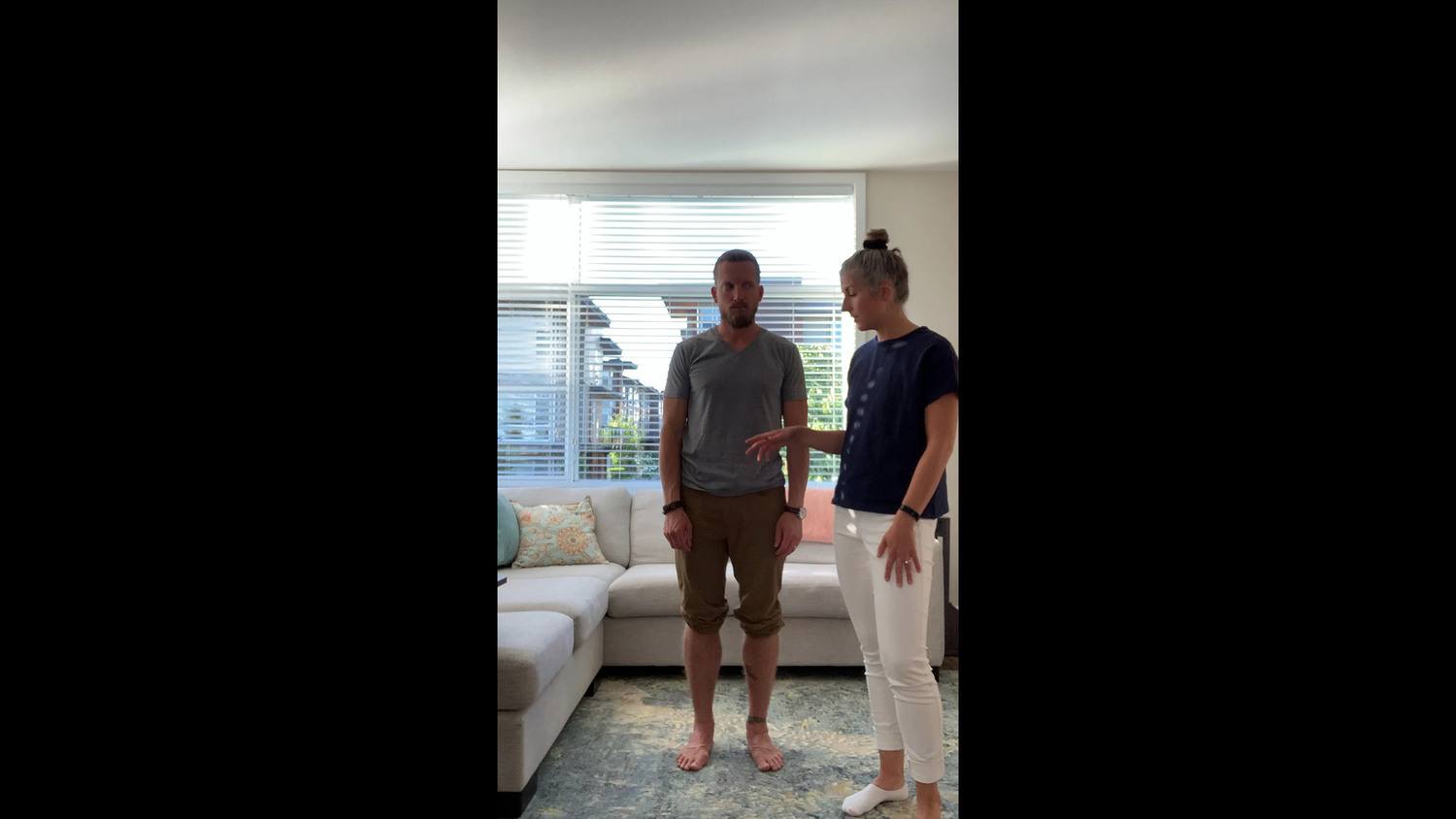
One leg standing was chosen because stance phase is a necessary component of running stride. It allows for weight acceptance and prepares the runner for push off. The right side was chosen over the left because the patient’s symptoms were more obvious and irritable on the right side. Non-optimal alignment, biomechanics and/or control can occur throughout the three functional units of the body during one leg standing and this is identified by observing and palpating body regions during this task.
Diane’s comment:
Sub-optimal performance of the left leg may create excessive loading of the right. The side of symptoms may not always be indicative of the primary site of impairment so both sides should be examined. I agree with starting on the right side, but I would have also assessed his strategy for L OLS particularly given the findings you noted in standing with the left foot.
Kjersti’s response:
Great point Diane, L OLS would have been ideal to assess as well.
Diane’s question:
In ISM how do you ‘know’ that the alignment, biomechanics and/or control findings are non-optimal for this task, or merely an incidental, non-relevant finding?
Kjersti’s response:
In the Integrated System’s Model, we are first noting all the areas of abnormal ABC’s (alignment, biomechanics and control) and then narrowing down to relevant findings. We can rule out irrelevant findings by correcting the alignment of one area (an incongruency) and noting the impact this correction has on the rest of the functional unit being tested. If correction of a specific area (i.e. the right hip) produces an improvement in multiple other areas (i.e. improves TPR of the pelvis, and a thoracic ring translated/rotated), then we know to focus on the area that corrects the alignment of other body regions. We can consider the other findings (TPR of the pelvis and thoracic ring translated/rotated) as an incidental or non-relevant because if we correct and treat the primary area (i.e. hip) these areas will improve as well. This way we can optimize and focus our treatment on 1-2 areas rather than getting bogged down with multiple findings.
Findings for R OLS
When Mr. M was asked to stand on his right leg, the following findings were noted:
Functional Unit #1 (FU #1)
Hips:
- Left femoral head was more anterior than the right femoral head due to the position of the pelvis in a right TPR/IPT, and the right femoral head started slightly more anterior relative to the acetabulum. With R OLS, the left femoral head remained centered in the acetabulum through left hip flexion and the right translated further anterior relative to the right acetabulum.
- With optimal alignment, biomechanics and control it is expected that the right femoral head should be centered in the acetabulum and remain centered as load is accepted on the right leg. The right hip demonstrated sub-optimal alignment and control for R OLS.
Pelvis:
- The pelvis started in a transverse plane rotation to the right (TPR R), with an associated intrapelvic torsion (IPT) to the right. This is considered non-optimal alignment at the start of the screening task as the pelvis is expected to start and remain neutral in the transverse plane.
- Upon right OLS, the right sacroiliac joint (SIJ), “unlocked”. An unlocking SIJ occurs when the sacral base counternutates relative to the innominate which is anteriorly rotated relative to the sacral base on that side.
Diane's question:
We do not talk about unlocking knees, ankles or shoulders, why unlocking SIJs? What is the history behind the use of this term?
Kjersti’s response:
Hungerford et al in 2004 showed that anterior rotation of the weight bearing innominate occurred in subjects with unilateral pelvic girdle pain. It is thought that anterior rotation of a weight bearing innominate relative to the sacrum is non-optimal as this loads the pelvis in the loose packed or “unlocked” position for the joints of the pelvis. So the term unlocking is based on the anatomy of the pelvis and the natural form and force closure mechanisms that occur during loading of this joint.
When load increased through the pelvic girdle, sacral nutation occurs. Nutation is the most secure position for transmitting load through the pelvis due to the wedge shape of the sacrum and the tightening of the interosseous, sacrospinous and sacrotuberus ligaments (Vleeming et al 1990). For example, in one leg standing the innominate on the weight bearing side either remains posteriorly rotated or posteriorly rotates relative to the sacrum, and the sacrum remains nutated relative to the innominate of the weight bearing side. Vleeming called this the ‘self-locking mechanism’ of the SIJ (1990). In an unlocking SIJ, counter-nutation of the sacrum relative to the weight bearing innominate as well as anterior rotation of the weight bearing innominate relative to the sacrum occurs.
Diane's question:
Is it possible to have a R IPT and a right unlocked SIJ at the same moment in time? If not, why not?
Kjersti’s response:
It is not possible to have a R IPT and a right unlocked SIJ at the same moment in time. In a right IPT, the left innominate rotates anteriorly, the sacrum rotates to the right and the right innominate rotates posteriorly. An unlocked R SIJ is not possible in an IPT because in an IPT the sacrum remains nutated relative to both innominates. The left side of the sacrum is nutated relative to the left innominate and the right side of the sacrum is nutated relative to the right innominate. Conversely, when there is an unlocked right SI joint the sacrum is counternutated relative to the right innominate. This cannot occur when there is an IPT because an IPT requires the sacrum to be nutated relative to both innominates.
Diane's question:
Reflecting back again on how ISM is criticized for too much emphasis on judging posture, or alignment, is it always non-optimal for the pelvis to be rotated in the transverse plane in standing? It is a common finding noted, but how do we determine if, for this individual, and for this task, it is OK or non-optimal. It isn’t ideal but is it non-optimal?
Kjersti’s response:
A TPR of the pelvis is a common finding and whether it is non-optimal or not is decided based upon the meaningful task, screening tasks and how the TPR of the pelvis changes or adapts through this movement. For Mr. M, in the task of right OLS, we discover that the right SIJ unlocks when in right one leg standing which is considered non-optimal. If in right OLS, the TPR of the pelvis moved back to neutral and the right SIJ did not unlock, the TPR of the pelvis we see in standing would not be ideal, but not necessarily non-optimal.
Thorax:
- The 3rd thoracic ring remained translated left/rotated right (no further change from start position, the thoracic ring started and remained in sub-optimal alignment for this task).
- The 4th and 6th thoracic rings remained translated right/rotated left (no further change from start position, the thoracic rings started and remained in sub-optimal alignment for this task).
- The 7th thoracic ring increased in left translation/right rotation (an increase from the suboptimal starting position).
- The 8th thoracic ring increased in right translation/left rotation (an increase from the suboptimal starting position).
Lumbar spine:
- No findings noted.
Driver for Functional Unit #1
Primary 8th thoracic ring driver, with a secondary right hip driver. This conclusion is supported by the following:
Correction of the 8th thoracic ring restored alignment and control of the 4th thoracic ring and the 7th thoracic ring which in turn corrected the 3rd thoracic ring, and the right transverse plane rotation of the pelvis. Correction of the R hip restored the control at the right SIJ (no unlocking).
Diane's question:
Did correction of the 8th thoracic ring change the alignment of the right hip, even partially? Did correction of the 8th thoracic ring restore control of the right SIJ, even partially?
Kjersti:
Correction of the 8th thoracic ring restored alignment of the right SIJ but not control of the SIJ. Correction of the 8th thoracic ring did not change the alignment of the right hip.
Diane's question:
Did correction of the right hip change the alignment of the 8th thoracic ring?
Kjersti:
Correcting the right hip did not change the alignment of the 8th thoracic ring.
Summary of driver for FU#1: The 8th thoracic ring was primary because it corrected all but the alignment of the R hip and the control at the R SIJ. The R hip was the secondary driver because it corrected the control at the R SIJ.
Functional Unit #2
Cranium: remained neutral through the task.
Cervical Spine:
- The 7th cervical vertebrae translated further to the right/rotated to the left (segment started and worsened its sub-optimal alignment for this task).
- The 2nd cervical vertebrae translated right/rotated left (starting position in standing was neutral).
- The 5th cervical vertebrae translated left/rotated right (starting position in standing was neutral).
Shoulder girdles:
- Shoulder girdles restored alignment from right rotation to neutral during right OLS, which in turn corrected the medial compression of the left clavicle into the While this body region started in suboptimal alignment, it corrected to neutral which was optimal for this task. The step deformity remained at the acromioclavicular joint as was expected due to previous trauma at this joint.
Thorax
- The second thoracic ring started in left translation/right rotation (suboptimal alignment) and corrected along with the shoulder girdles during right OLS (optimal alignment).
Driver for Functional Unit #2
Primary Driver: 7th cervical segment. Correcting the alignment of the 7th cervical vertebrae restored the alignment of the 5th and 2nd cervical vertebrae during the R OLS task. Correcting C2 and C5 did not change C7 during this task.
Functional Unit #3
Knees
- Optimal alignment and control noted bilaterally.
Right Foot
- The right talus moved further into plantarflexion, adduction and lateral tilt relative to the mortise, worsening alignment compared with the starting position in double leg standing. The calcaneus remained laterally rotated in relation to the talus and everted in relation to the lower leg. This is sub-optimal as in one leg standing the talus should not tilt in the mortice, the calcaneus should remain neutral relative to talus and the lower leg and the midfoot should remain in neutral alignment.
- As load was increased in R OLS, the navicular plantarflexed and externally rotated more and then less. Similarly, the cuboid moved into and out of internal rotation relative to the calcaneus and navicular. In one leg standing, the foot should start in neutral alignment (talus and calcaneus stacked in the mortice, mid and forefoot in neutral alignment) and maintain this position to prepare for moving into supination and push off.
- The first metatarsal moved in and out of further external rotation and abduction relative to the axis of the foot (medial cuneiform/2nd MT axis). This is suboptimal, the first metatarsal should maintain neutral alignment in right OLS.
Driver for Functional Unit #3
The right foot was the only site of impaired alignment and control in the right lower extremity for this R OLS task. Correcting the alignment of the left foot did not change the biomechanics or control of the right foot, and vice versa.
Summary of Functional Unit Drivers:
For each functional unit, the within unit drivers for the right one leg standing task were:
- FU#1: Primary Thorax (8th thoracic ring), Secondary right hip
- FU#2: Primary neck (7th cervical segment)
- FU#3: Right foot
Relationship between Drivers of Functional Units:
- Correcting the alignment of the 8th thoracic ring restored alignment of the 7th cervical vertebrae, however this did not correct the right foot.
- Correcting the alignment of both feet initially in double leg standing, restored alignment and control of the 8th thoracic ring.
- The correction of the right foot was done in standing by taking the right talus out of PF/Add (in relation to the calcaneus) and centering it in the mortice by decompressing the lateral aspect of the talar dome from the mortice. The right calcaneus was centered under the talus by applying an anterolateral glide to the posteromedial aspect of the calcaneous. A full correction of both the talocrural and subtalar joints for ROLS was possible on the right.
- The left foot correction was similar to the right however a full correction of the left talus, calcaneus and navicular was not possible. The left talus remained slightly PF/Add and in lateral tilt, the calcaneus slightly laterally rotated beneath the talus, and the navicular remained slightly externally rotated relative to the talus. The left calcaneus was corrected from a lateral rotation by applying an anterolateral force to the posteromedial aspect of the calcaneus. Normal alignment and biomechanics of the hindfoot in standing and one leg standing are that the talus should not be tilted relative to the mortise and the calcaneus should be stacked neutrally under the talus.
- While optimal alignment of the feet restored the biomechanics and control of the 8th ring, the right foot correction (even with the addition of a connect cue) was not maintained when loads were increased in the right OLS task.
- In standing, first Mr. M was asked to stand on his orthotics, and then both feet were corrected (as above). When both feet were corrected in standing, with the orthotics in place, the right foot position was maintained during the right OLS.
- Correcting the alignment of both feet, then supporting the correction with his orthotics (without shoes so foot alignment could be visualized) did not restore the alignment and control of the right hip.
- Correcting the alignment of the right hip restored the control of the right SIJ.
It was hypothesized that correcting the left foot in standing prior to the R OLS task, allowed for optimal control and alignment of thoracic ring 7 (which was driving TR 3). Without the additional correction of the left foot and thus TR 7 in standing, when load was placed on the right foot, the right foot ‘collapsed’ (talus PF, add/lat tilt, calcaneus Lat. Rot.) more, driving TR 8 into more right translation/left rotation, and in addition TR 7 translated further in the opposite direction to compensate.
With the left foot corrected in standing, TR 7 did not translate left/rotate right further in the task and therefore the right foot did not ‘collapse’ as much (talus PF, add/lat tilt). Then the right foot correction, maintained by the orthotic, improved alignment and control of the TR 8 as load was increased. With TR 8 aligned, the TR 7 did not translate left further to compensate.
Thoracic ring 8 was the primary driver in FU #1, so once this was corrected through the task, biomechanics in the rest of the thorax and FU #2 were restored.
No foot correction fully corrected the alignment of the right hip or control of the right SIJ. However, correcting the alignment of the right hip restored the control of the right SIJ.
Final drivers for the right OLS task: Primary left and right feet driver, secondary right hip.
Further Assessment of the Drivers
Further Assessment of Noted Drivers
Further assessment of a driver includes tests for active and passive mobility, active and passive control as well as vector analysis during all mobility testing. This series of tests helps to determine the dominant underlying system impairment of a driver (articular, neural, visceral or myofascial).
Vector Analysis uses a variety of tests to differentiate vectors in various systems that are impacting alignment, biomechanics and/or control of a driver. We use our hands during active and passive mobility testing to reach a hypothesis of the location and system producing the dominant vector. All of this is part of the umbrella term Vector Analysis.
This information combined with mobility and control testing, helps to determine what structures might be creating non optimal function of a primary driver or body region.
Definitions of Listening for the Analysis of Vectors:
- Active Listening: the patient actively moves through a range of motion or into more optimal alignment, until the first point of resistance is felt, then the therapist uses their hands to note the biomechanics produced and where the resistance is coming from. Active listening is done when the patient moves actively i.e. active mobility testing.
- Passive Listening #1: the therapist feels what happens when moving a body segment passively.
- Passive Listening #2: the therapist passively moves a body segment into neutral alignment, and then upon release, feels where the joint is drawn to rest by the vectors acting on it.
Diane’s question:
What system impairment is suggested if you cannot ‘place the body segment into neutral alignment’? How would you know that this body region is a driver if you can’t correct its alignment, provide better biomechanics and/or control during the task performance?
Kjersti’s response:
If the body segment cannot be placed into neutral alignment, an articular system impairment is suggested to be the cause. You cannot rule out this body region as a driver if you cannot obtain a correction that provides better biomechanics or control during the task. You would first need to address the articular system impairment and then retest.
Diane’s comment:
And, you cannot assess the impact of a complete correction of a body segment on another site of impairment until you can completely correct it. This fact becomes apparent later in this report when the drivers change after the release treatment of the feet.
Further Assessment of the Right Foot
- Active Mobility – From a seated position, the right talocrural joint was tested by asking Mr. M to move the right foot into active dorsiflexion and plantarflexion. There was a limitation of active dorsiflexion at the end of range present at this joint. This suggests that Mr. M had enough dorsiflexion range of motion for one leg standing but not for tasks such as running or squat. From a standing position, the ability of the right subtalar (ST) joint to supinate and pronate was tested through a body twist. The right ST joint started in a position of pronation, and the calcaneus was everted in standing relative to the lower leg because of the lateral talar tilt relative to the mortice. Through a body twist to the left the right ST joint maintained non optimal alignment and further pronated. The midfoot and forefoot on the right foot lengthened and fanned during the left body twist. Through a body twist to the right, the right ST joint demonstrated supination and resolution of the lateral talar tilt at the mortice joint (dorsiflexion and abduction of the talus relative to the calcaneus, decompression of the lateral talar dome from the mortice and calcaneus medially rotated relative to the talus). Through the right body twist, the right midfoot shortened and folded.
- Passive Mobility
- The right talocrural joint (TC) was passively moved into dorsiflexion, there was less passive joint mobility into full dorsiflexion of the talus. This was associated with a reduced posterior glide of the talus in the mortice and the end feel not super hard (articular) but more like over-active neuromyofascia.
- The right calcaneus was passively moved into a medial and lateral rotation at the subtalar joint (ST), less passive mobility of medial rotation of the calcaneus was noted. Mr. M has full active ROM of this subtalar joint in weight bearing, yet passive medial rotation of this joint is restricted. This cannot be an articular system impairment. The vector is likely neural or myofascial.
- Further arthrokinematic analysis of the two subtalar joints revealed that the anterolateral glide of the posterior subtalar joint was the cause of the restricted medial rotation; however the end feel of this glide was springy suggesting the impairment was more neural or myofascial or a combination of both.
- The right navicular was passively moved into external and internal rotation on a stabilized talus. There was an increased resistance to internal rotation of the navicular on the talus, however full passive range of motion was present. Again, an articular system impairment is ruled out by these findings.
- The first metatarsophalangeal joint mobility was tested by passively moving the first phalanx into neutral from abduction, and then while the first metatarsal was stabilized, the phalanx was moved into a dorsal and plantar motion as well as into full dorsiflexion and plantar flexion. Full range of motion was present at this joint.
- Active Control – Mr. M was unable to hold his hindfoot, midfoot, and forefoot corrected in right OLS without the use of an orthotic, suggesting impaired active control of the hindfoot, midfoot and forefoot.
- Passive Control – Not assessed as the story did not suggest a need for this.
Vector Analysis – For Mr. M’s right hindfoot, passive listening techniques during correction and release of the alignment of the right hindfoot were used to determine the underlying systems creating vectors of force that impacted mobility of the TC, ST and midfoot joints.
From a seated position the calcaneus appeared inverted relative to the lower leg and the talus was now medially tilted relative to the mortice and still plantar flexed. This suggests that the hindfoot in standing is everted to get the first ray on the ground.
Passive listening #2: First, the talus was distracted, abducted and dorsiflexed to center it in the mortice and the calcaneus was medially rotated relative to the talus by distracting and then applying an anterolateral force to the posteromedial aspect of the calcaneus.
Upon release of the correction of both the talocrural and subtalar joints, the calcaneus was felt to move first into lateral rotation. The direction of the pull was from the medial and posterior aspect of the calcaneus, and the vector was strong and short; not extending up into the lower leg.
The calcaneus was then specifically distracted from the talus with a focus on the anteromedial, posteromedial, anterolateral and posterolateral aspect of the two joint complex, which revealed that the posteromedial aspect of the posterior subtalar joint was most restricted. The posteromedial aspect of the calcaneus was distracted and upon release (passive listening #2) revealed the vector to be short and medial towards the talus. This test finding validates that of the first correct and release test.
Further Assessment of the Left Foot
- Active Mobility – From a seated position, Mr. M was asked to plantarflex and dorsiflex the left ankle to assess the active non-weight bearing mobility of the talocrural joint. A decrease in dorsiflexion of the left talocrural joint was noted. From a standing position, the ability of the left subtalar (ST) joint to supinate and pronate was tested through a body twist. The left ST joint started in a position of pronation, and the calcaneus was everted in standing relative to the lower leg because of the lateral talar tilt relative to the mortice. During a body twist to the right, the left foot moved more into a talar tilt at the talocrural joint, the subtalar joint remained in pronation, the mid and forefoot fanned. During a body twist to the left, the left talus remained in a talar tilt at the talocrural joint and the subtalar joint remained in pronation. The mid and forefoot stayed in a fanned position.
- Passive Mobility
- The left talocrural joint was passively moved into dorsiflexion and plantarflexion. There was an increased resistance to passive dorsiflexion; the end feel of this joint was not articular but more neuromyofascial (springy hard). Further analysis of the left TCJ revealed this restriction was associated with a reduced posterior glide of the talus in the ankle mortice again with a neuromyofascial (springy hard) end feel.
- The left subtalar joint (ST) was passively moved into a medial and lateral rotation, there was less passive mobility into medial rotation of the calcaneus with an articular end feel (hard stop).
- Further arthrokinematic analysis of the two subtalar joints revealed that the anterolateral glide of the posterior subtalar joint was the cause of the restricted medial rotation.
- The talonavicular joint was passively moved into internal and external rotation. There was an increased resistance to internal rotation of the navicular on the talus, however full passive range of motion was present; therefore the underlying system impairment is not articular.
- It was felt that there was adequate passive extension and flexion of the left first metatarsal.
- Active Control – Mr. M was able to hold his left hindfoot, midfoot, and forefoot in optimal alignment once it was passively corrected in standing. It was necessary to assess the ability of the left foot to maintain the correction in alignment as this was the starting position needed for the task of right OLS.
- Passive Control – Not assessed as the story did not suggest a need for this.
Vector Analysis – From a seated position the calcaneus appeared inverted relative to the lower leg and the talus was now medially tilted relative to the mortice and still plantar flexed. This suggests that the hindfoot in standing is everted to get the first ray on the ground. For Mr. M’s left foot, a passive listening technique during correction of the alignment of the left hindfoot was used. This started with his talus centered in the mortice and calcaneus being passively partially corrected to neutral alignment (full medial rotation of the calcaneus relative to the talus was not achievable due to the articular restriction at the posterior subtalar joint). The first bone to move upon release was the calcaneus. When traction was applied to the calcaneus (distraction of the subtalar joints), it was felt the posteromedial aspect was the most restricted. Then the posteromedial aspect of the calcaneus was distracted and a long medial vector up into the lower leg was felt upon release of the localized distraction. This finding suggests that in addition to the posterior subtalar joint restriction there was an overlying neural system impairment creating the long medial vector up the lower leg.
Diane’s question:
What is the mechanism by which the gastrocnemius and soleus can compress the posteromedial aspect of the subtalar joint when the muscles attach to the posterior calcaneus?
Kjersti’s response:
I think with this listening, the more dominant vector was the long posterior vector from the calcaneus into the calf, and I was also picking up an articular joint restriction on the posteromedial aspect of the subtalar joint which was drawing the calcaneus into lateral rotation, thus the medial part of the pull when the calcaneus was released.
Then, the talus position was corrected in the mortice. Upon release, the talus was felt to come anteriorly and to medially tilt relative to the mortice. While keeping the talus corrected in the mortice, palpation of the muscles in the posterior lower leg was done to determine the source of the vector. The muscles which were perturbing the alignment of the talus were the flexor hallucis longus, flexor digitorum and tibialis posterior.
Further Assessment of the Right Hip
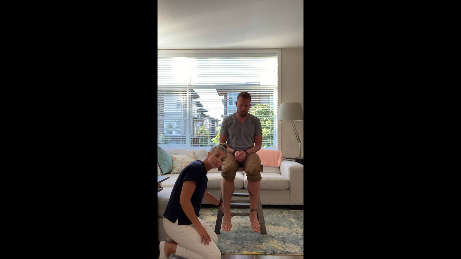
- Active Mobility – The right femoral head was palpated during active non-weight bearing flexion and extension of the right hip in supine. Right hip active range of motion was full and pain free, the femoral head remained centered throughout.
Diane’s question:
In the standing postural screen the right femoral head was noted to be anteriorly translated relative to the right acetabulum. Here, you note that the right femoral head is centered and remains centered through the full range of active flexion and extension. What does the clinical reasoning of these two findings tell you about this right hip joint?
Kjersti’s response:
The difference in the alignment of the femoral head in standing (anterior in relation to the acetabulum) vs supine (centered in the acetabulum) indicates first the vector acting on the hip joint is not articular because the femoral head position became neutral in supine. Also because optimal active control of the hip is possible in supine, we know so the motor control strategy used in standing differs from supine and that standing poses challenges to the right hip that result in the compensation strategy of the anterior shear of the femoral head in the acetabulum. This could mean the right hip positioning in standing is due to ‘compensation’ for another body region (non-optimal alignment of the right hip is the result of non-optimal control or alignment of another body region). Or that in standing where the motor control demands on the hip due to weight bearing increase, the patient can no longer control the position of the femoral head in the acetabulum.
Diane’s comment:
Excellent deductive reasoning!
- Passive Mobility – With Mr. M in supine, right hip passive range of motion was tested to determine the degree of functional range. Functional range of motion is defined as the range at which the femoral head stays centered in the acetabulum. The femoral head was taken from 30 degrees of flexion into full extension. It was found that the femoral head was able to remain centered throughout this movement.
- Active Control – During right one leg standing, Mr. M, started with the right hip slightly anterior in the acetabulum, and as loading on the right leg progressed, it translated further forward, demonstrating impairment in active control.
- Passive Control – Not tested, as the patient’s story did not suggest a need for it.
Vector Analysis – Because the femoral head remained centered in the supine position throughout range, vector analysis was completed in standing where the femoral head demonstrated non-optimal alignment.
Diane’s comment:
Perfect reasoning to do this in standing!
From the standing position, the anterior aspect of the femoral head was palpated, already slightly anterior in relation to the right acetabulum (suboptimal alignment). Mr. M was asked to flex both knees slightly until the femoral head became centered in the acetabulum, about 5 degrees of hip flexion. Then the femoral head was palpated as Mr. M returned to the standing position. The femoral head was felt to translate first anteriorly, then adduct slightly and then externally rotate. Then from a standing position, the femoral head was centered in the acetabulum, by first supporting the ischium posteriorly, and then repositioning the head of the femur posteriorly in the acetabulum. Upon release of this position, first a strong anterior medial vector down the midportion of the thigh was felt. This was palpated to be the adductor longus. Then a secondary vector that pulled the femur into extension and external rotation was felt. This was palpated to be the gluteus medius.
Diane’s question:
Which part of the gluteus medius, when acting alone, is responsible for extension and external rotation, the anterior or posterior? What does the other part do? How does this knowledge help you in deciding how to treat this neural vector?
Kjersti’s response:
The posterior fibers of the gluteus medius originate at the posterior aspect of the iliac crest and pass anteriorly and inferiorly to the greater trochanter of the femur. Thus the posterior fibers of gluteus medius, when acting in isolation contribute more to abduction, extension and external rotation while the anterior fibers abduct, flex and internally rotate the femur. Knowing that the posterior fibers of the gluteus medius act to extend and externally rotate the hip more, I would be interested in palpating these specific fibers for treatment of the vectors felt at the right hip.
Hypothesis
From this assessment, it is hypothesized that Mr. M’s meaningful complaint of adductor tightness when running long distances was related to impairments of alignment, biomechanics and control of his feet as well as a secondary driver coming from the right hip.
It was found that correcting both the right and left foot was necessary first in standing to restore the alignment and control of multiple thoracic rings during right OLS (primarily TR 7 translating/left rotating right and TR 8 translating right/rotating left). These improvements in the thorax didn’t result in complete alignment of the right hip, nor right SIJ control. Through further analysis it was found that correcting the alignment of the right hip restored control of the right SIJ in right OLS. Thus the right hip was a secondary driver. No vectors were found in passive mobility testing of the right hip, therefore the primary issue with the right hip is one of motor control.
Initial Treatment Session
Release
Right foot –
- Transverse friction massage over the tendons of the tarsal tunnel, to release the short medial vector between the calcaneus and the talus.
- Gastrocnemius and soleus muscle released with dry needling.
- A distraction force from the calcaneus to the talus was applied to release a medial ST joint capsule vector with some added friction massage over the tendons in the tarsal tunnel.
- The calcaneus was taken into an anterolateral glide on the talus to improve the restricted medial rotation of the left ST joint.
Diane’s comment:
Joint capsule vectors are short, tendon vectors with adhesions to the underlying capsule can also be short. How can a tendon limit the arthrokinematics of the joint? How did you differentiate that your friction massage was impacting the tendons and not the underlying joint capsule?
Kjersti’s response:
I was able to differentiate the structures I was affecting with the friction massage by palpating the tendons of tibialus posterior, flexor hallucis longus and flexor digitorium longus just under the medial malleolus and directing the friction massage here. I was able to differentiate the tendons from the joint capsule by altering the depth of tissue palpation. After I felt the more superficial vectors of tendon adhesions were released. I also released the medial STJ joint capsule by distracting the calcaneus from the talus in an inferior and anterolateral direction. In the explanation above I see that I combined the two techniques but the tendon release was done first followed by a joint capsule release. A tendon can limit the arthrokinematics of a joint if the tendon crossed two joints and is tight or tonic enough to compress the two joint surfaces together thus reducing the free movement of the joint surfaces.
Align
- Right hip – a cue to ‘soften at the front of the hip crease’ was given. An align cue was most appropriate for Mr. M because through vector analysis it was found the right hip was only translating anterior under load in standing, and the active/passive mobility and control were normal in supine. Release of the right hip was not required, and it was hypothesized the anterior shift of the right femoral head was due to a motor control deficit. With the alignment cue given, the right hip position was restored in standing.
- Left and right foot: Full alignment of the feet was not restored after the first session, so Mr. M was taught how to center the talus and calcaneus as much as possible in the standing position.
Connect
- A connect cue was given as follows, “imagine an elastic coming from the inside of the right foot (navicular tubercle) pulling up to the inner thigh”.
Diane’s question:
Why was a connect cue necessary for the loaded right foot?
Kjersti’s response:
The motor control strategy for holding the right foot in the corrected position when load was taken on the right foot was not sufficient, the foot muscles still buckled without the orthotic in place for additional support. So a connect cue was used to help support the foot in the corrected position by adding a layer of conscious control in asking the patient to actively engage stabilizing muscles. The connect cue was most effective when the patient was asked to imagine a line from the inside of the midfoot drawing up to the right hip.
Move
Maintain this alignment and connection while shifting the weight onto the right, stopping when alignment is no longer maintained.
Home practice:
- Self-release to the medial aspect of the gastrocnemius and soleus bilaterally using a lacrosse ball.
- Practice weight shift to the right foot with align cues for feet and connect cue from right foot to right hip.
- Continue use of orthotics for weight bearing activity.
Followup treatment sessions
Follow up treatment session #1
It was reasoned that the orthotics provided the additional mechanical support and facilitated better motor control strategies required to maintain the foot correction in the task. Even with the foot correction and the orthotics, the foot alignment wasn’t completely optimal on the right or the left so it was clear additional release and muscle training was required. The first follow up treatment session focused on further release of articular system impairments of the right subtalar joint (restricted anterolateral glide of the calcaneus on the talus, posterior joint) and the left and right talocrural joint (posterior glide of the talus in the mortice).
Reassessment then determined that control at the right SIJ was restored because the right hip was now centered. After the initial assessment, it was thought that the right hip was a secondary driver, as correcting the feet alone did not restore biomechanics and control of the right hip and SIJ. However once further release of both the feet was completed, the alignment of the right hip was also restored. Mr. M was still asked to add a connect cue from the right medial foot to the right hip before loading onto the right leg, because this cue still helped him to engage stabilizing muscles in the foot and lower leg. The left foot alignment was also not completely corrected, so the connect cue from the right foot to the R hip was needed to allow for the driver that wasn’t completely optimal.
In addition a motor control assessment of the foot intrinsic muscles was completed. The findings were as follows:
Right foot
- Flexor Hallucis Brevis: Grade 2
- Abductor Hallucis Brevis: Grade 1
- Abductor digiti minimi: Grade 2
- Flexor digitorum brevis: Grade 1
Left Foot
- Flexor Hallucis Brevis: Grade 1
- Abductor Hallucis Brevis: Grade 0
- Abductor digiti minimi: Grade 3
- Flexor digitorum brevis: Grade 1
For the muscles which tested in the 0/1 Grade range, the first step of motor control training was Stage 1 Cognitive retraining with the goal of waking up the muscle. Mr. M was asked to palpate the specific muscle and to avoid using long toe flexors or extensors to help. The goal was an isometric contraction for 5 seconds, 10 repetitions each to start. Once isolated activation of the muscle was possible Mr. M would be ready to progress to the next stage.
Next Steps
As further treatment sessions would progress, Mr. M would continue to build capacity of the foot intrinsics improving motor control in the feet. The right foot responded quickly with release, align and control principles however, the left foot still appeared in a position of subtalar pronation and talar tilt in standing. Mr. M noted that on longer runs, his adductor tightness had improved but it was his left ankle that was still bothering him. Occasionally he would notice an increase in left ankle crepitus and calf muscle tightness.
The control of the left foot was such that the left talus would fall into plantarflexion, adduction and lateral tilting in weight bearing even in orthotics and after release work because the foot muscles were not strong enough to maintain optimal positioning when in weightbearing. Mr. M was able to actively hold his foot when manually corrected in double leg stance but the left talus continued plantarflex, adduct and laterally tilt through single limb stance on the left. It was thought that supporting the talus would allow for more optimal control of the foot in weight bearing.
Mr. M was asked to book in to see a Pedorthist to reassess his orthotics.
The assessment from the Pedorthist resulted in new orthotics with increased support bilaterally for the talus and navicular. The left foot required an increased posting to support the left talus and navicular due to the more marked talar plantarflexion, adduction and lateral tilting on this side. Following this, the alignment and control of both feet were much improved in weight bearing and single limb stance. Mr. M was asked to continue release of the gastrocnemius, soleus muscles as well as to continue his foot intrinsic strengthening work.
The following stages for motor control training would continue as follows:
Stage 2: Associative – build capacity for the new integrated strategy – i.e. add load and movement such as, plantarflexion/dorsiflexion of the ankle while maintaining flexor digitorum brevis and flexor hallucis brevis activation (foot on a soft ball). Alternatively, maintaining abduction of the 5th or 1st toe with plantarflexion/dorsiflexion with the foot on a soft ball.
Stage 3: Automatic – integrate into meaningful tasks – i.e. right one leg standing, right single leg squat, or right one leg hop.
Due to the advanced loading task of running and the motor control deficit remaining in Mr. M’s feet, he was asked to follow up periodically for guidance and exercise progressions. With his feet well controlled, Mr. M will be able to enjoy a long healthy running career without excessive loading of his feet and hips.
Case Study Author

ISM Series Graduate ISM Certified Practitioner
15303 31st Ave #102, Surrey, BC V3Z 6X2
Clinical Mentorship in the Integrated Systems Model
Join Diane, and her team of highly skilled assistants, on this mentorship journey and immerse yourself in a series of education opportunities that will improve your clinical efficacy for treating the whole person using the updated Integrated Systems Model.
{"first_class":"1","title":"Heading","show_title":"0","post_type":"course","taxonomy":"","term":"","post_ids":"3346","course_style":"recent","featured_style":"custom_block_1","masonry":"0","grid_columns":"clear1 col-md-12","column_width":"200","gutter":"30","grid_number":"1","infinite":"0","pagination":"0","grid_excerpt_length":"100","grid_lightbox":"0","grid_link":"0","css_class":"block-small-thumb","container_css":"","custom_css":""}
We will come together for 3 sessions of 4 (4.5) days over a period of 6-8 months with lots of practical/clinical time to focus on acquiring the skills and clinical reasoning to put the ISM model into practice. Hours of online lecture and reading material and 12 hours of in-person lecture are...
More Info