Story
CR came to our clinic self-referred on 5/22/2019. She is a 33-year-old physical therapist from another outpatient facility. The reason for this visit is constant sacrococcygeal pain rated as a constant 5/10 since a fall down a flight of stairs while on a cruise ship two months prior. She slipped on a step and landed on her rear end and then slid down 3-5 more steps on her buttocks. She was able to get up and walk after the fall and enjoy the rest of the cruise despite the pain. She did not need medical attention following the fall. Since the fall her pain has limited her ability to sit comfortably.
Diane’s question:
When did she note the pain start in sitting? Immediately after the fall, within 24 hours, week later? What would the timeline of pain onset suggest with respect to etiology?
Paul's response:
CR had been partying all night when the fall occurred so she did not feel the full effects of the fall until the following day. She did have sacrococcygeal pain the next morning but was expecting it to resolve over the next several days. A fall can cause significant trauma including fracture (to the hip, pelvis, sacrum, vertebral body or end plate) disc herniation, muscular or ligamentous sprain and contusions. Immediate pain may indicate severe trauma. In this case she was able to continue to enjoy the rest of the cruise despite her symptoms which tends to rule out a fracture. Her symptoms remained basically unchanged and then progressed to include the leg after about four weeks. This could be caused by an annular tear with progressive migration of the nucleus or by the contraction of a healing scar in a muscle or ligament during the fibroblastic phase of healing stimulating nociception.
This is affecting her ability to sit when driving to work, sit at work doing her notes or sitting to eat. Over the past 4 weeks her pain is getting worse and she has begun experiencing increased right (R) calf and hamstring tightness with prolonged sitting and first thing in the morning. One of her coworkers tried to treat her with a muscle energy technique to the pelvis that normally helps her when she “is out” but this did not affect the coccygeal pain or her leg symptoms.
Past Medical History
She has chronic neck and back pain that occasionally flare up. She will occasionally get neck pain that radiates to the right shoulder and down the arm with intermittent numbness and tingling into the R upper extremity (UE) (she was unable to describe exactly in what distribution the paresthesia occurs). Her low back pain and right hamstring tightness will flare up any time she tries to exercise, is more active, or plays with her kids in the yard. She is not a runner but if she tries to run, she may get numbness and tingling in both feet. She also has chronic tightness in the right hamstring and R calf. She feels all these symptoms began when she played soccer in high school and had her legs taken out from her by an opposing player landing directly on her “tail bone”. She fractured her R ankle at age 12 and had her R knee scoped in 2003 (no specific trauma recalled just repetitive from soccer and this was before the traumatic fall onto the sacrum). She has two children age 7 and 4. She felt great when she was pregnant stating it improved her chronic neck and back symptoms.
Diane’s question:
CR seems to have had symptoms in her right hamstring and calf that pre-date this current event. Was there anything in her story that led you to belief there was any sensitization of the nervous system at this point?
Paul's response:
Yes, I believe she had a sensitized nervous system. She was able to describe her initial fall on the “tail bone” during the soccer match in great detail like it was yesterday. She carried a lot of anger and resentment to this injury and to the opposing player. She has diffuse symptoms with intermittent paresthesia that is easily exacerbated and worsened with activity.
Diane’s question:
Nociceptive pain is worsened with activity and eases with rest. What is the difference between nociplastic pain (not neuropathic or nociceptive) or what is also known as central/peripheral sensitization in pain behavior?
Paul's response:
Central sensitization presents with the following behaviors: Pain that persist beyond the known healing time of the specific tissue involved. Pain that spreads beyond the boundary of the initial injury. Pain that worsens over time. Pain that occurs with movements that “should not be painful“. Pain that is unpredictable and varies day to day ( Explain Pain – 2003 Butler & Mosely pages 82-83) “Central sensitization refers to reduced pain thresholds to non-noxious stimuli and increased pain evoked by noxious stimuli in the absence of peripheral sensitization” (Explain Pain Supercharged Butler & Mosely 2017 page 72).
Peripheral Sensitization is the hypersensitivity caused by the release of local inflammatory mediators released by damaged tissue. This would be the stimulation of nociception from a non-traumatizing stimulus to an injured tissue. An example would be light touch causing 8/10 pain on an acutely sprained ankle which is resting at a pain level of 2/10.
Nociplastic pain is a term that was described in the article “Do we need a third mechanistic descriptor for chronic pain states?” in the journal PAIN: July 2016 – Volume 157 – Issue 7 P 1382-1386. The article States that “neuropathic pain is a clinical description (and not a diagnosis), which requires a demonstrable lesion or disease that satisfies established neurological diagnostic criteria”. It also states that nociceptive pain is “pain that arises from actual or threatened damage to nonneural tissue and is due to the activation of nociceptors” in a “normally functioning somatosensory nervous system”. The article proposes that nociplastic pain is a term to “describe pain states characterized by clinical and psychophysical findings that suggest altered nociception, despite there being no clear evidence of actual or threatened tissue damage causing the activation of nociceptors or evidence for disease or lesion of the somatosensory system causing chronic pain”. The term is not a diagnosis nor is it a synonym for central sensitization or idiopathic pain. It states that patients who suffer from nociplastic pain “typically respond better to centrally than peripherally targeted therapies”. Unfortunately, they do not go into further detail of what this therapy would entail.
Diane’s comment:
Great summary of current terminology for pain mechanisms, thanks Paul!
Meaningful Complaint
Sacrococcygeal pain
Cognitive Beliefs
CR believes that her sacrum and coccyx are out of alignment due to her fall down the stairs. She is hoping I will be able to manipulate her pelvis to alleviate her symptoms.
Diane’s question:
What are your thoughts on SIJ or sacrococcygeal fixation? Is it possible for the joint ‘to go out’ – any evidence to support your belief? How did you address her cognitive belief on this topic and her expectation of you for this session?
Paul's response:
This is a great debate question when it comes to the spine. It is obvious that the peripheral joints can dislocate or “ go out” and then be then be put back in. I don’t like the term “go out“ when it comes to the spine and the current evidence of the neuroscience of pain evidence (e.g. collectively described in the book Explain Pain Supercharged Butler & Moseley (2017) would agree. I do believe the SIJ and sacrococcygeal joint can fixate or get stuck and need to be mobilized or manipulated to improve the alignment and biomechanics of the joint (ABC: alignment, biomechanics and control). Clinically, I have had too many people respond to this treatment when appropriate to make me think otherwise.
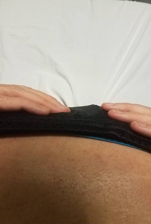
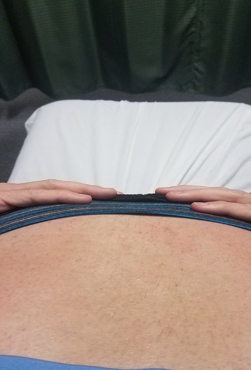
As you know dysfunctions never show up as well on pictures and this looked even worse in real life. It is an example of a fixation pre and post manipulation and is part of my evidence to support my belief. The following picture is of a client who presented with a left vertical sacral torsion. It is also debatable whether this dysfunction is possible. This is a non physiological fixation. The sacrum is incongruent with the pelvis along the vertical axis. This is a painful and debilitating dysfunction that in my experience will only resolve with a high velocity low amplitude thrust.

Left vertical sacral torsion Correction of vertical torsion post HVLAT I also believe the sacrococcygeal joint can fixate, usually in flexion with or without components of side bending and rotation. The coccyx just does not have any muscle that can help return it to neutral once fixated in flexion. Since this joint is a small syndesmosis it should respond to non thrust manual mobilization and release.
Diane’s comment:
Many researchers, and clinicians, would disagree that your experience is evidence to support the hypothesis that the SIJ or SCJ was truly ‘fixated’ or ‘stuck’. I have had the same clinical experience and wonder if the improvement in A, B, Cs secondary to a high velocity, low amplitude (HVLA) technique is more of a ‘neural reset’ and change in muscle tone that is causing excessive compression, as opposed to a change in the joints alignment. I agree that there is a condition in the pelvis/spine that responds favourably to HVLA technique but am not sure I understand the underlying mechanism of how it works! Great pictures of the condition though, and it is very debilitating and I doubt ever included in research studies.
In CR’s case, I knew she did not have a fixation of her SI joints as they unlocked during single leg stance. To address her cognitive belief that she may need a manipulation for this condition I specifically assessed her pelvis via palpation, SI joint pain provocation test, and SI joint passive mobility tests. In this case I believe these tests were warranted to help find the pain generator in the ischiococcygeus and to address her cognitive believe that her SI joint may be a source of pain and need manipulation.
Diane’s comment:
I’m not sure I would use a manipulation technique in this case if a different manual technique, or better yet, if a home release technique, would be effective. This would empower more self-management of her condition. Then again, if the manipulation was necessary then I wouldn’t withhold a treatment that would be beneficial; however, it may not address her cognitive belief of the joint ‘being out’.
Meaningful Tasks
Sitting
Screening Tasks
Standing postural screen and the squat
I only have 45 min for evaluations and 30 minutes for follow ups. I purposely limited my evaluation to two screening tasks as her medical history took longer than expected. She is coming today specifically for the unresolving sacrococcygeal pain that started two months ago from her fall but I could tell she would like a lot of her other issues addressed as well. She has had the prior symptoms for a long time and is not coming specifically for these symptoms although they are quite bothersome and concerning to her.
Diane’s comment:
Are you familiar with Sueki, Cleland & Wainner’s (2007-2013) Regional Interdependence Model? This model, much like the Integrated Systems Model, suggests that impairments that are remote from the site of symptoms may be contributing factors to the clinical picture. Therefore, although she is most concerned about the pain around the sacrococcygeal joint, I suspect you may find relationships between sites of impairment that are a factor here, let’s see!
Paul's comment:
I graduated in 1994 and at some point took Jim Meadows part A prep course. In his Module 3 Biomechanics: Clinical Application, he had a section on Quadrant Syndromes. So I always have treated in a “regional or integrated fashion”, not in an ISM way but in a facilitated segmental way. I was so shocked when Josh Cleland and his group came out with the Regional Interdependence Model as if it was a new Model when the Canadian System had been practicing this way for years.
And I do believe I will have distal impairment affecting the sacrococcygeal pain. I did end up treating it but I may have sped up the resolution had I addressed it sooner.
Standing postural screen
Functional unit # 1 (FU # 1 – 3rd thoracic ring to the hips)
Pelvis: The pelvis would change from a L TPR (left transverse plane rotation) with left IPT (congruent intrapelvic torsion) to a neutral pelvic alignment every 6 to 7 seconds. In my assessment of the IPT it was noted that both SI joints were locked in standing and they both unlocked or gave way in early single stance. There was an increase in tone in the right hamstring and right longissimus. Coccyx was not palpated in standing.
Diane's question:
Can you explain further what you mean biomechanically (osteokinematics between the sacrum and innominates) by the terms ‘locked’ and ‘unlocked’ since this term is not commonly used for other joints in the body.
Paul's response:
A locked SI joint is when the sacrum is nutated and the Ilia is in posterior rotation in relation to the sacrum. This is the closed pack position for the SI joint. This is where the SI joint is most stable and is efficient to transfer load. When the SI is unlocked, the ilia is in anterior rotation in relation to the sacrum which is said to be in couternutation. This would be a case of faulty alignment, biomechanics and control during loaded tasks.
Diane's question:
The increased tone noted in the right hamstring and right longissimus is significant in the presence of an ‘unlocked’ SIJ in single leg loading (early, mid or late). Why? Why would the brain choose to increase the recruitment of these two muscles?
Paul's response:
The longissimus attaches to the sacral base and assists in nutating the sacrum. The hamstring with its attachment to the ischial tuberosity (as well as the sacrotuberous ligment) will assist in posteriorly rotating the innominate. These muscles will often work together as a force couple to stabilize an unlocked SI. This would be the brain’s attempt to induce force closure due to faulty form closure.
Hips: The hips remained congruent with the TPR but the R appeared to be slightly more anterior compared to the L.
Diane's question:
A congruent hip position finding with a LTPR/LIPT of the pelvis is that the right hip would be more anterior to the L. Can you explain further what is meant above by “R appeared to be slightly more anterior compared to the L”. Shouldn’t it be if they hips were congruent to the pelvis? Not sure I understand this point.
Paul's response:
Yes, I agree that statement is not clear. My difficulty in giving a clear-cut statement on position was that her R hip was following the change in pelvic TPR from neutral to a L TPR. Sometimes the hip felt significantly anterior (in relation to the left hip) which is congruent with the TPR and then would follow the pelvis back to neutral changing my palpatory relationship to the left hip. Changing the focus of my palpation of the hip in relation to the ipsilateral acetabular fossa as opposed to the contralateral femoral head may have clarified the situation.
Diane's response:
Got it!
Thoracic Ring (TR):
- TR 10 translated right/rotated left
- TR 8 translated right/rotated left
- TR 7 translated left/rotated right
- TR 3 translated left/rotated right
Diane's question:
Which thoracic rings’ starting position are incongruent to that of the pelvis and why is it significant in an ISM approach to note incongruent body regions going forward?
Paul's response:
The pelvis TPR is to the left, so all rings that are translated right and rotated left are congruent with the pelvis. The thoracic rings that are incongruent are TR 7 and TR 3 because they are translated left and rotated right. Noting the incongruencies at this point in the assessment gives us an intra-regional and inter-regional starting position or reference point. Making corrections at these levels allows us to note the regional changes for better or worse in a meaningful task to assist in finding the driver for the task. The incongruencies may be sites of impairment or locations of failed ABC’s (alignment, biomechanics and control) depending on the meaningful task being assessed.
Functional unit # 2 (FU #2 – second thoracic ring to the cranium)
Upper thorax:
- TR2 oscillates into R and L rotation
Neck:
- C6 translated R rotated L
- C5 to C2 translated L rotated R
- Cranium: L ICT with congruent sphenoid
Diane's question:
What significance does an ‘oscillating second thoracic ring’ and pelvis (noted above) have for planning further assessment testing?
Paul's response:
The oscillating second ring and pelvis are signs that the nervous system is lacking mobility in its present state. The body is trying to find a position of ease for the nervous system. If I am able to find a correction in alignment that resolves the oscillation this would indicate that the nervous system is under less tension, with the improved alignment. This would then identify the driver (impaired body region for further assessment) for this impairment. Further assessment (not just vector analysis) of the driver would indicate what system impairments require treatment.
If all corrections worsen the alignment of another region, this indicates that the dura and perineural tissue is the primary system impairment requiring treatment and release before we can address the alignment, biomechanics and control of the musculoskeletal system for the meaningful task.
Diane's question:
Please identify the segments in FU#2 that are incongruent with each other and why this is important to note.
Paul's response:
C5 to C2 translated L rotated R are incongruent to C6 and the cranium and the pelvis. These two cervical segments should be corrected to note the impact on alignment of the other segments within functional unit #2 to determine the within unit driver.
Diane:
Perfect! So instead of just listing a whole bunch of non-neutral findings, what we are looking for in this postural screen is the incongruent sites of rotation in order to plan our next assessment step. If standing was the meaningful task, corrections would be done in standing. Since standing is NOT the meaningful task, these incongruent sites are assessed in the next screening task and those that don’t perform optimally and remain incongruent are corrected and the response assessed. This expedites and facilitates the speed with which the driver, or drivers, can be found.
Paul's comment:
In the course we spent so much time learning to find the incongruencies in standing as a means to “find a finding” that initially at the clinic I spent way too much time trying to find a driver in standing even though it may not be the task. We can only come to a “wow” moment when we induce a change in a meaningful task.
Diane's comment:
Well said.
Functional unit #3 (FU#3 – extremities)
Feet: The feet findings are subtle as there is no significant malalignment. That being said the calcaneus are mildly supinated bilaterally R > L and she tends to weight bear slightly on the outside of the right calcaneus. This is incongruent with the LTPR of the pelvis (right foot should pronate). The right talus is slightly anterior in the mortice. In a supinated foot the talus is dorsiflexed and abducted in relation to the calcaneus. So this is an incongruency within the hind foot (between the subtalar and talocrural joints).
Diane's comment:
The right foot is incongruent with the position of the pelvis but the right talus and calcaneus are congruent with a supinated hindfoot.
There is a mild “kink “ at the talonavicular joint, which may indicate slight ER of the navicular possibly from increase in tone in the tibialis posterior. In a supinated foot the navicular should internally rotate relative to the navicular and the cuboid. The forefoot is unremarkable, toes are straight and no malalignment noted.
All individual corrections worsened and or did not improve the alignment within and in between functional units 1, 2,and 3. Passive listening to C6 gave a very strong caudal dural vector.
Driver: Dural and perineural system
Diane’s comment and question:
If this is a standing postural screen to note the starting position of segments then finding a driver in standing is not a requirement. If standing is a meaningful task (and it isn’t – sitting is) then you would find the driver in standing. However, there is one finding in this standing screen that suggests a specific underlying system impairment that would be validated with trying to find a driver in standing. And that system impairment is altered dural/neural mobility, which you have noted above. The dura and neural system isn’t a driver – why? What is the ISM definition of a driver? Once answered we can amend the initial title of your report (Pelvic and Neural System Impairment Co Drivers)!
Paul's response:
The dura and neural system is not a driver, it is an impairment and classified as a neural system impairment since the dural and perineurium lines the nerves and are part of the neural system. A driver is a body region such as the pelvis, neck or ankle that demonstrates sub-optimal alignment, biomechanics and/or control and when corrected positively impacts other areas of impairment during the client’s meaningful task. Additionally, the correction has a positive impact on the client’s experience during the task. When corrected, a primary driver will result in complete correction of all other impaired regions for the task. You can also have a primary driver with a secondary driver. This is where you have the primary driver correcting most impaired regions and partial correction in another. You can also have co-drivers. Co-driver type 1 is when you make a correction and it worsens another area and vice versa. You need to correct both areas to improve the alignment, biomechanics and/or control for the task. Co-driver type 2 is when correction of either area improves the other body region for both the task and the client’s experience.
CR’s main system impairment is altered dural and neural mobility. The first clue is from her story. She has a history of R UE paresthesia, Right lower extremity leg pain and tightness with numbness in the feet with running. Symptoms are easily exacerbated with exercises and household tasks. She has now added an acute pelvic injury onto this chronic system impairment.
From my objective findings in standing there is an oscillation of position of the second thoracic ring. There are no other vectors besides the dural/neural system that would cause this phenomenon. This is also occurring at the pelvis with the L IPT moving back to neutral.
I would like to change the title of my report to “A Neural System Impairment in a Case of Prolonged Sacrococcygeal Pain.”
Diane's comment:
Perfect title change – and I added “with No MSK Driver” for clarity!
Squat
The squat is a useful screening task as this is the movement required to reach the sitting position which is CR’s meaningful task.
Diane's question:
When you ask someone to squat, they may do something totally different than the strategy they use to sit in a chair. How did you ask Mrs. CR to squat? i.e. What words do you use to simulate sitting in a chair vs a deadlift?
Paul's response:
I actually just asked CR to squat. She performed the squat as if she was going to exercise and do a body weight squat. For the initial evaluation I thought she performed the squat with enough ankle, knee and hip flexion to give me the information I needed for the meaningful task of sitting. If she did something totally wrong, I would have corrected her and asked her to squat as if to sit back in a chair. If someone was having pain with a deadlift type motion I would have then have them squat down as if to pick up a duffel bag or bag of groceries.
Motor recruitment patterns for sitting are variable and task specific not just for the type of surface you are sitting on but how you approach it – from the side, from the diagonal, from the right or the left. You will sit differently if the chair is at a table that you have to pull out verses one that is already ¾ pulled out at the table and you just have to slide in. Sitting in a car is very specific to the vehicle itself. If I needed to get more specific during her course of treatment, I would asked for her to show me how she sits down on a particular surface and perform my meaningful task assessment.
Functional unit #1
Pelvis: She started with the pelvis in neutral and then at 1/4 squat the pelvis rotated to the right and then the RSI unlocked.
Hips: Right hip stayed anterior and moved further anterior as the R SI unlocked. There was over activation of hip abductors bilaterally as she centered her patella over her second toe and then strongly activated her hip abductors and hip external rotators.
Diane's question:
Is the transverse plane rotation of the pelvis congruent or incongruent with the biomechanics of the right hip in this task?
Paul's response:
The R TRP is incongruent.
Diane's question:
Why is this significant?
Paul's response:
This is a site of impairment and we need to assess if centering the hip will affect the R TPR or SI unlocking.
Diane's comment:
That would be true if the right hip was congruent or incongruent with the RTPR of the pelvis. The significance of the incongruence between the right hip and the pelvis is that there are at least two things going on – one impairment is causing the pelvis to rotate to the right and the other is causing the right hip to remain anterior. Make sense?
Paul's comment:
Yes.
Thorax: TR8 got worse during the squat
Diane's question:
In what way – how did TR 8 get worse? What happened to TR 10, 7 and 3?
Paul's response:
TR8 went further into right translation and left rotation as did TR10 but not as significantly. TR7 and TR3 remained translated right and rotated left
Diane's question:
What is your FU#1 driver?
Paul's response:
I could not find a driver for FU #1. All corrections failed to improve the other areas of impairment and did not improve the performance, nor her experience of the squat. That being said I was still interested in the Right hip and TR8 as they were significantly malaligned.
Diane's comment:
BUT there was an oscillating pelvis and TR2 in standing – did not that finding alert you to the possibility of adverse dural/neural tension and thus help to explain why you couldn’t find a driver in FU#1?
Paul's comment:
Absolutely. From her history of multiple thoracic rings with dysfunction an oscillating pelvis and TR2, I was not surprised that I did not find a driver in FU#1. It is important at this point to continue with our assessment to find a driver and relationships to not get biased into thinking it is strictly a dural /neural tension condition. We need to make sure that these signs are not being caused by altered body alignment restricting the dural mobility.
Diane's comment:
Excellent point – stay on track, this is what an ISM approach helps us to do.
Functional unit # 2
Upper thorax: TR2’s behavior during the squat remained consistently inconsistent. At times it appeared rotated R and then rotated left
Neck: C6 increased in R translation and left rotation. C5 to C2 remained translated L rotated R
Cranium: L ICT
Driver: I could not find a driver in FU#2. All corrections performed increased impairments in the other areas and did not improve the performance of the squat. When C6 was corrected and released, a strong caudal vector was noted.
Priority between FU#1 and FU#2 drivers: No priority between FU#1 and FU#2 has been found. I feel there is a vector that bridges the functional units.
Functional unit #3
Feet: Both feet remained stiff and ridged in slight supination and the tibias externally rotated. Decrease in right ankle DF
Diane's question:
What is the relationship between the neuromuscular strategy noted above in her hips and this pattern of osteokinematics you are noting in her feet?
Paul's response:
The osteokinematics at the feet is congruent with the over activation of hip abductors and hip external rotators but not biomechanically efficient for the squat task. There should be conjunct IR of the tibia at the initiation of the squat and slight pronation of the foot. There was a lack of ankle DF on the right which then became an abnormal axis of rotation which may be causing the pelvis to go into a R TPR.
Priority of all unit drivers: I tried correcting the hindfoot during the squat motion but was unable to maintain the correction during the squat. I thought she would need a lot of release via verbal cueing to get her to change her motor pattern at the hips to allow me to maintain the correction. There was also a visible limitation on ankle DF that could be articular, neural or myofascial in need of vector analysis. With her history, I felt that the ankle limitation is a chronic dysfunction that she had prior to her fall and recent symptoms. In hindsight, I should have revisited the ankle much sooner in the treatment plan than I did.
Being mindful of my time constraints, I wanted to confirm my hypothesis of a dural/neural system impairment by assessing her slump test. If this was negative, I would have missed a driver and would have to reassess my functional units.
Diane’s comment:
Given her long history of right hamstring and calf tightness as well as the fracture of her right ankle, the posture of her right foot may be related to lack of mobility of the tibial nerve medial to the tarsal tunnel or lack of articular mobility at the talocrural joint. Further assessment of the dural/neural system and the foot would add clarity to this hypothesis, at a later date!
Paul's comment:
I agree.
Neurodynamic mobility test:
Seated slump was positive for lack of dural mobility bilaterally R> L . Mobility of the dura and neural system increased distally when the neck was extended. C6 and T8 increased in right translation and left rotation during slump. I did not palpate the other segments. There was a reproduction of her R leg symptoms and she was unsure if it affected her coccyx symptoms.
Diane’s question:
What was the impact of correcting any site of impairment on the mobility of the dura/neural system?
Paul's response:
C6 and T8 increased in right translation and left rotation most likely to decrease tension on the dural system. This is the equivalent of neck extension during the slump test. I did not assess the impact of any corrections. Any corrections during the slump should increase both the number of sites of impairment and/or worsen those already noted in other regions and increase the dural tension signs. This would be the equivalent of adding neck flexion to the slump.
Diane's question:
What we are trying to differentiate is
- whether the adverse dural/neural mobility is causing the body to shorten (thoracic rings and cervical segments to translate/rotate, coccyx to flex, cranium to twist (ICT)) or
- whether the multiple twists/rotations etc in her musculoskeletal system (MSK) are ‘taking up all the slack’, or available ability of the dural and neural system to elongate with body movement.
Two very different clinical scenarios. The first requires intervention to the dura and nervous system, the second requires unwinding the MSK system. From all your findings so far, which dural/neural system impairment does Ms. CR have and why?
Paul's response:
My lack of ability to find a driver by correcting the areas of dysfunction as well as the oscillation of TR2 and the pelvis lead me to believe that it was the body trying to shorten to take strain off the dural/neural system and would need treatment prior to addressing the ABC’s of the musculoskeletal system. I did treat the pelvis on my first visit contrary to my ISM findings of a primary neural/dural system impairment. I will elaborate on that on the question in a later section.
Diane's question:
At this point of the first draft of your case report I didn’t understand why you have next assessed the pelvis, since no correction of the pelvis was found to improve either the squat task, nor any other site of impairment? What made you decide to go to the pelvis for further assessment at this point? Which of your answers above would then make assessing the pelvis next, which you have done, make sense?
Paul’s initial response:
Good question Diane. I had a few reasons to go to the SIJ and pelvis at this point. There are points in an assessment where you can move into specific tests that do not pertain to the driver. This is done when you want to determine the location of a pain generator or to get information that addresses a cognitive belief. I wanted to assess her SI and pelvis in a manner that would satisfy her cognitive belief that she needed her SIJ manipulated. She also had a trauma to the pelvis so I wanted to perform the pain provocation test just to make sure she did not have an SI joint inflammatory response with an atypical pain distribution. The passive mobility tests were a smooth transition from the stress test and are a confirmation test to my palpation assessment.
Further Assessment of the Drivers, including Vector Analysis
Further Tests of the Pelvis
Pain provocation tests of the sacroiliac joint were negative
Active Mobility: The movement at the right and left SI joints where symmetrical during standing ipsilateral hip flexion.
Passive mobility test of the right sacroiliac joint: the arthrokinematics anteroposterior glide revealed a restriction of motion at all three components of the R SI joint. Passive listening on release of the glide revealed a medial vector at the inferior part of the right SIJ. This indicates possible overactivity of the ischiococcygeus and piriformis which can be confirmed through palpation.
Active control: R and left SI joints give way on single leg stance. During a squat the R SI joint unlocks at ¼ squat.
Passive stability test: SI joint stability test is not performed due to the restriction noted in the passive stability test. If the passive mobility test was not restricted and demonstrated expected motion or motion that is perceived as excessive then stability testing of the passive restraint system would be indicated.
Verbal consent was obtained for external palpation of the pelvis and pelvic floor musculature following a review of pelvic anatomy with the client.
Palpation of the ischiococcygeus may be performed in supine or prone. I chose the prone position as I also wanted to palpate the coccyx and sacrococcygeal joint which I find easier to do in prone.
Coccyx appeared flexed. There was increased tone in the ischiococcygeus and iliococcygeus bilaterally. Palpation of the right ischiococcygeus reproduced her sacrococcygeal pain. There was also tenderness in the right iliococcygeus and obturator internus but to a lesser extent. Increased tone was noted in the right piriformis on palpation and confirmed with a decrease in hip IR in prone on the R compared to the left.
I had CR contract her pelvic floor with a cue to stop the flow of urine. During this attempted contraction there was no palpable change in tone. When I had her attempt a pelvic floor contraction by lifting her anus to her pubic bone she had a reproduction of her sacrococcygeal symptoms. It also made her aware of her difficulty in differentiating her anterior pelvic floor and her posterior pelvic floor.
Diane’s comment and question:
Great examination Paul! Given your working hypothesis of adverse tension in the dural/neural system and the anatomy of attachment of the dura, did you have her extend her head and neck as you did in the slump and assess if there was any change in the tone of the ischiococcygeus or iliococcygeus? If not, what further information do you think this may have provided both for your ISM assessment and for CR’s cognitive belief of ‘things being out’ in her pelvis?
Paul's response:
There are many relationships during the slump test that I would have loved to explore. I only assessed the effects of neck flexion / extension on her symptoms and in my positioning, it was easy to palpate what was happening at C6 and T8 as noted. I would have liked to assess the alignment of the coccyx, the tone in the ischiococcygeus and iliococcygeus as well at the changes in local tenderness with not only neck flexion/extension but with knee extension/ankle DF/ankle EV. If I could have changed her local pain perception in the ischiococcygeus with active neck flexion and extension this would have been a great way to demonstrate that it was a dural / neural impairment rather than a pelvic dysfunction causing her symptoms. This could have been my “wow “moment. I unfortunately missed this opportunity to develop this potential relationship for the client on the initial visit. Further assessment of the slump test was performed on the second visit. Luckily, my slump findings confirmed my hypothesis and allowed me to confidently proceed with my plan for the initial and subsequent visits.
Diane’s comment:
Ah… the learning that comes with reflection – happens to all of us!
Palpation of the ischiococcygeus during the slump test should have demonstrated increase in tone and tenderness with neck flexion if the lengthening of the nervous system required for the slump exceeded her body’s ability to shorten (noted increase of translations at C6 and T8). Tenderness and tone should decrease with neck extension if the nervous system is contributing to the local symptoms from a cranial bias. Tone and tenderness from a localized contusion should remain fairly consistent. This was confirmed on second visit.
Hypothesis
CR has an acute sacrococcygeal contusion that may be taking longer to resolve due to her chronic history of a dural system impairment. Instead of progressively getting better as expected from a contusion, it is causing her to get progressively worse. During the fibroblastic phase of healing the collagen fibers go through a contraction phase and this may be causing a restriction in mobility in the sacral and coccygeal plexus and nerve fibers to the ischiococcygeus that is now exhausting her body’s ability to compensate for her neural system impairment. ”Elongation of the nervous system impacts the venous system first. At 8% elongation (of 18-22% capability) the intraneural veins become compressed and blocked. At 15% all blood flow through nerve is blocked. Compression of 30-55mmHg (.5 – 1 pound per square inch) blocks venous system drainage” (ISM class notes). Ischemia of any tissue is a very painful condition. CR is also having altered biomechanics of her SI joint during the squat task. Her SI joint unlocks at ¼ range during the squat task. Sitting with the sacrum in couternutation can alter perfusion to the sacral plexus (ISM personal class notes day 1). Since I cannot find any correction that maintains the SI joint in nutation during the squat task in FU 1-3 and I will need to treat the dural/neural system impairment prior to addressing the musculoskeletal system. This impairment may be the nervous systems as a whole or biased cranially as noted by the strong caudal neural vector palpated at C6 or biased distally in the tibial nerve at the ankle. This will be determined as the neural system is treated and assessed.
Initial Treatment Session
Diane’s question on Paul’s choice to treat the pelvis before the dura/neural system:
OK so what you have decided to do follows a conclusion that the pelvis is the driver for CR, yet ISM suggests that there is an underlying dural/neural system impairment that we have yet to determine if it is caused by the over-active pelvic floor or if the over-active pelvic floor is ‘in response’ to the adverse tension in the dura. Justify for me why you decided to treat the pelvis. ISM requires this!
Paul's response:
Why I decided to treat the pelvis when ISM did not determine it was the driver:
My initial treatment consisted of treating the pelvis. This goes against ISM philosophy as the pelvis was not found to be a driver but in this particular case it is warranted. In order to appropriately treat the dural/neural system in my subsequent visits, I need CR to have a functional pelvic floor contraction to mobilize the sacrococcygeal plexus. Presently she has altered recruitment of the pelvic floor musculature and a painful posterior pelvic floor contraction with inhibition. I have learned from prior experience that this is an important component when addressing the dural/neural system into the sacral plexus and the coccygeal plexus. I had a prior client that needed release of the dural/neural system. With this client, it took me a session with no results to realize that she was unable to contract the pelvic floor adequately to facilitate the needed release. Her pelvis was “asymptomatic” and I had made an assumption that she could contract the pelvic floor prior to treatment and this delayed treatment outcomes for this client. I have also discovered a pain generator in CR’s ischiococcygeus. From her history, I believe this is a result of direct trauma to the sacrum. If I am able to release any small nerve fiber entrapment in the hypertonic ischiococcygeus I may be able to get an immediate improvement in symptoms.
Diane’s comment:
There is no ‘right’ way to treat anyone, the only requirement is to have good clinical reasoning for your choices. While I may have started differently, I can follow Paul’s reasoning here.
Release: release with awareness in supine to the R ischiococcygeus and piriformis. This resulted in an immediate decrease in tone and tenderness to both muscles. She was educated in how to perform a release with awareness to the ischiococcygeus and piriformis using a small rubber ball. I then educated her in the use of an overball to connect to and move the pelvic floor. The overball is an effective biofeedback tool for her ability to recruit, relax and lengthen the pelvic floor muscles. With the overball she was educated in ”piston” breathing to assist in her ability to relax the posterior pelvic floor and get a more symmetrical contraction of her pelvic floor musculature.
Diane’s question:
What would the impact be on the coccyx position and the distal part of the dural with this PFM release?
Paul's response:
Release of the pelvic floor musculature should decrease the flexion moment on the coccyx and allow the dura to lengthen distally. This could result in an adverse treatment reaction if this is a reactive muscle spasm to decrease strain on the dura. One of the many functions of muscles is to protect nerves. If the local muscle spasm and fibrosis with nerve entrapment is from the localized contusion then this should allow the flexion moment on the coccyx to decrease and tension on the distal dura to decrease without adverse reactions.
She was educated in four-point hip flexion to mimic a squat and sitting position. A release cue was given to let the ischial tuberosities go wide to allow for functional lengthening of the ischiococcygeus. Four point was chosen over a standing squat due to her alignment issues. It takes her feet out of the equation and her spine is in an unloaded position which should minimize aggravating her dural system. Her pelvis did not rotate and her hips were centered. The initial home exercise program (HEP) did not include a specific align cue.
Diane’s question:
In your experience with the pelvic rock back exercise, does lumbar extension, or lumbar flexion facilitate widening laterally of the ischial tuberosities?
Paul's response:
I will use the pelvic rock back exercise in different ways depending on the situation. If I want the erector spinae to lengthen and the IVF to open I will allow the lumbar spine to flex. If I want to teach a hip hinge with the spine in lumbar neutral and facilitate widening of the ischial tuberosities then the exercise is performed in lumbar neutral lordosis. She was instructed to perform the exercise with a good hip hinge, neutral lordosis and a cue to allow the sit bones to go wide.
Followup treatment sessions
I was pleased that we were able to decrease her tenderness at the ischiococcygeus and that she was able to sit on the overball and perform piston breathing exercises without pain. She was also pleased that her symptoms were “muscular“ and that her pelvis was not “out”. Pending her re evaluation next visit my plan was to treat her dural and perineural system impairment.
Due to her work schedule and Memorial Day week her next visit was scheduled for June 4th.
Treatment number two June 4, 2019
Subjective
She reported about 50% compliance with her HEP. She thought the pain with sitting was about 70% better. She could drive to work without pain and only had pain with prolonged sitting on a hard surface. She felt benefit from the overball exercise. It made her realize how asymmetrically she sits at work when doing her notes. Today her biggest complaint was right leg tightness and pain that radiated into the hamstring and right calf after going camping with her kids this past weekend. She did a lot of walking, some hiking and had to load and unload the camper.
I was pleased from her initial treatment response. Her pain with sitting has improved. This indicates that her the initial treatment was to an “ actor” and not a “reactor”. She is having increased leg pain today but this was after a lot of activity on the weekend and tends to follow her pattern of irritation.
Objective
Palpation: mild tenderness to the ischiococcygeus and a significant decrease in tone
Slump: positive for dural tension on the R reproducing todays meaningful complaint. Palpation to the ischiococcygeus during slump test revealed an increase in local tenderness and tone with neck flexion and a decrease with neck extension. The same result occurred with ankle DF with the head in neutral. The addition of ankle eversion was not needed due to the positive finding with ankle DF.
LE myotomes and reflexes intact. Since she was having right leg pain today, I wanted make sure I was not dealing with nerve compression from a disc herniation at L5 with a potential associated nerve palsy. There are a group of individuals with leg pain that can move and function very well with an L5 disc protrusion despite having significant leg pain. This is due to the strong ligamentous attachment of L5 to the sacrum by the iliolumbar ligament and the limited amount of motion at L5. A quick neurological screen is warranted.
Standing screening task and weight shift: key findings
Functional unit #1:
Pelvis: remained inconsistent moving from L TPR to neutral. There was still over activation of the right hamstring. The SI joints were locked in standing ( IPT ) the right gave way immediately on single leg stance. The left no longer gave way on single leg stance.
Right hip: was anterior and congruent with left IPT as per her initial first treatment findings
Thorax: TR 10 translated right rotated left, TR 8 translated right rotated left, TR 7 translated left rotated right, TR 3 translated left rotated right as previous.
Functional unit #2:
T2 continued to oscillate from right to left rotation as described previously confirming my need to treat the dural/neural system today. Cervical and cranium unchanged from initial
Functional unit #3: unchanged from initial evaluation
Functional units 1, 2, 3 basically unchanged from initial evaluation
Squat:
Functional units 1,2 ,3 responded per initial evaluation with the following exception. The pelvis rotated R at ¼ squat, the right hip remained anterior but did not move further anterior as the SI gave way.
No musculoskeletal driver in any functional unit could be found.
Treatment
Release: ischiococcygeus was released prior to performing dural techniques. We had a good treatment outcome after the initial visit, there was still local tenderness and I wanted to minimize any painful inhibition to the pelvic floor in the later part of today’s treatment
I then proceeded with the following dural techniques. Cranium to C6, cranium to T8, Cranium to sacrum. I did not have time to continue correction from cranium to lower extremity or upper extremities during this session.
Diane’s question:
Dural tissue is non-contractile so what are you releasing? Do you have a plausible mechanism or hypothesis as to what is causing adverse dural tension and reduced mobility? Can you describe your release technique for the readers – say Cranium to C6 – what do you do to the cranium and how do you know you have reached C6?
Paul's response:
The dura is a strong fibrous tissue that encases and protects the CNS. It does have some longitudinal extensibility between the occiput and the sacrum. The dura can be damaged in trauma resulting in overstretching and microtears. The dura is innervated, pain sensitive and has a microvascular system. The epidural space is comprised of adipose tissue and a network of epidural veins. Rapid results in the treatment of neuralgias and radicular pain is usually the result of drainage of periradicular edema resulting from venous stasis (Jean-Pierre Barral, “Trauma “).
I believe in manual therapy, when we are treating the dural tube via passive external manual contacts we impose a traction force using Saint-Venant’s principle (Jean-Pierre Barral “Manual Therapy for the Peripheral Nerves”) . When a traction force or stretch is applied to one side of an elastic cylinder slowly the forces will transmit throughout that structure. This explains how we can treat otherwise inaccessible areas. The traction effect may be releasing restrictions within the dural tube itself due to creep of the tissue. This increase in length and space may result in a decrease of local venous stasis. This causes a decrease in nociception and a normalization of segmental tone and is the palpable release felt during the treatment. Conversely, our precise manual contacts may cause a dermo neuro modulation effect and segmental tonal release which then allows the traction forces to transmit to the dural tube and induce a treatment effect.
In CR’s case we have an acute dural restriction (microscaring at the sacrococcygeal plexus) that has compromised the adaptability of her chronic dural system impairment which may have a central and peripheral component. She may have neural tension at the tibial nerve from a prior ankle fracture in addition to a more central restriction from her prior sacral contusion that may have had far reaching impact on her neural system all the way to C6.
A simple way to explain this case is that CR’s body is a size 10 and her nervous system is a size 8. The recent fall and microscarring at the sacrococcygeal plexus have now put her nervous system at a size 7.5. This has exceeded her bodies adaptive potential. The force behind her altered alignment is her body’s mechanism to decrease tension on the nervous system. Her body is trying to get smaller in any way it can to match the size of her nervous system. We may never be able to get her nervous system to a size 10 but if we can give it a few more millimeters of length and get her to a size 9 it may make all the difference in the world to her symptoms and function.
The dural treatment began as follows:
With the client in lying, a passive local listening was performed at the rectus capitus posterior minor (RCPM). There was increased tone on the right RCPM and a caudal dural vector to C6 was palpated. To improve my connection to the myodural bridge accessible through the RCPM a release with awareness was performed followed by contract relax with awareness. Via the myodural bridge the RCPM behaves as a tensor for the dura mater and gives a way to mobilize and connect to the dural tube. Our distal connection to the dura is via the coccyx and pelvic floor. In order to maximize this connection, I wanted to decongest the epidural space cranium to coccyx to allow maximum dural mobility throughout the osseous canal.
The L ICT was corrected and then my left hand maintained the correction while I stabilized the atlantooccipital joint with left thumb and index finger. My right hand then palpated the interlaminar groove and quickly released any palpable neural “buds” ( NM 1 and NM2 Barral ) on my way to C6. A palpable connection was apparent from the RCPM to C6. A gentle longitudinal traction was maintained cranium to C6 through several cycles of expansion of neural system until a localized release was palpated.
I then returned to the RCPM and performed a local listening which palpated a vector to T8. I once again corrected the ICT and had CR roll slightly to the left so I could then palpate the lamina of T8 bilaterally and treated cranium to T8 as previously described.
The local listening at the RCPM was now going caudal to the sacrum and pelvic floor. I had CR perform a pelvic floor contraction and I could feel the cranium move into a L ICT and compress into the atlas. I opted to mobilize cranium to sacrum through a pelvic tilt. Since I did not have another therapist to assist with the technique via stabilization of the sacrum and coccyx, I needed the active participation from CR to perform the technique. I corrected the ICT, stabilized C1/2 and asked CR to slowly perform a posterior pelvic tilt until I felt the increase in tension in my hands. I then applied a moderately strong distraction force during the palpable expansion phase to engage the dural tube until I release of tension was felt. We both relaxed at this point and repeated the pelvic floor contraction. I could still feel a pull on the cranium so the technique was repeated 2 more times. I did not get a full correction today but noted about a 80% improvement in tension to the RCPM and cranium during her pelvic floor contraction.
Diane’s comment:
Well described Paul!
Post treatment: CR’s ability to flex the spine and neck during the slump test had significantly increased (it did not look limited pretreatment). She continued to have pain in the hamstring and calf during right knee extension with ankle DF which decreased with neck extension but the pain was not as pronounced post treatment.
Home Exercise Practice:
Release: to the ischiococcygeus with small ball as previous.
Align: educated in cranial correction of L ICT to be sustained during the performance of pelvic tilt 10 reps followed by ICT correction and 10 pelvic floor contraction 2-3 times a day.
Connect: to pelvic floor with overball as previous. Connect to pelvic floor during ICT correction.
Move: four-point hip flexion as previous and added seated pain free neural flossing to mobilize the neural system within the dural tube to prepare for tensioners at a later date. Four-point spinal flexion with breath to maintain the new spinal flexion gained.
Post treatment reflection
Her meaningful complaint of pain with sitting was improved (almost of no concern today because the tightness and pain down the leg was her dominant complaint) but still present and may need further pelvic floor assessment if persists. Her slump test remained positive post treatment. I did not get full dural mobility from cranium to sacrum and did not get into the lower extremities and upper extremity. My plan for the next session would be to continue with mobilization of the dural and perineural system.
Treatments 3 through 6
I continued to see CR once a week for the next 3 weeks. Treatments consisted of further release of her dural system impairment and was able to include the UE. It was only once her slump test was negative that her sitting was 100% pain free. She no longer had an “infinity” sign at TR2 (alternating rotation of the second thoracic ring). The following outlines the details of these treatment sessions.
Treatment 3 – June 10, 2019
Subjective
CR feels the sacrococcygeal pain is slightly better than last week and about 75% improved over all. She is having her normal post weekend leg soreness/tightness from her weekend of cleaning, yardwork and playing with the kids. She did not have any increase in soreness after our last treatment session.
Objective
Standing:
FU #1
Pelvis: remained inconsistent moving from L TPR to neutral. There was still over activation of the right hamstring. The SI joints were locked in standing ( IPT ) the right gave way immediately on single leg stance..
Right hip: was anterior and congruent with left IPT
Thorax: Omitted. I went straight to T2 to see if still moving into R and L rotation. Until the alternating pelvic IPT and T2 rotations are cleared I don’t consider any musculoskeletal causes of thoracic ring malalignment pertinent to my plan of care.
Functional unit #2:
T2 continues to oscillate from right to left rotation as described.
Cervical and cranium unchanged from last session.
Functional unit #3: unchanged from initial evaluation
Squat:
Functional units 1,2 ,3 responded per initial evaluation with the following exception. The pelvis rotated R at 1/3 squat, the right hip was anterior in relation to the pelvis as the SI gave way.
No MSK driver found for any functional unit.
Treatment
I palpated RCPM bilaterally and still had an increase tone on the right. Passive listening gave a caudal vector. I was able to quickly perform a repeat of last session and was able to quickly treat the suboccipitals, C6, T8 pelvis and pelvic floor. I no longer felt a pull on the cranium with the pelvic floor contraction. I now progressed my treatment from cranium to LE perineural tissue.
Maintaining a neutral cranium and moderately strong dural traction I had CR flex her R hip and knee to 90 and then dorsiflex and evert the foot as she straightened the knee. (Note: educate the client in the appropriate motions required for the release prior to getting yourself set up in your treatment position. This will save a lot of time. You do not want to have to repeat a good cranial correction with traction due to lack of education.) I had CR perform the movement slowly and precisely and had her stop when I felt any pull on the RCPM and occiput. The position was then held until I felt a release and decrease in tone in the RCPM. Ideally this is repeated until you no longer feel any pull on the cranium and the client only feels a hamstring stretch with the movement (as opposed to the nerve tension which can be easily discriminated by a client with adverse neural tension. They will enjoy the sensation of stretching a hamstring as opposed to their usual nerve pain). In this case I stopped once I had two to three repetitions that did not increase in ROM or change her symptoms on the right LE (about 8 repetitions). I then repeated the same motion to the left LE and was able to get any pull on the cranium to resolve after about 6 repetitions. I then returned to the right leg and felt an improvement in ROM and tension.
Home program:
Release: Continue with release of the ischiococcygeus. Release of the piriformis (Manual Therapy for the Peripheral Nerves by Barral states that “The sacral plexus is closely connected to the piriformis muscle by fascia and belongs to the same body compartment. Therefore, every stretch of the piriformis has an effect on the sacral plexus”)
Align: in supine hook lying CR is to perform a cranial correction of the L ICT
Connect: while maintaining the L ICT correction she is to connect to the RCPM by performing a gentle OA extension isometric. She is to connect to the pelvic floor by performing her pelvic floor contraction by lifting anus to pubic bone.
Move: she is to continue with her dural tensioning performed in the connect cue followed by hip flexion to 90, full ankle DF ( enough tension was performed and I did not feel the needed to add eversion at this time), followed by knee extension until a sensation of tension is felt in the hamstring or calf. Held for 5 seconds and repeated 5-10 times until there is a sensation of improved ROM and more of a muscle stretch is felt.
Post treatment reflection: I was pleased that we were able to get the sensation of hamstring muscle stretching as opposed to her usual neural tension. My next treatment plan is to continue to treat out the neural / dural system by progressing neural mobilization if needed to the anterior lower extremity and to the upper extremity.
Treatment 4 – June 17 2019
Subjective: CR states she had been doing very well this past week at work and that she only had a few episodes of her sacrococcygeal pain during the week which she states occurred if she sat in poor alignment at work. CR went bowling with her kids on Sunday afternoon so today she is flared up once again. She is having right sided neck pain and intermittent paresthesia in the R UE to the hand in no particular distribution. It was worse upon waking this morning. It cleared with movement but she felt it again while driving to work and then while sitting at work. She has had these symptoms before as noted in the evaluation. Her sacrococcygeal pain was worse today as well 2/10.
Paul’s note: Although CR is flared up today, I cannot let the client or myself get discouraged or taken off the ISM treatment plan. We have been making good progress. Bowling is a very challenging task for the entire nervous system followed by sitting on hard chairs at the bowling alley. It is not surprising she is feeling worse today.
Standing:
FU#1
Pelvis: moved from neutral to L IPT
Hips: Right hip anterior when in neutral pelvic alignment. R hip was centered when in L pelvic IPT. Increase tone in the right hamstring and posterior hip rotators palpable in standing.
Thoracic: lower thoracic spine as a unit is translated left rotated right. T3 translated R rotated L.
FU#2
Thoracic: T2 is alternating from right rotation to neutral
Cervical: C2 to C6 is translated left rotated R
Cranium: L ICT
Scapula: elevated and protracted with anterior clavicular rotation
FU#3
Unchanged as per initial evaluation
Squat:
FU # 1 The right pelvis gave way at ¼ squat, the right hip was anterior and moved further anterior as the SI gave way. Lower thoracic increased in left translation right rotation in relation to standing alignment. T3 increased in R translation left rotation.
FU #2 C2 to C6 remained translated left rotated right, L ICT
FU #3 as per initial evaluation
No MSK driver found for any functional unit.
Treatment
Since we continue to have subjective and objective signs of adverse dural/neural tension I will continue with the prior ISM plan of care. Local listening with cranial correction at the RCPM gave a caudal and lateral listening gave a vector toward the right brachial plexus. CR was educated in UE movement that bias the median, radial and ulnar nerves. Every motion of the R UE caused an increase in tension to the cranium and RCPM. Correction of FU#2 Cranium C2-C6 worsened the alignment of the shoulder and T2. Passive listening gave a vector to the right anterior and middle scalene. She also had an apical breathing pattern that was not present previously.
To calm the nervous system and decrease tone in the scalenes I educated CR in diaphragmatic breathing and performed a release with awareness to the right anterior and middle scalenes. This was followed by contract relax to the anterior and middle scalene to decrease tension on the brachial plexus. A palpable neural “ bud “ at C6 was released with a double listening at the brachial plexus which was released following Barral NM2 course work. Correction of scapular alignment gave a vector to the pec minor which was released with a release with awareness in side lying followed a localized manual release. I now felt as I had adequate release to the scalenes and clavicular fascia to perform UE perineural mobilization from cranium to the upper extremity.
In this technique I maintain my cranial correction with moderately strong dural traction. CR then supinates the forearm and abducts the shoulder to girdle until a pull is felt on the cranium. This is held until a release is felt. The abduction is repeated until we reached 90 degrees. At this point we add wrist extension until the pull on the cranium resolves followed by forearm pronation with wrist flexion. Once this is clear we then add wrist extension and elbow flexion. Once the right side was cleared and I did not feel a pull on the cranium I was able to quickly clear the LE through the same movements.
I then released her piriformis and ischiococcygeus with a release with awareness and had her perform RACM from last treatment session.
Home program: was to continue with the RACM from last session. She was then to align her right scapula into retraction and posterior depression and perform and active release to scalenes. No connect cue for the UE was needed. To maintain the mobility gained she was instructed to move by performing wall and ceiling pushes.
Post treatment reflection: the flare up from bowling was not unexpected as this activity put her through a lot of her triggers. We have been able to work through neural mobilization to the LE and UE which has been part of the treatment plan for the dural / perineural system as a whole.
Treatment 5 – June 24 2019
Subjective: CR is feeling really good today. She did not do any unusual activity this past weekend which has helped. She feels her sacrococcygeal pain is about 90% better. It is only noticeable if she really thinks about it and is sitting for long periods.
Objective:
Standing:
Overall this is the best her standing posture has looked and is a marked improvement from last week.
FU#1
Pelvis: neutral pelvis, no TPR or IPT, pelvis no longer alternating in rotation; however, the R SI still gave way on single leg stance.
Hip: Right hip is mildly anterior. Mild increase tone in the right hamstring
Thoracic: neutral alignment
FU#2: neutral cervical spine and no ICT
FU#3: feet remain supinated as previous
Squat:
FU#1
Pelvis: at ½ squat the pelvis rotates to the right and the R SI gives way
Hips: Right hip centered during the squat
Thoracic spine: remained neutral
FU#2
She maintained good alignment of the cervical spine and cranium during the squat until the pelvis rotated right and the R SI joint gave way. At this point the C6 rotated to the left.
FU#3
Feet remained stiff and ridged in supination and lacks tibial IR. The pelvis rotates to the right ankle reaches its limit of DF
Diane’s comment:
There are no drivers noted – can you add a comment about why no corrections were done at this point as you are now able to assess the MSK system.
Paul's response:
In hindsight, this is where I should go into assessment of the musculoskeletal system and find a driver for the functional task if she had been new patient today. In this case, I have been biased towards her neural dural system and I wanted to make sure her it was maximally corrected. Sitting, was our meaningful task and in my last treatment session I did not check her dural neural mobility in sitting. I felt this was the last piece that needed to be assessed for completeness prior to moving on to the MSK system.
Seated slump: During the following meaningful task assessment I assessed and treated at the same time.
I was able to get CR sitting on a neutral pelvis without a TPR or IPT by having her sit back and limit her ankle DF. I maintained a neutral cranium while I had CR perform a posterior pelvic tilt until I felt her cranium want to twist into a L ICT. I maintained the position without allowing any of the thoracic rings to translate/rotate. Once in this position I had CR perform ankle DF with EV the right leg while adding knee extension until I felt the cranium want to twist. This was maintained until I felt a release. I was then able to repeat the motions in slightly more spinal flexion while avoiding any change in alignment of the thorax. Once I achieved a good slump position without any cranial torsion or translation/rotation of the thoracic rings, I repeated the motion with the left leg.
I now felt throughout the last several sessions we were able to clear posterior dural and perineural system. As well as into the upper extremity. For completeness I assessed her anterior dural and perineural system. First by performing prone knee flexion with while maintaining a neutral cranium. This was clear so I had CR go into prone on elbows and perform knee flexion. I did not feel a cranial twist occur.
Home program:
Align: Achieve a neutral pelvic sitting posture (getting into sitting by avoiding excessive ankle DF so that the R SI did not give way) and maintain a neutral cranium.
Release: maintain and release the dural perineural system by performing the following movement of ankle DF with eversion followed by knee extension in mild posterior pelvic tilt. No connect cue needed.
Treatment 6 – July 1 2019
Subjective: CR has had another good week. She feels her sacrococcygeal has resolved for her meaningful task of sitting. She has not had any leg pain or neck pain but has had another fairly easy weekend and has not really tested herself.
Screening task:
Standing:
FU#1: Pelvis: neutral pelvis, no TPR or IPT. R SI give way on single leg stance.
Hip: Right hip is centered in the pelvis. Very mild increase tone in the right hamstring
Thoracic: neutral alignment
FU#2: neutral cervical spine and no ICT. Second thoracic ring is centered and stable (ie. no longer moving from one rotation to another)
FU#3: Feet remained stiff and ridged in slight supination R > L and she tends to weight bear slightly on the outside of the right calcaneus. The right talus is slightly anterior in the mortice. In a supinated foot the talus is dorsiflexed and abducted in relation to the calcaneus. This is an incongruency within the hind foot (between the subtalar and talocrural joints). There is a mild “kink “at the talonavicular joint, which may indicate slight ER of the navicular possibly from increase in tone in the tibialis posterior. In a supinated foot the navicular should internally rotate relative to the navicular and the cuboid. The forefoot is unremarkable, toes are straight and no malalignment noted.
Diane’s comment:
These findings suggest that there may now be a relevant MSK driver to address.
Squat:
FU#1: Pelvis: rotates to the R at mid squat, R SI joint unlocked at mid squat.
Hip: remains centered and congruent with the pelvis during the squat. There was over activation of the hip abductors during the squat.
Thorax: remains neutral
Driver: Since the thorax and right hip demonstrated optimal alignment and biomechanics for the squat task the driver for FU#1 is the pelvis.
FU#2: C6 rotates L after the pelvis rotates right and the R SI unlocks
Diane’s question:
Driver: Any ICT in a squat? Name the FU#2 driver here. Only thing reported is C6. What about TR2 or the cranium?
Paul's response:
TR2 remained neutral and congruent with the pelvis. There was no ICT, the cranium was congruent with C6. Correction of C6 during the squat resulted in a L ICT and no improvement at the pelvis. There is no driver found in FU #2.
Diane’s comment:
Still a missing piece here. The neck and cranium are reported to be neutral in the standing start position and during the squat C6 rotates and ‘the cranium follows C6’. As in the whole head rotates at the AA joint but there is no ICT? You note, no ICT in the squat so I’m assuming only the neck is rotating – correct?
It would be important to note whether holding the cranium corrected during the squat (i.e. prevent the L ICT) prevented the C6 from losing alignment and control. If C6 still translated/rotated with the cranium corrected, this would further support your decision that there was no driver in FU #2.
FU#3
Feet remained stiff and ridged in slight supination R > L and she tends to weight bear slightly on the outside of the right calcaneus. The tibias externally rotated. Decrease in right ankle DF noted at the point in the squat that the right SI joint gives way.
Priority of unit drivers: FU #1 is the pelvis, FU#2 has no driver, FU # 3 is the right foot. The right foot is the primary driver.
I was unable to maintain a correction of the right talus and hind foot during the squat to see if this would improve the pelvic R rotation and prevent the R SI joint from giving way. Therefore, both the left and right calcaneus were placed on a 2 x 4 wood block placing the ankles in PF and the squat repeated. This eliminated the alignment issues in FU #1 and FU#2 during the squat ( no R pelvic rotation and the R SI joint did not give way, cervical spine did not rotate L). This indicates a need to treat the limited ankle DF to improve her squat mechanics.
Correcting C6 had no effect on the pelvis and increased left rotation of the upper cervical spine and L ICT.
Diane’s comment:
I’m still curious if maintaining the alignment of the cranium would have controlled C6 in the squat task.
Correcting pelvis decreased squat depth at the limit of right ankle DF.
Priority driver of all functional units: Right foot
Paul’s note: It may take 4 to 6 visits to treat and normalize an impaired dural/perineural system. I am no longer having the dural perineural system dominating over the articular and myofascial systems. I may now treat drivers that specifically impact the meaningful task of sitting.
Right foot assessment:
Active mobility: Talocrural joint: Mild limitation on active ankle DF of about 3 degrees compared to the left. Subtalar joint: Mild limitation of right calcaneal lateral rotation of about 2 degrees is present compared to the left.
Passive mobility: Talocrural joint: pathomechanical end feel noted on talar swing test limiting the medial aspect of the talus to go back into the mortice (dorsal glide of talus relative to the mortice). The calf musculature was palpated during passive ankle DF and there were long vectors present in the tibialis posterior, medial gastrocnemius and local tenderness in the tibial nerve proximal to the medial malleoli. Subtalar joint: The anteromedial subtalar joint was limited in its ability to distract and glide laterally with a capsular end feel compared to the left ankle. The posterolateral subtalar joint was not restricted. Talonavicular joint: There was limited plantar glide of the navicular on the talus with a pathomechanical end feel. Palpation of the foot demonstrated a local area of tenderness in the plantar aspect of the foot proximal to the medial plantar nerve between the calcaneus and the adductor hallucis.
Passive stability test of the talocrural and talonavicular joint were negative.
Treatment:
Due to CR’s history of dural/perineural tension I specifically treated the tibial nerve and the medial plantar nerve. Lack of movement at the medial plantar nerve can mimic a navicular fixation. The tenderness in the tibial nerve was localized proximal to the medial malleoli. This was treated by “induction” by palpating on either side of the painful lesion. Once this was released, I maintaining contact on the two points of the medial plantar nerve and applied a stretch locally followed by dorsiflexion of the foot. Release of the medial plantar nerve was performed in a similar manner. Palpation on either of the tender spot of the medial plantar nerve was treated by “induction” followed by local stretch via toe extension and ankle DF. Following release of the medial plantar nerve the plantar glide of the navicular was normalized.
Ankle DF on passive talar glide has improved but was still limited so release of the of the long vectors present in the R tibialis posterior and the medial gastrocnemius was performed (neural system impairment). Post release, the remaining articular restrictions at the talocrural joint and anteromedial talocalcaneal (subtalar) joint were treated with a traction manipulation and a calcaneal flip manipulation respectively.
Home program:
Release of the right tibialis posterior and medial gastrocnemius with a foam roll and release of the medial arch with a small rubber ball. Proper alignment of the foot and knee was emphasized during stretching to the gastrocnemius and soleus to maintain ankle DF gained. She was also instructed and supplied with a whole body “ neural yoga “ program to maintain the mobility (move) of the dural and perineural system. No connect cue was needed.
Treatment 7 – July 8, 2019
Subjective: CR was able to play volleyball and corn hole on July 4th and perform her weekend house cleaning and yard work without right leg pain for the first time in years. Her meaningful complaint of sacrococcygeal pain with sitting has completely resolved.
Objective:
Standing:
FU #1 : pelvis: no TPR or IPT. R SI joint unlocked on single leg stance with increase in hamstring tone noted during single leg stance
Hips: centered
Thorax: neutral
FU #2: T2 remains in neutral alignment
Cervical spine: neutral alignment
Cranium: no cranial torsion present
Squat:
FU #1
Pelvis: At ¾ squat depth the pelvis rotated to the R (at the functional limit of her Right ankle DF) and the right SI gave way.
Hips: remained centered although there was over activation of the hip abductors and right hamstring.
Thorax: neutral alignment maintained
Driver: pelvis
FU #2 remained in good alignment as noted above until the pelvis rotated right and followed by left rotation at C6
Driver: Correction of C6 during the squat caused a L ICT during the squat and no improvement in alignment at the pelvis. No driver found in FU#2
FU#3 the tibias did not IR at the initiation of the squat and the feet did not pronate as expected. There was excessive activation of the hip abductors. Right ankle DF was limited at ¾ of her chosen squat form but significantly improved from prior treatment sessions.
Driver: verbal correction was given to have her decrease the activation of the hip abductors. This resulted in normalization of tibial rotation and foot pronation in the squat. No manual correction can increase her ankle DF during her chosen squat. Verbal correction to bring her hips and pelvis back at a 45 degree angle allowed for an increase in squat depth with pelvic control by limiting ankle. Driver for FU# 3 is the ankle.
Priority of all unit drivers for the squat task: Changing the amount of dorsiflexion required of the ankle for this squat task restored pelvic control (but did it correct C6 Paul?), FU #3 – the foot (ankle)
Special tests
Slump test is negative for any cranial torsions or dural signs but she has a slight sensation that the right hamstring is tighter than the left but the overall ROM is good and was more subjective than objective.
Passive mobility test R SI joint: Right sacroiliac joint demonstrated no restriction and is not compressed.
Articular stability test of the Right SI joint: The Right SI joint was placed in the closed pack position and demonstrated mild laxity to anteroposterior glide on the R compared to the left.
Note: the following special tests were performed as I was trying to explain why the R SI joint was still giving way on single leg stance and why she still had increased tone in the R hamstring after maximally clearing the dural perineural system.
Diane’s comment:
To me, if no corrections provided pelvic control in her squat task – you haven’t mentioned any, then this is a pelvic driven pelvis and a full motor control evaluation of her deep system is now warranted. The deep sacral fibres of multifidus (dmf) provide a force couple to the pelvic floor and I wouldn’t be surprised if the right side of the dmf wasn’t inhibited by the dural pull. This could cause the unlocking of the right SIJ in the squat.
Paul's response:
That is a good point Diane. I did not consider that. I made the assumption that the ankle was driving the pelvis. Decreasing ankle DF during the squat task provided pelvic control. This can be achieved by altering her squat mechanics, limiting squat depth or using a 2 x 4 under her heels when exercising. With her chosen squat mechanics there is a lack of flexibility at the ankle causing a relative hypermobility at the R SI joint. Further evaluation of the pelvis could give us insight why this relative inflexibility is causing the pelvis to unlock at this point in the squat and what control strategies could be utilized to further stabilize the pelvis during her chosen squat.
Diane’s comment:
Right!! I missed that piece (reducing the ankle DF position) from before (had to go back and look again) so I added it above to take away the question about why the pelvis is still not a driver. Did C6 improve with the ankles on a block/squat task?
Treatment
Release: during the squat she was instructed to release and relax the hip abductors and to make this more a gluteus maximus movement.
Align: her alignment cue was to use the least amount of energy in the hips to maintain knees over her toes.
Connect: her connect cue was to connect to her pelvic floor and allow eccentric separation of the ischial tuberosities while keeping her hip abductors quiet and feet relaxed.
Move: Squat. By relaxing the hip and feet she was able to squat with good mechanics ¾ range (to the limit of her present R ankle DF) with bilateral tibial IR and pronation.
She was to continue to try and increase her ankle DF with her ROM exercise. If she was going to perform squats as an exercise then she must limit herself a ¾ squat or alter her squat form by making sure she brings her hips back at a 45 degree ankle and make sure her knees do not go past the plane of her toes which limits ankle DF. If she wanted to go deeper she could raise her heels on a 2 x 4 to maintain good pelvic alignment.
During single leg stance she was able to delay the SI joint giving way with activation of her pelvic floor and transverse abdominus. This is a good sign that if she continues with a pelvic stability program and is able to increase the endurance of this musculature that she may be able to control the stability of the R SI joint. She had always been apprehensive to initiate exercise as this would often flare up her symptoms.
Diane’s comment:
Only thing missing here is a motor control assessment to determine the relationship between her pelvic floor, transversus abdominis and multifidus – that may have revealed your underlying system impairment for her pelvic control.
As a physical therapist, she now felt comfortable with her mechanics and her ability to progress her exercise program. She was pleased with her progress. Her meaningful complaint had completely resolved. She was no longer experiencing flare ups from her “usual” weekend activities which had been a distressing trend the first few weeks of treatment. She was only planning a follow up if needed.
Post treatment reflection
The Integrated System Model gave me a frame work to better manage this client. I am not a pelvic floor therapist but the skills obtained in the ISM coursework allowed me to confidently perform external pelvic floor assessments when indicated. If her symptoms had persisted after clearing the dural system and vector analysis continued to implicate the pelvic floor then referral to a pelvic floor therapist would be indicated.
She had an acute injury that instead of getting better as expected was actually getting worse. The initial improvement after the first treatment to the ischiococcygeus only got her to about 70 % improvement and it was not until the neural signs had normalized did she get full resolution of her symptoms. C6 initially had a strong dural vector and once this was released, she never once complained of her episodic neck and arm symptoms. The entire length of her dural and perineural system needed release. Once this was maximized, she no longer had the multiple sites of impairment within her thorax noted on the initial evaluation. She presented with a clinical instability of the right SI joint (Diane’s comment: multifidus atrophy can give you a false positive on this test). At her present normal activity level it was non symptomatic. The restriction in ankle DF was most likely long-standing dating back to her ankle fracture as a child and may have contributed to her poor LE mechanics (lack on tibial IR during squat) resulting in a knee right knee arthroscopy at a young age.
Further treatment was discontinued at this point per her request.
Diane’s question:
What do you think her prognosis is for recurrent symptoms if she fails to restore optimal right foot biomechanics and pelvic control?
Paul's response:
I believe that we have resolved her sacrococcygeal pain and neural system impairment. If CR does not restore her foot biomechanics and pelvic control then she is at high risk for recurrent SI joint episodes. She has a mild instability at the R SI joint and a hypomobility in ankle DF. She needs to manage this condition through activity modification of limiting ankle DF in her squat or maintaining a high level of neuromuscular control at the pelvis via exercises to maximize her lumbopelvic stability.
Skills Demonstration
Synopsis of Video Case Study:
This synopsis is for the following videos which are not the same person as the report.
Story
In the following skills demonstration videos the client is a 47 year old female with a meaningful complaint of R SI joint pain with prolonged walking.
Past history: As a young teenager she was unrestrained rear seat passage in a MVA. She did not need hospitalization but states she had on and off low back pain since. In her later teens she had chiropractic care that aggravated her condition. X rays at the time diagnosed her with a spondylolisthesis. She had a concussion playing flag football in college. In 1998 she had an episode of severe L hip pain while running hills with her training group. This persisted for many months and had a hip MRI to rule out AVN which was negative. She had two uncomplicated vaginal deliveries in 2000 and 2003. In 2008 while performing a double leg “jackknife” move in a pilates class she suffered a herniated disc at L5/S1 with severe L leg radiculopathy with paresthesia and slight myotomal weakness in L5/S1 distribution. After many months of conservative care without resolution she consulted with a neurosurgeon who recommended lumbar fusion. (MRI showed a large herniated disc and grade II+ spondylolisthesis. Microdiscectomy was not recommended for fear of destabilizing the spondylolisthesis) The client opted not to have back surgery and her radiculopathy spontaneously resolved after about 1 year but reports she had lingering dural tension for several years following. Since then, she gets low back pain with strenuous activity around the home. Over the last several years she has low back pain in the morning upon waking which clears with walking after about 30 minutes. The walking has become part of her daily routine to manage her back pain. She is now doing a 5 mile loop and over the last several months has developed R SI pain after 3 – 3.5 miles of walking. This is her present concern as she has never had right sided back pain before and she uses her walking to manage her “usual” back pain and morning stiffness. She also reports R knee pain with squatting but is less concerned with the knee as it is not affected during her walks.
The screening tasks for walking include single leg stance, stepping forward and thoracic rotation. In video number 1, I will demonstrate how I found the driver for the screening task of single leg stance.
Video #1: Finding the driver and determining the influence of one site of impairment on another. Co-driver 10th thoracic ring and C6 (thorax and neck)
There were a few things that I found very interesting while finding the driver.
- FU1 is congruent as a whole but is in a position of altered alignment and is incongruent with the R foot FU3 and the cervical spine FU2.
- The severity of the R translation and left rotation at C6
- Correcting the R TPR IPT made the experience of standing worse in her low back. She did not like this correction at all and I could feel some spasm trying to prevent the correction.
- She presented with the classic posture of a pericardial vector as described in page 134 of Diane’s Thoracic spine book. See figure 3.71
- She also presented another common postural cranial pattern as described on page 90 figure 3.35 and figure 3.28
Video #2: Determining the underlying impairment of the driver using vector analysis and listening. Visceral pericardial vector via the vertebropericardial and phrenpericardial ligaments.
Video #3: Initial treatment of the pericardial vector
Things of interest in this video is the significant loss of balance upon release of the corrections in single leg stance and stride standing. The client was able to significantly feel the impact and value of the corrections of the co drivers.
What is not shown on the video is that this is also the driver for her squat task. She had R anterior knee pain at ½ squat that resolved with the co correction of 10th thoracic ring and C6. It was another ‘wow’ moment.
Case Study Author
Paul Rouleau PT, OCS, COMT, FAAOMPT
Orthopedic Physical Therapy Associates LLC
ISM Certified Practitioner ISM Series Graduate
4720 Cleveland Heights Boulevard, Suite 101, Lakeland, FL, USA
Clinical Mentorship in the Integrated Systems Model
Join Diane, and her team of highly skilled assistants, on this mentorship journey and immerse yourself in a series of education opportunities that will improve your clinical efficacy for treating the whole person using the updated Integrated Systems Model.
{"first_class":"1","title":"Heading","show_title":"0","post_type":"course","taxonomy":"","term":"","post_ids":"3346","course_style":"recent","featured_style":"custom_block_1","masonry":"0","grid_columns":"clear1 col-md-12","column_width":"200","gutter":"30","grid_number":"1","infinite":"0","pagination":"0","grid_excerpt_length":"100","grid_lightbox":"0","grid_link":"0","css_class":"block-small-thumb","container_css":"","custom_css":""}
We will come together for 3 sessions of 4 (4.5) days over a period of 6-8 months with lots of practical/clinical time to focus on acquiring the skills and clinical reasoning to put the ISM model into practice. Hours of online lecture and reading material and 12 hours of in-person lecture are...
More Info