Story
Mrs. A was referred for physical therapy at Within Physical Therapy by her family Doctor. Mrs. A was assessed on February 18, 2016.
Mrs. A is a 49-year-old female employed in a supervisory role at a large corporate retail store. She reported her primary concern was chronic lower back pain with left hip and sciatica pain. She reported secondary concerns involving neck and shoulder tension bilaterally with left shoulder pain. The subjective concerns were reported to start following a motor vehicle accident in 2003. “Whiplash” was diagnosed and SGI-sponsored physical therapy treatment was carried out with eventually the treatment ceasing despite ongoing symptoms.
In 2007, the left shoulder underwent a rotator cuff repair with acromioplasty. Cortisone injections for ongoing back pain were sought but brought no symptom relief, and so in 2009 Mrs. A underwent back surgery (an L5/S1 foraminectomy). Back surgery was reported to have somewhat aided left lower extremity parasthesias but back symptoms continued to persist. Physical therapy sought elsewhere post surgeries was reported to bring no gain and had since been long discontinued by the time of Mrs. A’s initial assessment at Within Physical Therapy.
Mrs. A’s priority at the time of the assessment was to aid diffuse lower back and left hip / lateral crus symptoms described as being “achy”, “tight”, “throbbing”, “under pressure”, and “tired”. Symptoms were reportedly constant and varied in intensity depending upon what activity was done. Symptom aggravators included working (prolonged static standing, prolonged walking, prolonged standing to work the cashier desk involving neck movement and use of the arms, and lifting load), cleaning the house, stooping, golfing, meal preparation, and prolonged sitting. Symptom relievers included taking two up to four Tyelenol-3 per day, taking Naproxen as needed to fill in between Tyelenol-3 doses, using icy-hot liniment, and laying down to rest. Mrs. A reported she organized her work schedule (32 hours per week) to avoid working beyond two shifts in a row to control symptoms.
Mrs. A’s past history is significant for an ORIF to the left hip in 1978 (when she was age 12) due to a dislocated hip which is thought to have pre-existed from infancy. The ORIF was subsequently removed in 1979. Mrs. A also underwent a navel hernia repair as an infant. Mrs. A was in motor vehicle accidents in 1983 and in 1991 in addition to the accident in 2003. She fractured her right large toe at age 17. She reported she fractured her tailbone during child delivery at age 21. She has current medical issues related to acid reflux, hypotension, and thyroid problems.
The motor vehicle accident in 1983 involved a single roll-over onto the vehicle’s roof at slow speed. Mrs. A was the passenger, and in this accident she was not wearing a seat belt and ended up in the back seat of the vehicle. The back was reportedly injured as well as the right shoulder (which required sling use for approximately 2 weeks), and stitches were required for a knee.
The motor vehicle accident in 1991 involved an impact to the front left side of the vehicle in which Mrs. A was the driver and she was wearing her seat belt. No reported injury occurred in this accident.
The motor vehicle accident in 2003 involved being stopped on the highway for road construction, and a large truck carrying horses did not stop and at high speed ran directly into Mrs. A’s vehicle causing her to hit the vehicle stopped ahead of her. Mrs. A was again the driver and was again wearing her seatbelt. The left shoulder, back, and left hip/ sciatica symptoms started after this accident.
Meaningful Complaint
Mrs. A’s meaningful complaint at the time of assessment was diffuse lower back and left hip / lateral crus symptoms described as being “achy”, “tight”, “throbbing”, “under pressure”, and “tired”.
Cognitive Belief
Mrs. A expressed in the assessment that she believed she physically could not work 40 hours per week as the mid-week day off was required to recuperate. She also clearly indicated she felt the bulk of her symptoms were related to the car accident in 2003.
Diane’s question
As she was telling her story did you get any sense of elevated emotional concerns (anxiety, fears, depression etc.?)
Lynne's response
I mostly noted that Mrs. A routinely referred to the 2003 MVA as “the bad one” and she was rather matter-of-fact when explaining the nature of each MVA. Nothing about the dialogue flagged for depressive nor anxious tendencies, but she did express fear related to her current job and her continued ability to do that job.
Diane’s question
Given the length of time since the MVA that she believes caused her problems (2003), one would suspect that some sensitization of the nervous system would be present. How long did it take for any of the symptom aggravating activities to increase her symptoms and how long would it take for rest to relieve these symptoms?
Her symptoms do not suggest a nociceptive source. What do the words “achy”, “tight”, “throbbing”, “under pressure”, and “tired” make you think in terms of what the underlying system impairment could be here?
Lynne's response
Mrs. A reported that symptoms related to her work would generally develop within hours of starting her day, and continually worsen towards the end of the day. While rest in the evenings of a workday was reportedly somewhat helpful, Mrs. A reported the day off work mid work week was required to allow sufficient rest and a full recovery before starting work again.
Mrs. A’s choice of words to describe her symptoms appear to fit within the fascial and/or vascular system impairment categories. Specifically, the words “throbbing” and “under pressure” may suggest a vascular impairment while the words “tight” and “achiness” may suggest a fascial impairment.
Meaningful Tasks
Based on Mrs. A’s history and expressed goals, the meaningful tasks identified at the time of the initial assessment included
- working in standing, using her arms and looking down (cashier and meal prep)
- walking, vacuuming
- golfing
- sitting
Screening Tasks
- Standing and using the arms while looking down – This relates to Mrs. A’s cashier position at work (which was her priority task) as well as meal preparation.
- Thorax rotation – This relates to Mrs. A’s goals of walking, house cleaning (vacuuming), working the cashier position, and golfing.
- Single leg stance – This again relates to Mrs. Ae’s goal of addressing walking.
- Squat – This relates to Mrs. Ae’s goal of sitting.
Screening task – Standing
A standing postural screen is necessary to know a patient’s habitual default posture or alignment in standing. The purpose of further screening tasks beyond standing is to know whether the patient can move out of their non-optimal starting standing position into movement with optimal alignment, biomechanics, and control. To determine if movement with optimal alignment, biomechanics, and control has occurred, we need to know the starting standing position. Mrs. A also had a goal of addressing static standing to further justify screening this position.
As such in the default standing position, Mrs. A demonstrated the following:
Pelvis
- Left intrapelvic torsion with an incongruent right transverse plane pelvic rotation
Hip
- Anterior left femoral head (congruent to the right transverse plane rotation, but incongruent to the left intrapelvic torsion)
Foot
- Right calcaneal varus and an externally rotated navicular (congruent to the right transverse plane pelvic rotation, but incongruent to the left intrapelvic torsion)
Thorax
- The upper, middle, and lower thorax were in right transverse plane rotation (congruent with the right transverse plane pelvic rotation, but incongruent to the left intrapelvic torsion)
- 1st thoracic ring right rotated, ring 2 left translated/ right rotated, ring 3 right translated/ left rotated, ring 5 left translated/ right rotated, ring 6 left translated/ right rotated, and ring 9 right translated/ left rotated (the rings in right rotation are congruent to the right transverse plane pelvic rotation but are incongruent to the left intrapelvic torsion while the rings in left rotation are congruent with the left intrapelvic torsion but are incongruent to the right transverse plane pelvic rotation)
Terminology question
What is the difference between a rib shift and a ring shift? And if the left 1st rib is anteriorly rotated, what position should the 1st thoracic ring be in? Is the thorax really rotated relative to the pelvis or merely rotated in space but not rotated relative to the pelvis?
Response
A thoracic ring describes the unit of the right and left rib as well as each rib’s attachments both anteriorly to the sternum through the costocartilage and posteriorly to the spine through the costotransverse and/or costovertebral joints. Our body has 10 thoracic rings (rings 1 through 10) while ribs 11 and 12 are not considered rings as they have no anterior attachments. The first thoracic ring is unique in that, unlike the other rings that attach to two vertebrae, the first ring only has spinal attachments to T1.
When the first ring is right rotated the left 1st rib is anteriorly rotated and the right is posteriorly rotated. There were four thoracic rings rotated right and two thoracic rings rotated left, and the net rotation of the thorax was right rotation through the upper, middle, and lower thorax relative to the pelvis. The net right thorax rotation indicated that the vectors related to the rings that were left translated/ right rotated were stronger than the vectors related to the rings that were right translated/ left rotated.
Question
How does the anatomy of the 1st thoracic ring impact its ability to translate during rotation?
Response
This ring cannot translate and the ribs of this ring purely rotate anteriorly/ posteriorly due to the ring only have spinal attachments at T1.
Question
You mention that the rotation direction of the pelvis and rings were congruent, my question is, was the thorax even more rotated to the right than the pelvis?
Response
Yes. The thorax was more right rotated than the pelvis in the transverse plane.
Shoulder Girdle
- anteriorly tipped left scapula with congruent anterior rotation of the left clavicle (this position is congruent with global right thorax rotation)
Question
Where was the right clavicle and how would you describe the resultant shoulder girdle torsion? Is this consistent with the related thoracic rings?
Response
The right clavicle was relatively posteriorly rotated relative to the left clavicle. The resultant shoulder girdle torsion was thus right rotated which was congruent with the noted upper thorax right rotation.
Cervical
- Several segmental cervical spine shifts were noted: C1 right shifted/ left rotated, C2 right shifted/ left rotated, C3 left shifted/ left rotated, and C4 right shifted/ right rotated (the global left rotation in the cervical spine is incongruent to the globally right rotated thorax)
Clinical reasoning question
You mention C4 was right shifted/ right rotated. Is this correct? If so, there has to be degenerative changes here to allow the segment to translate and rotate to the same side?
Response
I do not anticipate there were degenerative changes in the cervical spine, as the noted cervical spine shifts fully resolved, and maintained resolved, upon treating Mrs. A’s first drivers of the right foot and rings 2 and 5. The coupling pattern for the cervical spine proper is to translate opposite to the congruent rotation/ side flexion, however, I now realize C1, C2, and C3 were congruent to each other while C4 was not. I suspect this is why you question if there is degenerative change at C4? That is a new concept to me, but makes sense now.
Being all the cervical spine shifts resolved upon treatment of the first driver and maintained well, what I’ve noted at C4 must be in error.Diane’s further comment
You note that C4 is right shifted (translated) and right rotated. The congruent segmental biomechanics of a cervical segment is for the joint to translate in the opposite direction to the rotation/sideflexion. If this finding is correct there has to be an anatomical change in the anatomy of the segment to allow this incongruent finding. This is why I suggested there may be some degeneration of the disc or something else to allow non-physiological translation/rotation. This could also clear with correction of the driver so it doesn’t mean your findings were wrong at all.
Cranium
- Right intracranial torsion with congruent right sphenoid rotation (which is incongruent to the globally left rotated cervical spine but congruent to the right globally rotated thorax)
Terminology clarification question
You mentioned Ms. A had a left IPT and right pelvic TPR and that this was incongruent however this is the same region. Can you consider how you could expand your definition of incongruence to include this possibility?
Response
To be congruent is to match with or move in the same direction as that region or another region. For example, a right transverse plane pelvic rotation would be congruent with both a right intrapelvic torsion and with the left femoral head being forward in the acetabulum. A right intracranial torsion would be congruent with the sphenoid resting in right rotation as evident through a right eye being more deeply set than the left eye. To be incongruent is to not match or not move in the same direction as that region or within the same region. For example, a left intrapelvic torsion is incongruent if the pelvis is rotated in the transverse plane to the right (intrasegmental), or if the left femoral head is anterior in the acetabulum (interregional).
Diane’s further comment
This is a perfect definition of regional congruence. Can you now describe what is meant by intrasegmental congruent biomechanics? For example, what directions of translation, rotation and sideflexion are congruent for one cervical segment – say C4-5?
Response
For C4-5, intrasegmental congruent biomechanics are C4 translation in the opposite direction to the segments coupled ipsilateral pattern of side flexion/rotation. So, for example, if C4 was left translated, it should also be sideflexed and rotated to the right with respect to C5.
Diane
Thanks for clarifying intrasegmental and interregional congruence and incongruence!
Several corrections in the standing screen were trialed. Temporary corrections targeted to isolated body regions or segmental shifts can be helpful in determining potential drivers in that the driver(s) will give the most optimal alignment globally. Options can be that there is a primary driver (one region that corrects the alignment globally), primary and secondary drivers (one region that corrects the majority of alignment globally along with another secondary region that brings the remaining regions into optimal alignment), or co-drivers (two regions that contribute equally to bringing optimal alignment globally). Since static standing was a significant component of Mrs. A’s goals, trialing corrections in static standing had relevance. While trialing corrections in static standing will not correlate to tasks that move beyond static standing, there was relevance in Mrs. A’s case due to her meaningful task of prolonged standing for work.
The corrections trialed with Mrs. A included:
Foot
- Correcting the right hindfoot (specifically the subtalar joint) resolved the left intrapelvic torsion, left anterior femoral head, thoracic ring mal-alignments at 1, 2, 5, 6, and 9, and C1 but did not at all change or bring improvement to the right transverse plane pelvic rotation, thoracic ring 3, left scapula nor clavicle, C2, 3, 4, right intracranial torsion, nor right sphenoid rotation
Hip
- Correcting the left femoral head worsened the right hindfoot and ring 3
Thorax
- Correcting thoracic rings 5/6 did not change the pelvis nor left hip and worsened remaining rings
- Correcting any individual ring worsened the bulk of the remaining rings of interest
Shoulder Girdle
- Correcting the left scapula and clavicle did not change the pelvis nor left hip and worsened the rings
Cranium
- Correcting the cranium did not change the cervical spine shifts, worsened ring 3, did not change the remaining rib/ ring shifts, and did not change the pelvis
Cervical Spine
- Correcting the cervical spine shifts worsened the appearance of the right eye (sphenoid rotation)
At this point, I hypothesized Mrs. A may be a right foot primary or co-driver, and had one or more potential other drivers to decipher for static standing. The right foot correction corrected approximately 50% of overall alignment, but no one other correction brought about the remaining 50% of optimal alignment sought. While on a corrected right foot, correcting any one other remaining area seemed to worsen one or more other remaining areas, bringing the hypothesis that two other remaining areas would need to be corrected concurrently to bring about the remaining 50% of optimal alignment sought.
Question
Please define what a primary driver is again and outline why I state that her foot could not be a primary driver. Your hypothesis is correct but the terminology for drivers chosen is inaccurate.
Response
A primary driver is a region that upon correction brings alignment as well as optimal biomechanics and control to all of the remaining sites of non-optimal alignment, biomechanics, and control. Being Mrs. A’s foot correction brought only approximately 50% of optimal alignment globally, the foot could not be a primary driver, but could certainly be a co-driver or a secondary driver.
Diane
Perfect!
Screening task – Standing and using the arms (shoulder flexion) while looking down (neck flexion)
For this task to be optimal, the pelvis and hips should remain neutral with no pelvic torsion and with the femoral heads centered in the acetabulum. The sacroiliac joints should remain dynamically stable with the innominates posteriorly rotated relative to the sacrum throughout the task. The feet should be in a neutral hindfoot position with no calcaneal varus/ valgus and with neutral pronation being shared through the remainder of the foot. The thorax would be expected to move into some flexion free of any segmental ring shifts. The scapulae should upwardly rotate free of anterior tilting while the humeral heads remain seated posteriorly in the glenoid fossa through shoulder flexion. Clavicles should posteriorly rotate and be free of any medial translation. The cervical spine should flex free of any segmental shifts, torsions, or hinges. Any deviation from the above would constitute failed load transfer for that region/segment.
During Mrs. Ae’s performance of this task, the following relevant findings were observed:
Shoulder Girdle
- Left scapula and clavicle move out of an anterior tilt and anterior rotation respectively into a posterior scapular tilt and posterior clavicle rotation constituting moving out of a non-optimal starting position into movement with optimal alignment, biomechanics, and control
Pelvis & Hip
- The pelvis and left hip did not change through the task and remained in non-optimal alignment through the task
Thorax
- The earliest failed load transfer occurred at ring 3 with the ring translating further to the right/ left rotating
- Ring 9 failed third with the ring translating further to the right/ left rotating through the task
Cranium
- The cranium failed second in the task in that the right intracranial torsion worsened
Foot
- The right foot remained in hindfoot inversion with an externally rotated navicular and was not observed to pronate through the task
Impact of Correcting the Various sites of Failed Load Transfer:
- On a corrected right foot, several starting mal-alignments resolved with thoracic ring shifts remaining only at rings 2 and 5, the pelvis remained in a right transverse plane rotation, and the cranium partially corrected
- Correcting the right foot improved the task in that the right foot correction resolved the alignment and biomechanics at rings 3 and 9 in full, but a mild right intracranial torsion remained throughout the task
- On a corrected right foot, rings 2 and 5 quickly worsen in their non-optimal alignment, biomechanics and control concurrently during the task
- Correcting rings 2 and 5 while on the corrected right foot resolved the right transverse plane rotation of the pelvis and the right intracranial torsion and optimal alignment, biomechanics, and control occurred through the entire task
Mrs. A’s experience with these corrections was that her left arm felt much lighter to flex.
Current Working Hypothesis
For the standing arm and neck flexion task, Mrs. A had a co-driver in her thorax (rings 2 & 5) and another between her foot and thorax.
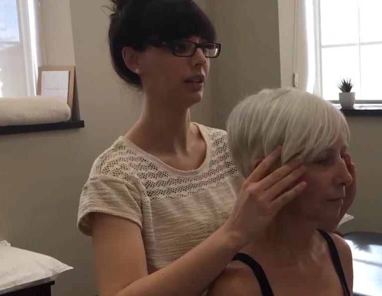
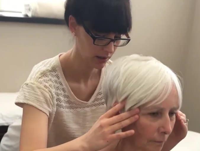
At this point in the assessment, a general listening assessment (Barral Institute) was done. This is a technique involving pressure from the therapist’s hand at the patient’s superior cranium in an inferior direction, and upon release of the pressure the therapist pays attention to what vector presents within the patient. For Mrs. A, this technique was of interest because rings 2 and 5 failing together could indicate a vector connecting these two sites of failed load transfer. General listening revealed a vector of pull which appeared to be coming from the left lung in the upper chest region which in the ISM would be considered a visceral system impairment.
Question
If you did not have general listening skills (taught in the Barral Institute courses) what other assessment technique taught in ISM courses would have revealed that thoracic rings 2 & 5 were demonstrating non-optimal A, B & C’s for this task due to an intra-thoracic visceral vector and what would that vector have felt like that differentiated it from a neuromuscular vector outside of the thorax?
Response
Vector analysis is an assessment technique utilized to determine what vector(s) is/are the primary impairment(s) at a driver(s) in question and this is described later in the case study. The lung being an intra-thoracic visceral vector felt like a strong pull on the rings from inside the thoracic cavity during the co-correction of thoracic rings 2 & 5 as well as on release of this correction. An external neuromuscular vector would instead have felt like the therapist was resisting a pull from outside of the thorax in order to maintain the aligned rings. The pull from either the outside or inside the thorax would be local and specific to the visceral, articular, and/or neuromuscular vector(s) respectively influencing the ring.
Screening task – Seated thorax rotation
In seated thorax rotation, all of the thoracic rings should rotate congruently. In a right rotation task, the thorax as a whole should left translate/ right rotate with no sites of failed load transfer. Failed load transfer may be seen as an isolated segmental ring shift either through translation to the right (coupled with left rotation which would be incongruent to the task) or in excess translation to the left (coupled with right rotation which would be congruent to the task). Failed load transfer may also be seen as more than one ring translating/rotating concurrently in the task as seen with “glued rings” or “ring sandwiches”. Glued rings specifically indicate an intercostal neuromuscular vector is “gluing” two rings together and causing the two rings to fail concurrently in a task. Ring sandwiches more generally describes a group of 3 or more rings that are failing concurrently in a task due to a variety of possible vectors.
Question
There is another feature of a ‘glued ring’ that is important. When you correct one thoracic ring that is glued to an adjacent one, what happens to the one you are not correcting if the ring is truly glued?
Response
In a glued rings scenario, the ring you are not correcting will worsen immediately in translation/rotation as soon as you start to induce correction on the other ring.
Mrs. A had decreased ability to rotate her thorax to the left and she reported much greater effort to rotate in this direction. The rings had several sites of failed load transfer in that optimal alignment, biomechanics, and control were not occurring during left rotation. Specifically, the most notable sites of failed load transfers included rings 1 through 3 and 5. Rings 1 and 2 rotated further to the right (incongruent to the task) at the same time that ring 3 rotated to the left (congruent to the task) and ring 5 rotated to the right (incongruent to the task).
Correction of the right foot prior to Mrs. A sitting and rotating the thorax, improved the range, effort, alignment, biomechanics, and control of this task. Specifically, while the same sites of failed load transfer occurred now at solely rings 2 and 5, the failure of these two rings occurred later in the task and were less severe than initially seen. With further correction of thoracic rings 2 and 5 during the task, the task became optimal for alignment, biomechanics, and control globally with Mrs. A experiencing further improved ability to left rotate her thorax. She reported that her left rotation ability now felt equal to the right and her range was observably equal.
Current Working Hypothesis
Similar to the first task, Mrs. A had co-drivers within her thorax and between her thorax and right foot during seated rotation of her thorax.
Screening task – Right single leg stance
Optimally in a right single leg stance, the right sacroiliac joint should remain controlled with the innominate posteriorly rotated relative to the sacrum. The right femoral head should remain centered in the acetabulum with no anterior translation nor rotation. The thorax should translate as a unit to the right free of ring shifts. The ankle and foot should pronate. The cervical spine should globally remain in neutral rotation and flexion/extension with no segmental kinks nor hinges. The shoulder girdles should remain neutral with the scapulae remaining in neutral anterior/posterior tilt. The clavicles should remain in a neutral anterior and posterior rotation free of compressive forces. The cranium should remain neutral with no rotation nor intracranial torsion.
In right single leg stance, Mrs. A demonstrated failure to control the right sacroiliac joint. The thorax showed various sites of failed load transfer. Specifically, rings 1 through 3 and 5 failed to maintain optimal alignment and control. Rings 1 and 2 right rotated (incongruent to the task) at the same time that ring 3 left rotated (congruent to the task) and ring 5 right rotated (incongruent to the task). The right hindfoot remained inverted through the task and did not pronate. Mrs. A reported her balance ability on the right leg was far more challenging than the left.
Again, upon correction of the right hindfoot (the subtalar joint), the same sites of failed load transfer occurred now at solely rings 2 and 5, but the failures occured later in the task and were less severe than initially seen. With correction of the right hindfoot, Mrs. A also was able to control the right sacroiliac joint.
Additional correction of thoracic rings 2 and 5 along with the right foot restored optimal performance of all regions throughout the entirety of the task. Mrs. A then reported her balance felt largely improved with the concurrent corrections.
Current Working Hypothesis
For the right one leg standing task, Mrs. A had co-drivers within her thorax (rings 2 & 5) and between her thorax and right foot, similar to the previous two examined screening tasks.
Screening task – Squat
In an optimal squat, the sacroiliac joints should remain stable with no unlocking occurring. The hips should remain centered in the acetabulum while the pelvis anteriorly tilts over the femoral heads. There should be no ring shifts throughout the thorax. The lower extremities should symmetrically flex while the feet pronate. The cervical spine should remain neutral with no kinks nor translations/ rotations. The shoulder girdle should remain neutral with the scapula remaining in neutral anterior/posterior tilt and the clavicles remaining in neutral anterior/posterior rotation. The cranium should remain neutral with no intracranial torsion.
In Mrs. A’s squat, the right sacroiliac lost control as did the thoracic rings 1-3 and 5. Rings 1 and 2 right rotated at the same time that ring 3 left rotated and ring 5 right rotated. The right hindfoot remained inverted in the task and did not pronate. Mrs. A reportedly had no symptoms during this task.
Correction of the right hindfoot (the subtalar joint), improved the performance of the thoracic rings 1-3 and 5 as well as the right sacroiliac joint. The thoracic rings 1-3 and 5 lost alignment and control later in the task when the right foot was corrected.
Additional correction of thoracic rings 2 and 5 along with the right foot restored optimal performance of all regions throughout the entirety of the task.
So, as seen in all tasks, co-correction of the right foot and thoracic rings 2 and 5 resulted in optimal performance of all previously noted sites of failed load transfer.
Current Working Hypothesis
For the squat task, Mrs. A had co-drivers within her thorax (rings 2 & 5) and between her thorax and right foot.
Screening Tasks Summary
Based on the findings across all of the above screening tasks, Mrs. A was determined to have two co-drivers; one within her thorax (rings 2 & 5) and another between the thorax and the right foot.
Vector analysis of the Drivers (right foot, thoracic rings 2 & 5) for all Tasks Screened
Vector analysis is a technique used to determine the underlying system impairment of the driver(s) in question. Potential underlying system impairments can involve the articular, neural, myofascial and dural or visceral systems.
Vector Analysis – Right Foot Driver
Vector analysis of the right foot first revealed the following neural system impairments:
- Overactive tibialis posterior and flexor digitorum longus.
- Beneath these vectors was also an underlying articular impairment involving restriction of the right anterior/medial subtalar joint.
Question
Can you describe the specific arthrokinematic restriction for the right subtalar joint?
Response
No, I am not sure – I specifically addressed solely the “gap technique” as taught in the ISM and noted the anterior/ medial restriction and went straight to frictioning it. I would guess being Mrs. A’s hindfoot rested in inversion that she was restricted into eversion, but whether that was from the anterior or posterior subtalar joint or both I cAot say for certain. If Mrs. A’s anterior subtalar joint was restricted into eversion, it would have presented as limited lateral glide of the anterior calcaneal facet on the talus versus if Mrs. A’s posterior subtalar joint was restricted into eversion, it would have presented as a reduced medial glide of the posterior calcaneal facet on the talus. Based on where the gap technique showed the most restriction, I’d assume Mrs. A had a restriction at the anterior calcaneal facet gliding laterally on the talus.
Diane
Great reflective biomechanical reasoning Lynne. If the ‘gap’ or distraction was most limited at the posterior/medial aspect of the subtalar joint and osteokinematically the calcaneus was held inverted then the likely arthrokinematic restriction would be a restriction of a medial glide of the posterior subtalar joint. A restricted anterior joint would create a ‘gap’ restriction of the antero/medial subtalar joint. Below, when you describe the ‘gap’ restriction after releasing the neural vectors, you state that the ‘gap’ was more restricted at the antero/medial part of the joint. If the calcaneus was still somewhat inverted relative to the talus and the reason for this inversion was the articular system impairment you note below, then your arthrokinematic description of the anterior calcaneal facet being restricted in gliding laterally on the talus is correct. This is a great example of how arthrokinematic joint restrictions are not always reflective of stiff joints!
Question
What is the difference in the ‘felt sense’ as a practitioner between a neural system impairment such as tibialus posterior and flexor digitorum longus and an articular system impairment?
Response
A neural system impairment (such as those created by tibialis posterior and flexor digitorum longus vectors) feels like resistance to the practitioner’s hands upon correcting the region in question (in this case the subtalar joint). The felt resistance is not rigid and allows for the desired correction to be made. The felt resistance is also local and specific to what vector is exactly influencing the region you are correcting. For example, with Mrs. A’s tibialis posterior vector, upon everting the subtalar joint to neutral, the vector resisting the correction was long and was localized exactly to an overactive region of the tibialis posterior specifically at the proximal posterior/ medial calf just below the knee. Comparatively, Mrs. A’s flexor digitorum longus vector was also long but revealed an overactive region of the muscle specifically at the mid posterior calf, rather distal to the tibialis posterior vector. The tibialis posterior vector presented more strongly upon the subtalar correction coupled with ankle dorsiflexion/ eversion while the flexor digitorum longus vector presented more strongly upon the subtalar correction coupled with extension of the proximal and distal interphalangeal joints and metatarsophalangeal joints.
Conversely, an articular system impairment presents to the practitioner as a short and rigid resistance that does not allow for the desired correction to be fully made. In Mrs. A’s case upon attempting to evert the subtalar joint to neutral, after release of the neuromuscular vectors, a deeper short vector local to the anterior/ medial subtalar joint presented as a rigid resistance to the correction. With also attempting a gapping at each pole of the subtalar joint (anterior/medial, posterior/medial, anterior/ lateral, posterior/lateral), the anterior/ medial pole resisted the opening or decompression sought through the gap assessment technique whereas the remaining other poles of the subtalar joint gapped as desired.
Diane's comment
It appears that the location of the ‘gap’ restriction changed after releasing the more superficial neural vectors. This is quite common.
Vector Analysis – Thorax Ring Co-Drivers
Vector analysis of thoracic rings 2 and 5 revealed an underlying visceral impairment involving the left lung tissues, and confirmed results of the general listening done earlier.
Question
What is the difference in the ‘felt sense’ as a practitioner of a thorax visceral system impairment on vector analysis of thoracic rings 2 & 5 and a neuromuscular vector of say the intercostals and how did you determine that rings 2 & 5 were ‘connected’ by this vector? You explain ‘the how’ very nicely below but what does this feel like in your hands?
Response
When correcting thoracic rings 2 and 5, Mrs. A’s thorax visceral impairment created a sense of resistance that was coming from within the thorax cavity localized to a specific region inside the lateral left chest about 3 inches below the mid-clavicular line. I determined that rings 2 and 5 were connected by this vector in a couple of ways. One, with individually correcting each ring, the same local region within the left chest 3 inches below the mid-clavicular line was felt as the resistance point to that ring correction. Two, I noted that upon correcting one ring, I worsened the alignment of the other ring.
If instead Mrs. A had an intercostal vector presenting as a neuromuscular system impairment, upon correction of the ring I would have met resistance external to the thorax, and that external resistance would have been local and specific to an overactive trigger point at that intercostal. For example, if Mrs. A had an anterior right intercostal vector presenting on ring 5, the resistance would have been specific to the right and anterior intercostal of ring 5 versus a mid-axillary line intercostal or posterior intercostal.
Further question from Diane
It is possible to have mid-axillary line intercostal or posterior intercostal overactivation so the fact that the vector was anterior doesn’t differentiate a visceral ‘inside’ the thorax impairment from the intercostal. Can you explain how the depth of the vector listening would be different between the two possible vectors?
Response
Correct. I was more stating that the intercostal vector external to the thorax would specifically create resistance exactly local to the vector allowing the practitioner to differentiate an anterior from mid-axillary line from posterior intercostal. To further expand on the felt difference between a visceral and intercostal vector, the depth felt on the intercostal vector would be largely superficial (shallow) compared to the depth felt on a visceral vector which feels like you are being drawn deeply and well into the thorax cavity versus being drawn to the external shell of the thorax as with the intercostal vector.
Diane’s response
The depth of the pull or ‘listening upon release’ was exactly what I was after with this question.
Anatomically, the connection of rings 2 and 5 to the lung may be explained by the lung’s fissures and pleura. The oblique fissure of the lungs are at the level of T4 while the 5th thoracic ring would encompass T4/5, the 5th ribs, and their attachments (costo-vertebral and costo-transverse joints). The lung fissures, as well as the rest of the lungs, are lined with pleura. Pleura itself is divided into four regions: costal, mediastinal, diaphragmatic, and the pleural dome. Of particular interest, the costal pleura is firmly attached to ribs and the sternum.
Treatment
Initial treatment
The first two sessions entailed the assessment and treatment of the initial drivers.
Right Foot Driver
Release: The right tibialis posterior and right flexor digitorum longus, and right (posterior/medial corrected following the question below) anterior/ medial subtalar joint were released utilizing release with awareness and frictioning respectively.
Question
What was the intention of the friction of the posterior/medial subtalar joint? My understanding is that this part of the joint was compressed by the neural vectors and that the location of the compression changed to the antero/medial part of the joint after release and that this is where the articular system impairment was (antero/medial joint).
Response
Yes, error corrected on my end.
Align: Tape was also used to decompress the anterior medial subtalar joint and facilitate a neutral hind and midfoot.
Thoracic Rings 2 & 5 Co-Driver
Release: Thoracic rings 2 and 5 were released using the stack and breathe ISM technique (Lee LJ 2003) and manual stretching of the left lung into left shoulder horizontal extension and scapular retraction.
Home Practice (connect and move): Mrs. A was sent home with homework including mindfulness of appropriate footwear, caring for her foot tape, self-aligning her right subtalar joint and foot with an ankle stacking cue, self-aligning her upper rings with a breastbone lift cue, and lateral costal breathing.
Question
What is appropriate footwear for Mrs. A?
Response
Mrs. A’s work shoes were notably worn into her inversion and supination pattern and were several years old. I advised she purchase new footwear and be mindful of replacing her work shoes at least yearly. We also discussed that outside of work Mrs. A reportedly “lived in” crocs or flip flops or Sanucks, none of which offered appropriate support for her feet, so I advised supportive shoes or sandals.
At the end of this initial treatment, Mrs. A could raise her left arm with reportedly and observably less effort than initially seen, fully rotate her left thorax, balance on the right leg reportedly and observably easier than initially seen, and most importantly, all of these tasks occurred with optimal alignment, biomechanics, and control globally across all regions post treatment.
Final Working Hypothesis
The motor vehicle accidents in 1983, 1991, and 2003 likely contributed to Mrs. A’s overall mal-alignment and dysfunction of her thorax. Vehicle accidents are common mechanisms of injury to create lung traumas due to the nature of force and torque created by the seatbelt, and this is likely why rings 2 and 5 (with a shared left lung vector) were found to be co-drivers of her condition. The right foot may have become impaired secondary to initial compensation and subsequent mal-adaptation that occurred following the fracture of the right great toe at age 17 or from simply adapting in a congruent mAer to other areas of non-optimal alignment such as the right pelvis rotation in the transverse plane. The left shoulder rotator cuff repair in 2007 and the L5/S1 foraminectomy in 2009 may have potentially been additional traumas that triggered further compensation and adaptation responses in addition to the car accidents and toe fracture.
Mrs. A likely functioned with global non-optimal alignment, biomechanics, and control for several years which would explain why the rotator cuff/ foraminectomy procedures did not completely rectify her symptoms.
It is also possible that Mrs. A’s vascular symptoms (diffuse lower back and left hip / lateral crus “tightness”, “throbbing”, feeling “under pressure”, and “tiredness”) arose from compression within the vascular system related to her ring drivers. Rings 2 and 5 shared a common impairment of the left lung. Being the left lung was restricted, it is likely that optimal left diaphragmatic breathing could not occur due to the inability of the left lung to fully expand. If the left diaphragm subsequently had tension due to non-optimal expansion, the aorta and vena cava may have been under compression creating the vascular symptoms reported in the lower back, left hip, and left crus.
Follow-up treatments
March 2, 2016
After the initial assessment and treatment on February 18th and 24th, 2016 Mrs. A was seen for follow-up March 2nd. At this time Mrs. A reported “some pressure” in her frontal bone region of the cranium. The lower back and left hip were reportedly relieved while the left shoulder was reported to feel “fatigued”.
Question
Mrs. A’s original meaningful complaints included the words “achy”, “tight”, “throbbing”, “under pressure”, and “tired”. Were all these symptoms cleared in her lower back and left hip at this point? Is the frontal bone pressure a new symptom or had she experienced this previously? Is this now the meaningful complaint?
Response
The frontal bone pressure was indeed a new symptom and was now the meaningful complaint. This symptom’s onset was two days following our initial treatment. Otherwise, Mrs. A reported that while her low back felt “tired” she had no left hip symptoms, and the left crus “achy”, “tight”, “throbbing”, “under pressure”, and “tired” symptoms “were definitely diminished”. Mrs. A’s meaningful task continued to be using her arms while standing at work, but her meaningful complaint was now frontal bone pressure.
Screening task related to current meaningful task – Standing
Objectively, Mrs. A had improvements in her standing screen as follows:
- mild pelvic transverse plane rotation to the right
- mild anterior left anterior femoral head (congruent to the pelvis rotation)
- left clavicle anteriorly rotated (congruent to the pelvis and left hip findings)
- right intracranial torsion with congruent right sphenoid rotation (congruent to all the prior findings).
Screening task related to current meaningful task – Bilateral shoulder flexion in standing
Bilateral shoulder flexion in standing produced a painful catch in the left shoulder and left chest prior to reaching 90 degrees of flexion.
The sole site of failed load transfer was the cranium in this task. The thoracic rings showed no sites of failed load transfer, nor did the pelvis, left hip, left scapula, nor left clavicle. Upon correcting the cranium, the transverse plane rotation of the pelvis, anterior left femoral head, and left clavicle corrected in their alignment and continued to perform optimally through the task. Correcting the cranium also made the left arm “feel lighter” in the task. The cranium was the primary driver for this task.
Vector Analysis – Cranial Driver
Dural vectors, including the left posterior cerebellar tentorium and mid to lower thorax region of the dural tube, were noted.
Treatment
Both dural vectors (intra-cranial and spinal) were manually released in full, and a self cranial alignment cue was added to the home management program.
Question
On vector analysis of the cranium, you noted two different regions of the dura that were impaired:
- Left posterior cerebellar tentorium
- Spinal dura
When correcting and/or releasing the correction of the right ICT can you please describe what you felt in your hands that left you with the impression that these two dural regions needed to be released?
Response
Upon release of the corrected intracranial torsion (with congruent sphenoid rotation), I initially felt a pull inside the cranium on the left side. The pull felt like I was being drawn into the cranium into a slide ending inside the cranium at a spot local and horizontal to the left ear. I then felt the pull continue as I was drawn down the dural tube to the level of the mid/ low thorax.
My additional hypothesis at this time was that Mrs. A had a significant amount of non-optimal alignment in standing prior to our initial treatments. That, in combination, with whiplash mechanisms of injury and a restricted left lung likely led to the dural shortening seen. After the initial drivers and their vectors had been released, it would be plausible that the deeper dural system needed to be released back to an optimal length, and this is consistent with the cranium presenting as the primary driver and having dural system impairments during this second session.
March 9, 2016
In a subsequent follow-up on March 9th, 2016 Mrs. A reported she had 3 full days without taking any Tylenol-3. Mrs. A reported the back, left hip, and left shoulder were still symptom-free and that walking felt normal. She did report having some pressure again in the frontal region of the cranium as well as in the mid thoracic spine. Mrs. A reported she felt so well that she did an abundance of household tasks related to cleaning and baking that she had not been able to do for years. She also reported working 6 full days in a row due to staffing issues at work and was amazed she tolerated this as she had never previously been able to work beyond 2 days in a row. Mrs. A wished to address her meaningful concern of ongoing frontal pressure within her meaningful task of standing using the arms.
Question
What underlying mechanism or system could explain persistent ‘pressure’ in the frontal region that would involve the 2nd thoracic ring and her ICT?
Response
Vasculature, specifically the vertebrobasilar system, could explain Mrs. A’s persistent frontal pressure symptoms. This system also has connections to the 2nd thoracic ring (via the subclavian artery) and noted intracranial torsion (via the basilar artery). The subclavian arteries lie posterior to the clavicles and upper rings and in turn branch to form the vertebral arteries that run vertical up along the cervical spine, which then join to form the basilar artery that supplies the brain. Mrs. A’s left clavicle resting in anterior rotation along with her cranium resting in right intracranial torsion may have compressed upon blood supply causing her “feeling pressured” frontal symptoms.
Further comment from Diane
Biologically, you present a plausible mechanism. Another one would be venous drainage. A right rotated 2nd thoracic ring potentially could increase compression in the region of the brachiocephalic/jugular vein drainage into the superior vena cava. The back flow up the jugular brain could create poor drainage of the entire brain, especially on the right side. The increased pressure in the right transverse sinus could be responsible for the perceived increased tension in the right cerebellar tentorium.
Lynne’s response
Okay, I wouldn’t have thought of this. But it was left cerebellar tentorium. I had a right/ left error there. Me and my rights/ lefts is a constant issue.
Diane’s response
Me too! I can still hypothesize that the venous system is confluent – especially in the transverse sinus and could impact both the left and right sides!
Screening task related to current meaningful task – Standing neck flexion and bilateral shoulder flexion in standing
Objectively, Mrs. A’s standing screen showed a compressed ring 7 on the right and right sphenoid rotation although no intracranial torsion was noted. During the standing neck flexion, bilateral shoulder flexion task Mrs. A reported mid back fatigue within a few seconds. Objectively during this task, ring 7 and the noted sphenoid rotation, now along with right intracranial torsion, worsened in the task at exactly the same time. Correcting either ring 7 or the cranium or both concurrently worsened symptoms in that the mid back felt worse and low back pain was additionally triggered. Being no correction brought improvement for Mrs. A, I began to consider Mrs. A still had a dural driver.
I then transitioned from the ISM to listening techniques (specifically a general listen) to confirm that Mrs. A was a dural driver. The results of the general listening technique was a force pulling me into the left cranium as well as down the dural tube. With a falx spring technique (as taught through the ISM), dural tension was initially noted at the left petrous and then posterior tentorium. Following release of these cranial dura vectors, the falx spring test pulled me down the dural tube all the way to the coccyx. Following this, with manually feeling down the dural tube, dural tension was noted at the level of the left clavicle, mid thorax (ring 7), and coccyx.
Treatment
Manual dural releases of the cranium and dural tube.
March 17 & 29, 2016
Subsequent follow-up treatments continued to identify the dura as the main driver. While the dural restrictions were no longer present at the left clavicle, ring 7, nor cranium, there were still dural restrictions related to the coccyx (again noted via general listening as well as the falx spring test) and I further assessed to find out why. Through both the feel of the restriction down the dural tube as well as results of the falx spring test, overactive muscles at the left coccyx and pelvic floor were found.
It is possible that these pelvic floor and coccyx gripping strategies may have followed the sacral fracture from child delivery, and may have exacerbated in pain response years later with respect to the back, left hip, and left sciatica symptoms.
Treatment addressed the overactive coccyx muscles with manual release, and the pelvic floor gripping strategies with reverse Kegel cueing. Cueing reverse Kegels along with releasing left of coccyx muscles resolved the dural tension noted at the coccyx in this session.
Question
What was the impact of correcting the cranium on recruitment/relaxation of her PFM?
Response
Mrs. A wasn’t a cranial driver at this time and the cranium specifically no longer held dural tension, so I didn’t think to assess this. Are you suggesting that because Mrs. A was a dural driver, that imparting a correction somewhere within the dural system (such as the cranium) may have relaxed her pelvic floor?
Further question/ response from Diane
Yes. It is extremely common to find over-activation of the PFM which subsequently restrict or deviate the coccyx and thus create an ascending pull up the dura. Conversely, ‘twists in the cranium’ either from internal or external cranial vectors can create dural tension (as you’ve noted) all the way down to the coccyx to which the PFM react. In the first instance manual release of the PFM will release the dura. In the latter, releasing the cranial/spinal dura will release the PFM. Did you have to do a manual release of the pelvic floor for the dura to relax?
Response from Lynne
No, I’m not certified to do internal release work involving the pelvic floor (Saskatchewan College of Physical Therapy rules) so treatment was solely reverse kegels cueing along with manual release (specifically release with awareness and frictioning) of the left ischiococcygeus.
Further response from Diane
We now teach external PFM palpation techniques on the ISM Series so that you don’t have to go internal to understand the relationship between PFM reactors and actors – check out the videos on Part 2 prep and support. Dr. Holly Herman has put together some nice tutorials on how to do this incorporating ISM principles into the PFM assessment.
Further question from Diane
Did you consider that the pelvic floor was perhaps reacting to the pull on the dura?
Response
No, I didn’t consider this – I immediately thought the tension in the pelvic floor was causing or at least contributing to the pull on the dura. This thought was guided by the results of the falx spring test as well as general listening. My clinical reasoning at the time was to release wherever possible to further decompress the dura being Mrs. A was a dural driver, and I chose to focus on the left ischicoccygeus and pelvic floor because of the results of the falx spring and general listen.
Further question from Diane
So how did you differentiate the two? That the muscles were causing the dural tension and not that dural tension was resulting in overactivation of the PFM?
Response
Quite honestly, I simply interpreted the results of the falx spring and the general listen to mean these overactive muscles were causing or at least contributing to dural tension. But I cAot say for certain I definitively know if dura versus the overactive pelvic floor muscles were the causer or reactor, or if both dura and pelvic floor were co-contributing to Mrs. A’s dysfunction.
What would you suggest or how do you determine the “causer” versus the “reactor”?
Diane’s response
If you correct the cranium, thorax, pelvis and there is still over-activation of the PFM – release the floor. If you correct the cranium, thorax, pelvis and the PFM relax – don’t worry about the PFM.
Lynne’s comment
With further reflection, despite the treatment done in these sessions, Mrs. A continued to be a dural driver in subsequent sessions, so it is possible the pelvic floor was reacting to the dura, or that both the pelvic floor and the dura needed to be treated concurrently, or that dural tension for some other reason or reasons (emotion, stress, poor breathing etc) later developed again post this treatment.
And on the other hand, if the pelvic floor was solely reacting to tight dura, wouldn’t it have relaxed within the first few sessions where I specifically treated the dura?
With more reflection, could the same principle of dura perhaps causing overactive pelvic floor muscles be applied to the sub-occiptals and diaphragm? These are regions I commonly note as palpably tense in dural drivers, so was wondering your thoughts on this.
Further comment Diane
Yes yes and yes. Great clinical reasoning Lynne! Whenever you have to ‘do the same thing thing’ across multiple sessions, you should wonder if you are treating a reactor and have a clinical reasoning process to differentiate if a vector is reacting to another or causing the problem in the first place.
April 2016
In late April, 2016, Mrs. A’s father passed away, and her back symptoms reportedly flared. Then, she had a colonoscopy in mid May 2016 and some complications requiring hospitalization ensued.
May 17, June 7 – 2016
When seen in follow-up on May 17 and June 7, 2016, Mrs. A reported having no lower back nor hip/ lower extremity concerns. Dural treatment was again completed due to Mrs. A continuing to be a dural driver.
In June, 2016, outcome measures done showed some objective improvement in that pain rated on the Functional Pain Scale (where 0 means “no pain or discomfort” and 10 means “Worst imaginable pain / Requires immediate emergency hospitalization”) was initially rated at 4 up to 7 for the low back, left hip, and left lower extremity, and at present was rated 2 up to 4.
Meaningful task ability as rated on the Patient Specific Functional Scale (where 0 means “unable to perform activity in any capacity” and 10 means “Able to perform activity at pre-injury or pre-problem level”) was initially rated at 5 for Mrs. A’s priority task of standing and repetitively using the arms, while at present this task was rated at 8.
Conclusion
Mrs. A likely had long-standing altered mobility in her dural system that her musculoskeletal system adapted to via shortening, shifting, and twisting. The progression of releasing Mrs. A’s various drivers shows how the dural systems adaptations change frequently with appropriate treatment. Towards the end of Mrs. A’s treatment, and following discussions between Diane and I on core engagement in this case study but not included in this report, it became clear that long-standing non-optimal engagement of Mrs. A’s left lumbar spine deep fiber multifidi and gluteus medius presented functional challenges which are at present showing improvement with proper core engagement and progressions.
While Mrs. A’s case study comes to an end, we have agreed that Mrs. A may need to attend for infrequent visits on an “as needed” basis for further core progressions and dural treatment.

Clinical Mentorship in the Integrated Systems Model
Join Diane, and her team of highly skilled assistants, on this mentorship journey and immerse yourself in a series of education opportunities that will improve your clinical efficacy for treating the whole person using the updated Integrated Systems Model.
{"first_class":"1","title":"Heading","show_title":"0","post_type":"course","taxonomy":"","term":"","post_ids":"3346","course_style":"recent","featured_style":"custom_block_1","masonry":"0","grid_columns":"clear1 col-md-12","column_width":"200","gutter":"30","grid_number":"1","infinite":"0","pagination":"0","grid_excerpt_length":"100","grid_lightbox":"0","grid_link":"0","css_class":"block-small-thumb","container_css":"","custom_css":""}
We will come together for 3 sessions of 4 (4.5) days over a period of 6-8 months with lots of practical/clinical time to focus on acquiring the skills and clinical reasoning to put the ISM model into practice. Hours of online lecture and reading material and 12 hours of in-person lecture are...
More Info