Story
Ms. A is a 33 year old female, who presented to clinic on January 10, 2023, reporting pain in her right “sit bone” with workouts. She describes the pain as “nervy”. This pain has been present, on and off, for many years (~10 years), and has worsened recently. Her goal is to increase weightlifting, but she is discouraged because she feels this sensation with lower body exercises (ie: deadlifts, squats, running) and it feels like it could get worse if she increases her workouts, like she’s done in the past. She reports occasional tingling sensation in hamstring area after a workout, and tension in the hamstring with sitting. She has had success with IMS needling, and other physio treatments but never lasting.
PMHx
- Multiple WAD injuries (3x)
- Separated right AC as a teen
- Lumbar disc injury in 2014 (when symptoms started)
- Crohn’s Disease – controlled with Humira, but flares with stress
- Multiple sphincterotomies
- Scar tissue around organs
Meaningful Complaint
Ms A’s meaningful complaint is pain and discomfort in the right ischium and into the posterior thigh with hip hinge motions. She is an avid exerciser and notices this sensation with lower body workouts and running. She also notices a numb sensation over her right hamstrings at times when the pain is worse. She is currently training for an endurance race this summer.
- Diane’s question:
The International Association for the Study of Pain (IASP) has recently updated its classification to three phenotypes for pain: nociceptive, neuropathic, nociplastic. How would you classify Ms. A’s pain and what findings from the story so far justify this classification.
- Lynn’s response:
Her pain would be classified as neuropathic pain, as the sensation comes on in a neural tension position (i.e.: hip hinge), as well as involves a numb sensation. The tract along which she describes her pain is the sciatic nerve pathway. Neuropathic pain is defined as “pain caused by a lesion or disease of the somatosensory nervous system” by the IASP.
Cognitive Beliefs
Ms. A feels like this problem is with her fascia around glutes and hamstrings because it seems to temporarily improve with foam rolling. After foam rolling, she finds she can contract her glut muscles appropriately. She also feels like her sciatic nerve is involved and that the disc injury from 2014 caused nerve tension.
- Diane’s question:
Her beliefs are biologically plausible. How will you organize your assessment to validate or negate these beliefs and why is this important to do?
- Lynn’s response:
This assessment will include a slump test and a straight leg test to test the sensitivity and mobility of the nervous system, particularly the sciatic nerve. These tests will help identify the “bio” piece of the biopsychosocial model. It can help build confidence in patients, as the current medical model focuses so heavily on the anatomy and physiology, and excluding it from the assessment could make the client feel unheard or unseen.
Meaningful Tasks
Ms. A reported that her main motivation for coming for treatment is that she’d like to get stronger to support her running training and the issue arises when she tries to increase her weightlifting. I determined that her meaningful tasks are weightlifting and running.
- Diane’s comment:
I would suggest her ‘why’ is ‘I want to be stronger to support my running’ and her barrier to achieving this (her main meaningful task to get there) is weightlifting. It’s always important to understand the ‘why’ that underpins the ‘what’ as this then frames our conversations and her motivation.
- Lynn’s comment:
I agree. The ‘why’ is most important in the biopsychosocial model of care.
Screening Tasks
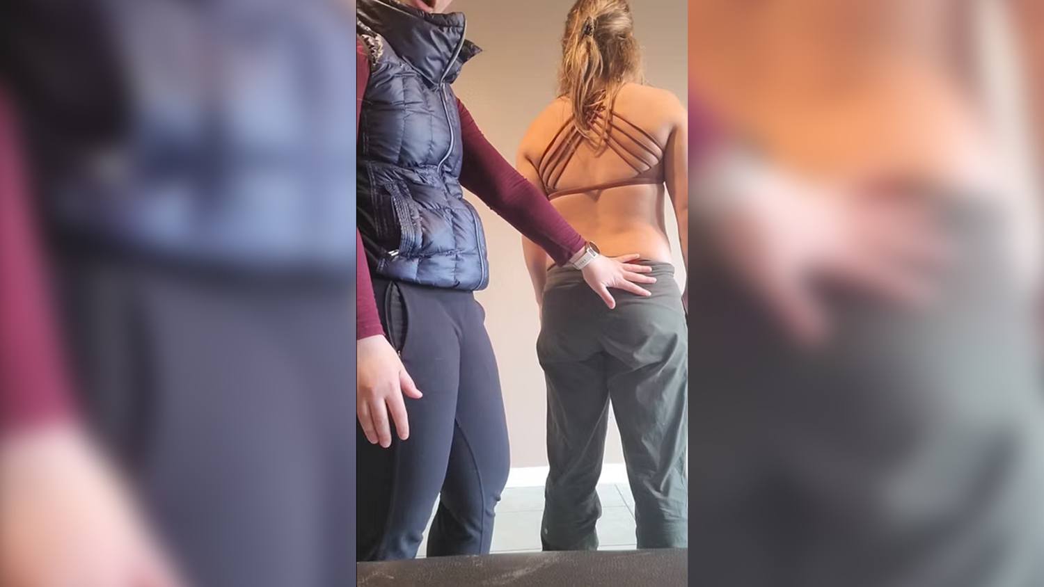
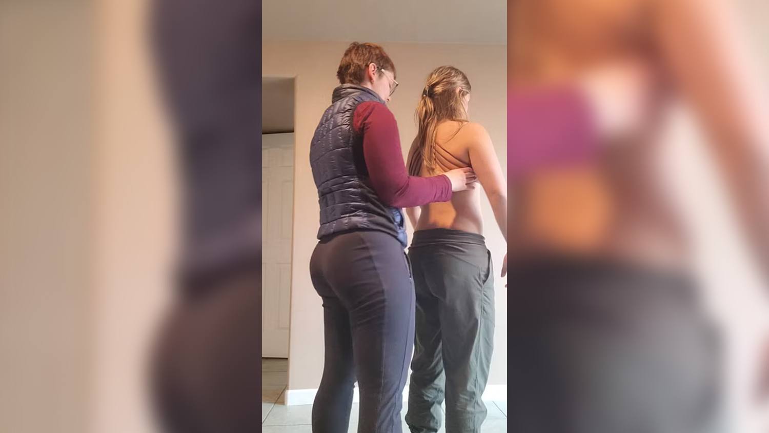
Since weightlifting is her primary concern and her symptoms seem to have a dural component, I chose a hip hinge task as it lends itself to many weightlifting moves (deadlifts, squats, lunges). At a later date, I would also assess a single leg stance on the right for running as a meaningful task.
- Diane’s question:
What makes you think this is dural as opposed to neural? Does dura cause numbness or ‘nervy’ sensations?
- Lynn’s response:
Dural tension can only exist in the central nervous system, as the dura only extends from the cranium, around the brain, to the filum terminale at the level of S2. Peripheral neural tension would be more accurate, although neural tension will transmit to the dura through the perineurium which is continuous with the dura.
- Diane’s comment:
Exactly, well clarified – her symptoms have a neural component.
Functional Unit #1
Standing Postural Screen Including a Summary of Incongruencies
Because the position of the meaningful task begins in standing, standing was chosen as a start screen position. Most of the patient’s complaints involved an upright position (ie: weightlifting, running) and it is helpful to determine where the alignment of the body segments are relative to each other at the start of the task, so one can see when, and if, they change during the task, as the task is the most meaningful part of the assessment.
Start Screen Findings: Standing
Pelvis: left TPR, left SIJ unlocked in standing, right does not appear to unlock
Hips: Left femoral head is forward relative to pelvis
- Diane’s question:
Can you clarify what you mean when you say the left femoral head is forward ‘relative to pelvis’? Is this an anterior shear of the left hip joint and you mean ‘left acetabulum’ or do you mean the left femoral head is more forward than the right femoral head (but then this wouldn’t be about the pelvis, would it?)
- Lynn’s response:
To clarify, the left femoral head is forward in the acetabulum, incongruent to the left TPR of the pelvis. This is an anterior shear at the joint.
Thorax:
- Vertebrosternal region is translated right, rotated left
- Vertebrochondral region
- TR5, TR7 translated left, rotated right
- TR6 translated right, rotated left which is incongruent with the rings above and below. This is called, in ISM, a thoracic ring sandwich.
- TR5 and TR7 are incongruent with most of the thorax, where they are rotated right and translated left, while most of the thorax is rotated left.
Lumbar: L4 is rotated right, incongruent with the rotation of pelvis
- Diane’s question:
You note that the pelvis starts in a LTPR with the left SIJ unlocked. Which direction is the sacrum rotated to? Where is the left innominate relative to the left side of the sacrum and where is the right innominate relative to the right side of the sacrum? You note also that L4 is rotated to the right, incongruent to the ‘pelvis’ but which bones of the pelvis is this rotation incongruent to? Where would you expect L5 and L4 to be in this situation, in fact where was L5?
- Lynn’s response:
In a LTPR of the pelvis, the left innominate is posteriorly rotated relative to the left side of the sacrum, and the right innominate is anteriorly rotated relative to the right side of the sacrum. In this case the sacrum is nutated on the left/right rotated, but the left innominate is posteriorly rotated, causing an unlocked SIJ on the left side.
- Diane’s comment:
In a LTPR of the pelvis the left side of the pelvis may be either locked or unlocked and therefore the relative position of the left innominate to the sacrum will vary, so I disagree with your statement above that in a LTPR of the pelvis, the left innominate is posteriorly rotated relative to the left side of the sacrum. You told us that the left SIJ was unlocked. Therefore where was the left innominate relative to the left side of the sacrum (regardless of the TPR of the pelvic ring)?
I also disagree with where you state the right innominate is relative to the right side of the sacrum because this SIJ is NOT unlocked. In a LTPR, the right innominate is anteriorly rotated relative to the LEFT INNOMINATE, but not the right side of the sacrum. Have a think about the relative biomechanics and try again! The last sentence above is also TOTALLY wrong. A hint which will help with your answer about L4 below – the sacrum is left rotated in space – it follows the TPR always unless there is a joint fixation.
- Lynn’s response:
In a LTPR, the left innominate is posteriorly rotated relative to the right innominate and the right innominate is anteriorly rotated relative to the left. The left innominate is anteriorly rotated to the left side of the sacrum in an unlocking SI joint. The sacrum is rotated left in the transverse plane.
L4 is rotated to the right, which is incongruent to the pelvis and sacrum. L5 is congruent with the sacrum, rotated left in the transverse plane.
- Diane’s comment:
Well clarified Lynn!
Screening Task: Hip Hinge
FU#1 Biomechanics with No Corrections
Pelvis: The LTPR of the pelvis increased. Both SIJs unlock, the right side early in the task and the left side unlocks further at about 75% (since it was already unlocked)
- Diane’s question:
The left SIJ was already unlocked in standing as you note. How can it unlock further in this task?
- Lynn’s response:
The left SIJ was already unlocked, so it would be more accurate to say that the left innominate rotated further anteriorly relative to the sacrum at about 75% of the range of the task.
- Diane’s comment:
I have always felt that once the SIJ unlocks, it can’t unlock further (the joint only has 4 degrees of motion); however, strategies for loading once movement begins can change. I wonder if another hypothesis here could be that when she initiated the hip hinge, her motor control strategy for loading stabilized the left SIJ from the standing position until 75% of the range at which point it then unlocked?
- Lynn’s response:
That would make sense since the unlocking was present in standing, based off findings of unloading the limb and then reloading to centre, but then when she initiated movement, she was able to initiate pelvic control until 75%.
Hips: The left femoral head stays forward relative to the right, but no additional shearing is present
- Diane’s question:
In the standing start screen you noted that the left femoral head was anterior to its acetabulum, and there is no mention where it was in space relative to the right femoral head. Because this is incongruent to the LTPR of the pelvis, the left femoral head is not necessarily anterior to the right. Therefore, saying that it stays forward relative to the right comes out of the blue because the right side has yet to be mentioned. It sounds like it is no longer anteriorly translated relative to the left acetabulum though. Can you help to clarify the start position and behavior of both hips a little more clear with this comment in mind?
- Lynn’s response:
The left femoral head started anterior to the right femoral head and anterior to the left acetabulum. In the task, the right femoral head stays centered in the acetabulum, and the left starts forward in the acetabulum but does not translate further forward, nor does it centre in the acetabulum during the task.
- Diane’s comment:
So it is still sheared in the task, since it started ‘sheared’ relative to the acetabulum and didn’t center, correct?
- Lynn’s response:
Yes. The femoral head stays forward in the acetabulum which is a shear, but it doesn’t translate any further forward during the task.
Thorax: The priority rings in the thorax (TR5-6-7 – sandwich) get worse
The lower thorax follows the pelvis into further left rotation
Lumbar: L4 rotates right further
FU#1 Driver Analysis- Primary thorax, secondary pelvis
In this task, correcting the left hip did not correct the biomechanics of the pelvis or thorax in the task but gave the patient a slightly better experience. The pelvis, with a corrected TPR and some force closure, gave the client the best experience but did not correct the alignment of the sandwiched rings in the thorax, in fact, made the translation of the sandwiched ring (TR6) worse, though it did correct the hip alignment. The thorax correction of the sandwiched TR5-6-7 corrected the lower thorax, the TPR of the pelvis, and the alignment of the hip. It did not fully correct the pelvis, though, as the right SIJ still unlocked, though later in the task (~50%) and the left SIJ lost control at the very end range of the hip hinge.
The primary driver in FU#1 is the thorax, even though it doesn’t give the very best experience in the task, because it improves the biomechanics of the most things and doesn’t make any other regions worse. The pelvis is a secondary driver, though, as the force closure in the correction seemed to improve the sensation in the task.
- Diane’s question:
The pelvis is a secondary driver mainly because there is still loss of control of both SIJs in the task with the thorax driver corrected. Did you happen to do a co-correction of both the thorax (TR5,6,7) and the pelvis? This is not easy to do and often requires a second set of hands or an SI belt! So the hypothesis is made by deduction.
- Lynn’s response:
I was not able to co-correct the two drivers in this session, because, as you say, you would need another set of hands. In the past, I have tried to correct the pelvis with my hands, and then hold the correction by bracing their pelvis with mine, while correcting the thorax. It doesn’t allow for a very good correction of either driver, so I typically use deduction. However, when I just corrected the pelvis, it did make the task feel better for the patient even if it didn’t correct the thorax, so that added to the strength of the hypothesis of the pelvis as a secondary driver.
A quick screen of the head and neck was performed in the hip hinge position with the FU#1 driver corrected to see if it was necessary to look for another driver in FU#2. With the thoracic rings (TR5-6-7) corrected, the sensation near the ischium was still present, though diminished, and right rotation of the head and neck felt stiffer. Therefore, more investigation was required, as the driver of FU#1 made FU#2 worse.
- Diane’s comment:
You have two drivers in FU#1. Both should have been corrected to test the impact of the co-correction on the biomechanics of the head and neck. If the pelvic floor was attempting to control the SIJs this pull could have impacted the position of the coccyx and imparted tension to the dura and thus the sciatic nerve. With compression of the pelvis, the pelvic floor may have relaxed releasing this tension and possibly some of the sciatica. Remember to always correct ALL unit drivers when doing a quick screen of another unit. Typically, I use an SI belt to force close the pelvis at least, in this situation.
Functional Unit #2
Start Screen Findings: Standing
Cranial region: Left ICT, right rotated sphenoid, incongruent with the ICT, C1 translated right, rotated left, which is congruent with the ICT of the cranium.
Cervical region:
C2 translated right, rotated left
C7 translated left, rotated right incongruent with upper neck and cranium
Shoulder girdle: Rotated left, congruent scapulae incongruent with most thoracic rings
Thorax: TR1 left rotated incongruent with C7
TR2 left translated, right rotated incongruent with C1, and to the shoulder girdle
- Diane’s comment:
Here is another ‘sandwich’ – C7, TR1, TR2
In FU#2 the most significant findings in the starting screen position were:
- the incongruent sphenoid to the cranium,
- C7 being incongruent to the rest of the neck and cranium, especially C2, and
- the shoulder girdle being incongruent to most of the thoracic rings, including TR2, though it was congruent to TR1.
Screening Task: Hip Hinge
FU#2 Biomechanics with No Corrections
Cranial region: the cranium position corrects, along with C1
- Diane’s question:
It is really very unusual for an incongruent sphenoid/ICT to BOTH correct in a screening task. This is saying that two vectors (something rotating the sphenoid and something twisting the temporal bones and occiput) were no longer present in the task. What is your hypothesis that this could, and did, occur?
- Lynn’s response:
In this case, there was a fascial system impairment in the viscera, I suspect, related to the client’s Crohn’s disease. When the body position changes, gravity affects the visceral fascia in a different plane, which could have slackened the deep cervical fascia that connects to the sphenoid and then down into the thorax. The correction of the ICT of the temporal and occipital bones, was likely more related to the compensation of C2, which is the driver of FU#2. The other factor, which is related, is the dural system, which, if there is an impairment, could unwind the cranium as the dura pulls on another part of the MSK system.
- Diane’s comment:
Nicely hypothesized Lynn!!
C2 translates further to the right and rotates left, while C7 translates further to the left and rotates right (ie: alignment gets worse), these two vertebra are incongruent to each other
The alignment of the shoulder girdle does not change
The upper thoracic rings (TR1, TR2) both correct
- Diane’s question:
Another interesting finding here. TR1 and TR2 are incongruent and both correct in this task. LICT and left rotated TR1, Right rotated sphenoid and right rotated TR2. Something connecting TR1 to the posterior cranium and something else connecting the sphenoid to TR2 ‘slackened’ in the hip hinge position. What’s your hypothesis here?
- Lynn’s response:
Because of the chronic nature of this issue, there are several adaptations in the MSK system to compensate for the shortened dural system. The dural connection is likely the factor in the TR2 and sphenoid relationship, and the left upper fibres of trapezius and the left cervical erector spinae were the connection between TR1 and the posterior cranium.
FU#2 Drivers for the Hip Hinge Task
In the task, the only components that do not self-correct in alignment are in the neck, specifically C2 and C7, and the shoulder girdle. The two vertebra were corrected separately, and C2 was found to correct C7 and all of the other elements of FU#2.
- Diane’s comment:
In your CRF you said that correcting C2 corrected the shoulder girdles and above you state that everything else in FU#2 either auto-corrected in the task or corrected with C2. There is no need to test any other correction in this unit.
Prioritizing FU#1 and FU#2 Drivers
The next step was to prioritize the FU#1 and FU#2 drivers. In the task, correction of the thorax (TR5-6-7) and the neck (C2) each made the other better, and both improved the feeling of the task. The right SIJ was still unlocking at the very end of the task, therefore I hypothesized that the pelvis is still a secondary driver.
- Diane’s comment:
You may have found there was a relationship between the pelvis and C2 (strong dural connection pelvic floor to C2). I wonder if you had controlled the pelvis (compression with a SIJ belt) and then corrected the three thoracic rings (TR5-6-7) if C2 would have auto-corrected?
- Lynn’s response:
I didn’t try this, but I also think this patient could hold the pelvis corrected manually while I correct the rings, so it would be a great next assessment step.
My hypotheses are:
- a slight motor control deficit at the pelvis, which makes the pelvis a Secondary Driver, or
- a dural vector in lower spine, with also neural tension in the right leg specifically, that is causing the pelvis to unlock at the end of the task, to avoid a pull on the sciatic nerve at the end range of the hip hinge, which makes the dural system a Secondary Driver. This was assessed at a second visit.
- Diane’s comment:
The dural system is NEVER a driver because it isn’t a MSK body region. The dura can be a system impairment but not a driver, by definition of drivers. There is a video in the ISM flow (new one) that discusses this in further depth.
Functional Unit #3
A quick screen of the feet in both the standing start screen and the hip hinge task was performed. Even though there were some alignment issues at the feet, correcting the drivers in FU#1 and FU#2 (co-drivers) corrected the feet in the task and therefore, no further examination was required.
- Diane’s comment:
This is your examination and it was required!
Overall Drivers for Task
In the hip hinge task, correcting both the thorax (TR5-6-7) and the neck (C2) together created the best patient experience for the task and almost completely restored pelvic control, so I determined that they were Co-Drivers. In this case, they correct each other, but do not completely correct the task, so there is still a secondary driver present.
- Diane’s question:
How does knowing the type of driver (co-driver vs primary and secondary) influence treatment priority in ISM?
- Lynn’s response:
A primary driver, on its own, will correct all other body regions, so it is the only region that needs to be treated. When there is a secondary driver, the primary driver or drivers are treated first, then the secondary driver, because the primary driver will correct the most body regions and bring back the most control, even if it’s not complete.
With the co-driver where both require correction to improve the task and patient experience, both should be treated in the same session, where possible, because correction of one makes the other worse so it could cause a flare up. If correction of either driver makes the other better, it doesn’t matter which is treated first. Again, if there is a secondary driver present, it would be treated next.
- Diane’s comment:
Well explained!
In this case, the secondary driver could be the pelvis, specifically, the active control of the pelvis needs to be assessed. There is likely some dural tension present, but from the story, there could also be some scar tissue at the pelvis from past surgeries of the rectum, which could be causing neuromuscular, myofascial, or dural vectors.
Further Analysis of Drivers
The Cervical Region Driver (C2)
The first driver assessed was the neck (C2).
Active Mobility
- Diane’s question:
Active mobility testing should be in relationship to the requirements of the meaningful task. What are the biomechanical and control requirements of C2 in a hip hinge task? What should happen to C2? In the start screen it is left rotated/right translated.
Specifically, and in relationship to the requirements of the hip hinge task, tell me ONE and only one, range of motion requirement you need to test.
- Lynn’s response:
Because the start position of C2 is in left rotation, but in the hip hinge task, it should be in neutral, right rotation should have been assessed.
The active ROM of the neck was assessed to determine the mobility at C2. Notable findings were decreased upper cervical flexion on right compared to the left. With breathing, C2 also was pulled into further right translation.
Passive Mobility
For the hip hinge task, all the vertebrae in the neck should be in neutral. Since C2 started in left rotation/right translation, it had to be corrected manually for the task. The PROM of C2 in right rotation was slightly decreased compared to the left, but it was possible to correct C2 on C3 to neutral by derotating and translating the segment.
In Passive Listening #1, where the segment is corrected and quality of the movement is felt (ie: how difficult is the segment to correct), it was noted that traction of C1-2 to allow space for the translation part of the correction was more difficult than the lateral translation of C2 on C3. This could mean than there is likely a myofascial or capsular vector on the left and a neuromuscular vector on the right. In Passive Listening #2, where the corrected segment is let go and the direction, quantity, and quality of movement back to the resting position is felt, the vectors noted were the right longus colli, middle scalenes, left upper fibres of trapezius, and left splenius capitis and semispinalis capitis.
Passive Control
No ligamentous laxity was present in the neck, and there was little in the story to recommend that this would be a factor
Active Control
In the task, there was increased activation of anterior neck muscles (SCM, scalenes) as soon as head crossed the shoulders in sagittal plane, especially on the right. This would cause C2 to translate right, and rotate left, as was noted in the task.
After release, control of C2 came back until the very end of the task. Therefore, a control cue for the neck was given as part of the home care program to improve motor control of the driver.
- Diane’s question:
After release, was C2 in a neutral start position and remain there until the very end of the task?
- Lynn’s response:
Yes. She was able to maintain neutral rotation (no rotation) of C2 until about 95% of range of motion of the hip hinge task.
The Thorax Driver (TR5-6-7)
The second driver assessed was the thorax, specifically the “thoracic ring sandwich” created by rings TR5, 6, 7 where thoracic rings 5 and 7 were translated left and rotated right and the ring 6 was translated right and rotated left. Prior to assessing the active mobility of each individual ring, the Ring Stack and Breathe technique was applied to decrease the neuromuscular vectors in the muscles of respiration (intercostals, diaphragm). This corrected the TR6-7 part of the sandwich, leaving TR5 and TR6 parts to assess.
Active Mobility
There was decreased flexion of the right T5-6, and decreased excursion of the right ribs with breath. Notably, there was decreased superior glide of right rib on exhalation. Once the ring stack and breathe technique was performed, these findings were eliminated. They are also not important to the hip hinge task. After TR5 returned to neutral, TR6 was the priority ring in the sandwich, and the only one that didn’t correct with ring stack and breathe, so I should have assessed right rotation, as the thoracic rings, like the neck, should be neutral in the hip hinge task.
- Diane’s comment:
Above, you have performed an arthrokinematic assessment for the right costotransverse joint, yet there is no indication from your assessment so far that there is an articular restriction because you can completely correct the thoracic ring sandwich at TR5-7 to the position required for the hip hinge task. An Active Mobility test for a 3 ring sandwich is merely to palpate the thoracic rings and test, in this case the ability of each ring to rotate back to neutral. I suspect TR5 and 7 were limited in left rotation and TR6 in right rotation. This is an active mobility test. Even if you had just tested the noted priority ring of the sandwich (TR6 which was rotated left/translated right, the direction to test would have been rotation right, not left, to bring it back to neutral.
Passive Mobility
TR5-6 were the harder segments to correct in the sandwich, so the Ring Stack and Breathe technique was performed, as described above to remove some of the neuromuscular vectors so that passive mobility testing could be assessed. At that point, TR6 was able to rotate right back to neutral, though there were still some vectors present.
In Passive Listening #1, it was noted that the rings were fairly easy to correct. It didn’t feel like there were any capsular vectors that were difficult to correct. Neuromuscular vectors feel easy to correct and one only needs light correction, and visceral vectors are need slightly more pressure to correct.
In Passive Listening #2, the vectors felt at TR6 on release of the correction were neuromuscular and visceral. The neuromuscular vectors pulled TR6 on the right, and were likely right serratus anterior and external oblique where they interdigitate at ribs 5-7.
The visceral vector at TR6 was quite strong to the right, so a general and local listening for visceral vectors was performed. The general listening technique (Barral), performed by applying gentle pressure to the top of the head in standing, produced an anterior sway in the body. The local listening (Barral), performed in supine, showed a vector in the small intestine, specifically the region of the duodenum.
- Diane’s question:
These vectors are impacting thoracic rings that are above the diaphragm, yet the small intestine is below. How do you hypothesize that the small intestine can be the appropriate vector relevant to TR5, 6, 7?
- Lynn’s response:
Since the visceral fascia below the diaphragm is continuous with the fascia of the diaphragm, and the central tendon of the diaphragm attaches to the pericardium, which has posterior attachments from T5-7.
When Ms. A was in standing, the strongest neuromuscular TR6 vectors, seemed to be the right serratus anterior and external oblique where they interdigitate at ribs 5-7, but interestingly, in supine, the rib angle showed that the left external oblique and right internal oblique had the most resting activation. This is inconsistent with TR6, which was the priority ring in this assessment. This showed that the visceral vector was very strong, since it changed the position of the thorax in standing versus supine. After the visceral vector was released, the standing and supine vectors for TR6 were the same (i.e.: left EO/SA, right longissimus)
The other notable vector of TR6 was the left longissimus. It was determined that it was the left longissimus over the other erector spinae muscles because the right SI joint was the side with poorer motor control. Since the origin of the longissimus through the erector spinae aponeurosis is more medial than iliocostalis, it nutates the sacrum to assist in force closure of the pelvis.
Passive Control
With the neuromuscular and visceral vectors of TR6 released, there was no ligament laxity present in the thoracic region assessed.
Active Control
Active Control was assessed in standing, after vector release, using isometric flexion and extension of the arms, starting at 90 degrees of flexion. This position was chosen because even though there were no weights in the screening task, holding weights in that position is a normal part of the task. TR6 translated to the right/rotated left, when the flexed arms were loaded into extension, isometrically. The client was able to maintain control with cueing with moderate resistance, so motor control exercises will be important to build capacity and endurance of the thoracic stabilizers. Active Control was also assessed in standing versus in the hip hinge task because of the dural vectors that weren’t released yet, and it was more appropriate to work on some motor control in neutral than with a dural pull.
Hypothesis
The Primary Driver of this task is a Co-Driver including the neck and thorax, with a secondary pelvis driver. Most of the vectors are neuromuscular, which indicates motor control issues, more than articular faults. Because the pain comes on in a dural tension position, it could be classified as neuropathic pain, considering the original injury at the lumbar spine. However, due to its chronicity, it could also be classified as nociplastic pain. A slump test and a straight-leg raise test were performed at a later assessment, when the pelvis was reviewed as a secondary driver.
- Diane’s comment:
I’m not certain that the nociplastic phenotype of pain can be assigned only based on chronicity. As long as the pain can be provoked by mechanical loading, it remains either nociceptive or neuropathic or a blend.
Treatment
Initial Treatment Session
Release
The vectors of the C2 and TR6 were released through multiple techniques. The first technique was Ring Stack and Breathe. In this technique, the driver in question is manually corrected and the patient is instructed to breathe deeply, in and out, three times. Then the patient is instructed to rotate to the side of the translation. In this case, both the neck (C2) and thorax (TR6) were translated to the right/rotated left.
Ring Stack and Breathe was performed separately at each the neck (C2) and thorax (TR6). Following this, the visceral vector of the thorax was released in supine, using a visceral release technique that involves holding the vector organ, in this case, the duodenum, then holding light pressure at the neurovascular bundle (superior mesenteric ganglion), sympathetic innervation (T6-8 intervertebral foramen), and parasympathetic innervation (vagus nerve) of that organ. The holds are applied until a shift is felt at the organ and it gains back some of its regular motility (this is a Barral technique). Finally, the rest of the neuromuscular vectors of the neck and thorax were released using Release with Awareness (RWA) – an ISM technique. This technique involves shortening the affected vector muscle and applying light to medium pressure and asking the client to relax or release the tension at the muscle. After a change is felt in the tissue, a stretch is applied. This technique was used on the right longus colli, middle scalenes, left upper fibres of trapezius, and left splenius capitis and semispinalis capitis in the neck, and the right serratus anterior and external oblique, and left longissimus muscle. These techniques were taught to the client using a RAD roller release tool, to perform as part of their home care program.
Align
The Align Cue that worked best for Ms. A was to create space in the mid-thorax, especially around TR5 and TR6 and imagine a paper fan opening. This helped to hold the alignment of the priority rings of the thorax.
Connect
The Connect Cue needed to maintain control of the neck was to imagine a guy wire between the sternal notch and the spinous process of C2, and then imagine something is gently pulling the end of the wire up. She was able to use this cue to maintain control of C2 until the very end range of the hip hinge task.
Move
Since the pelvic control doesn’t fully come back at the end range of the task, the pelvis needs to be assessed further at a different session. In order to work on building motor control and coordination of the task, the hip hinge task was given in four-point kneeling, rather than standing, where pelvic control can be maintained through the task.
Home Exercise Program Summary
- RWA left side neck, upper fibres of trapezius, right serratus anterior, external obliques, left iliocostalis thoracis
- Postural cue: create space in rib cage, throughout the day in various positions
- Motor control cueing of neck 3×10, 3x/day in standing
- Four-point hip hinge 3×10, with neck cue
Follow Up Treatment Sessions
In a subsequent session, Ms. A reported that she was still having a pulling sensation in the right ischium with full hip hinge, but has been working on motor control in sitting and 4-point, but also continuing to perform her regular workouts. The slump test and straight leg raise tests for dural tension in the right side were assessed and both were positive on the right, reproducing the original symptoms. When the primary co-drivers, C2 and TR6, were corrected, the sensation only came on at the very end of the range in the dural tension test. After releasing the vectors, which were the same as the previous session, in the neck and thorax, and performing a slump slider exercise with the drivers manually corrected, the sensation of pulling into the ischium was completely eliminated and the right SIJ had complete control through the whole hip hinge task. The slump tensioner exercise to gently lengthen the dural system was added to the HEP, with instructions to complete it after the release, align, and connect exercises.
Future sessions will focus on increasing the level of difficulty for the motor control tasks to continue to build capacity and endurance of the local stabilizers of the neck and thorax. Additionally, weights could be added to the hip hinge task to assess the effect of the shoulder girdle further.
Case Study Author

Lynn Orsak
NeuMovement Vernon
ISM Series Graduate ISM Certified Practitioner
5100 Anderson Way, Vernon, BC, Canada
25054528482505452848
Clinical Mentorship in the Integrated Systems Model
Join Diane, and her team of highly skilled assistants, on this mentorship journey and immerse yourself in a series of education opportunities that will improve your clinical efficacy for treating the whole person using the updated Integrated Systems Model.
{"first_class":"1","title":"Heading","show_title":"0","post_type":"course","taxonomy":"","term":"","post_ids":"3346","course_style":"recent","featured_style":"custom_block_1","masonry":"0","grid_columns":"clear1 col-md-12","column_width":"200","gutter":"30","grid_number":"1","infinite":"0","pagination":"0","grid_excerpt_length":"100","grid_lightbox":"0","grid_link":"0","css_class":"block-small-thumb","container_css":"","custom_css":""}
The Live 2-Part ISM Series is designed for practitioners who have purchased and completed Part 1 of the online ISM Series and are ready to further develop and refine their clinical reasoning and hands-on ISM skills through mentored, in-person learning. Part 2 of the online ISM Series must be...
More Info