Story
Ms. PR was a 59-year-old woman who attended for physiotherapy assessment in spring 2018. She reported that she had a longstanding issue with her left hip, and in speaking with a family member who had a similar issue, learned about pelvic floor physiotherapy and wondered if this may help her.
Ms. PR stated she had been doing yoga for a long time and thought at some point she may have hyperextended her hip. Her symptoms started with a pinching sensation in her left groin when jogging and over the past number of years (she was unsure when symptoms initially started) has increased in intensity and irritability to the point it hurt even with walking. She said that after walking for about 30 minutes she starts to limp, and she feels a deep pinching in her left groin and an ache into her upper left thigh. She said she also feels very sensitive to touch along her pubic bone and described a sensation like her left leg was going to detach from her pelvis. She did not report any numbness or tingling.
To date, Ms. PR reported she had tried massage therapy and seeing a TCM practitioner for acupuncture. She said she also occasionally sees a chiropractor for low back pain.
Her current activities include work (sitting at a desk, works from home), yoga (is a trained yoga teacher), going to gym 4 times per week (treadmill up to 20 minutes) and weights. She stated that because of the hip pain she avoids taking big steps on the treadmill when walking and during yoga she avoids warrior 3 pose and is very cautious with any one leg stance poses on her left side due to pain and a sense of instability/poor balance. She also reported difficulty with poses such as child’s pose.
Ms. PR stated that she gets a bit of dribbling after voiding and needs to make sure she is completely empty before standing. She denied any stress urinary incontinence, but intermittent urgency and sensation like she was getting a UTI. She had not had any children.
Diane
Given the length of time she has had these symptoms, what role do you think sensitization of the nervous system is having on her experience and what findings from the story support your answer?
Leigh
Given the length of time Ms. PR has been experiencing her symptoms, it is very likely that a component of her symptoms is driven by central nervous system sensitization. Following known timelines for tissue healing, any ongoing symptoms beyond approximately 3 months following an injury cannot be attributed only to tissue. Even in an acute injury less than 3 months old, the nervous system is responsible for producing pain and can immediately respond by increasing sensitivity levels of receptors at tissue level, synapses at spinal cord and in the brain, all in an effort to protect and allow time to heal. Because of the role of the nervous system in producing pain, it is important for health care professionals to spend time educating patients about pain even from early stages of their injuries, so they have an accurate understanding of where their symptoms are coming from. Beyond 3 months or so, while there may be ongoing inputs to the brain from peripheral tissues that may include nociception, these signals are less likely coming from ongoing tissue injury, and more likely from tissue that is unhealthy, compressed, overactive or tight for example. The brain will interpret these signals as either safe or dangerous, and if after it takes into consideration all the other inputs coming into the brain from eyes, ears, past experiences, environment (the list can go on and on!), it determines these signals are dangerous, it will produce pain as an output to continue protecting the body. The brain itself can also be responsible for maintaining ongoing pain, as it can start to interpret signals that are not nociceptive, touch for example, as something that could potentially be dangerous, and thus it produces pain. In Ms. PR’s case, this is happening with her with the sensitivity to touch she has along her pubic bone. Touch should feel like touch! The majority of her symptoms are produced with specific movements, which would suggest they are mechanical in nature, but given the timeframe, even accurate nociceptive input to the brain is likely being “ramped up” by a degree of central sensitization.
Past medical history
Ms. PR stated her medical history is significant for endometriosis. She had an ablation but did not find this helpful. She had a vaginal hysterectomy 18 years ago, and her ovaries removed 8 years ago. She stated she is very thankful for the surgery and has found significant symptom improvement. She also reported having her gall bladder removed “a long time ago” (central abdominal scar). She reported a history of low back pain. She stated she had had bilateral bunion surgeries a number of years ago and had old orthotics that she had not been using. She had not been taking any medications until the day prior to our assessment when she was put on a proton pump to help settle stomach acidity. As a result of her hip pain, she has had an ultrasound and CT scan to rule out a hernia (negative findings).
Meaningful Complaint
At the time of initial assessment, Ms. PR identified her main meaningful complaint as left groin pain she described as a pinching sensation, which also radiates as an ache sensation into her left upper thigh. This was reported to be aggravated by sitting or walking more than 30 minutes.
Meaningful Tasks
Based on Ms. PR’s subjective history and her goals, the meaningful tasks identified included: sitting, walking longer than 30 minutes (with goal of running), and one leg stance/balance (as this is a component of yoga poses she finds challenging). The screening tasks chosen at initial assessment were squat and left one leg stance. These were chosen because to achieve sitting one must move through a squat, and the hip flexion component of squat relates to yoga poses moving through squat or crouch. One leg stance is required for gait and one leg balance.
Cognitive Belief
Ms. PR believed at some point she hyperextended her left hip and caused an injury to her hip. She also stated she wondered if her pain had something to do with her pelvic floor, as a niece had spoken to her about a similar problem and her issues were related to her pelvic floor.
Diane
Great story-taking! Screening tasks chosen for evaluation relate well to her functional goals. In every patient’s story, there are three dimensions:
-
- Bio (physical dimension)
- Psycho (thoughts/beliefs/emotions)
- Social (work/family/relationships)
I was drawn to this statement in the story – she “described a sensation like her left leg was going to detach from her pelvis”. A statement like this could be a flag for many changes in movement behaviour – why and how would you pursue this comment with further questions to see if this ‘belief’ could present as a possible future barrier to recovery.
-
Leigh
I agree completely. When we hear statements that are dramatic and often fear based (something is tearing, something will fall off, pain kills me, it goes out, etc.), note should be made of these statements as potential barriers to treatment. Patients may completely avoid the movement or posture that they are fearful of, not necessarily because of symptoms, but because of fear of what may happen. Follow up questions asking specifically why they believe this would guide further follow up testing or education. Often patients will correct themselves in “I know that won’t happen but…” However, even if they “know” it won’t happen, their nervous system hears them saying the statement or thinking it, and this can be ramping up central sensitization. In Ms. PRs case, she moved well and was not limited by fear but by the onset of her symptoms, so I was less worried about this in her case. If further questioning or observation of movement revealed that she was avoiding tasks or had more fear of movement statements in her history, I could then support her through those movements and see what happens, I could also complete hip stability testing to test her concern that her hip could detach from her body.
Screening Tasks
Standing posture screen
All people have habits, and in clinical practice it is rare to find individuals without asymmetries right and left. Knowing how a person stands and habitually holds regions of their body allows an assessor to know where these regions are starting from, and from there they can evaluate the quality and quantity of movement. This also allows an assessor to know if there are regions of the body that are starting in non-optimal alignment before a movement task is initiated. Once a starting position is known, further evaluation of alignment, biomechanics and/or control can occur during a movement task.
Standing:
Functional Unit 1
Pelvis: L transverse plane rotation (TPR), L sacroiliac joint (SIJ) unlocked
Hips: R femoral head anterior > L
Thorax: 4th thoracic ring translated left, rotated right.
Functional Unit 2
Thorax: 2nd thoracic ring translated right, rotated left
Cervical spine: C6 and C7 translated right, rotated left.
Cranium: no finding documented.
Shoulder girdle: no finding documented
Functional Unit 3
Feet: Relative to the lower leg and the mortise (i.e. the inferior tibiofibular joint), the left calcaneus was everted and the left talus was plantar flexed, adducted and laterally tilted. Relative to the talus, the left navicular was plantarflexed and externally rotated. This was initially interpreted as an “over pronated foot”. On reflection of video #1 and following language clarification with Diane Lee & Cathy Rogers, it is noted that at the subtalar joint itself, the talus was dorsiflexed/abducted relative to the talus such that the calcaneus was actually medially rotated relative to the talus (initially I assumed the calcaneus was laterally rotated as it would be in pronation). Medial rotation of the calcaneus at the subtalar joint causes the talus to be dorsiflexed/abducted relative to the calcaneus, which in turn was plantarflexed/adducted and laterally tilted relative to the mortise. The subtalar joint was in a position of supination; however, the lateral talar tilt made the hindfoot on initial glance appear pronated. Specific positional testing of each bone of the hindfoot relative to each other in non-weight bearing is required to make this determination. There was splaying (abduction away from the 2nd ray) of her 3rd through 5th metatarsals with loss of the transverse arch.
Right foot: Her right calcaneus was laterally rotated relative to the talus, and talus was plantarflexed/adducted relative to the calcaneus – this is a pronated hindfoot.
Knees: no findings documented
The following glossary of terms will be required to understand the terminology updated for biomechanics of the hindfoot in this case report.
Pronation
Osteokinematics: Pronation is initiated with heel strike during which the ground reaction forces stop the forward momentum of the calcaneus and the talus glides anteriorly on the calcaneus. Following this anterior glide, the talus then plantarflexes relative to the mortise and plantarflexes and adducts relative to the calcaneus. Simultaneously, the calcaneus laterally rotates relative to the talus.
Arthrokinematics: Plantarflexion of the talus in the mortise requires an anterior glide of the talus relative to the tibia and fibula. Pronation of the subtalar joint requires a relative lateral glide of calcaneus on talus at the anterior joint(s) and a posteromedial glide of calcaneus on talus at the posterior joint.
Pronation persists through midstance.
Supination
Osteokinematics: From midstance to heel lift the hindfoot supinates secondary to external rotation of the lower leg and the ground reaction forces. As the lower leg advances over the talus, the talus dorsiflexes in the mortise and dorsiflexes and abducts relative to the calcaneus. Simultaneously, the calcaneus medially rotates relative to the talus.
Arthrokinematics: Dorsiflexion of the talus at the mortise requires a posterior glide of the talus relative to the tibia and fibula. Supination of the subtalar joint requires a relative medial glide of calcaneus on talus at the anterior joint(s) and an anterolateral glide of calcaneus on talus at the posterior joint.
Inversion/Eversion
Inversion/eversion is a physiological motion of the entire foot that can occur in non weight-bearing (NWB). Inversion: the sole of the foot turns medially. Eversion: the sole of the foot turns laterally
In weight-bearing (WB), the ground prevents the entire foot from inverting or everting and instead produces a non-optimal tilt of the hindfoot relative to the lower leg, resulting in tilting of the talus in the mortise.
Tilt
Lateral tilt of talus: Lateral aspect of talar dome more compressed in mortise, medial part distracted in mortise, inferior surface of talus points laterally. A lateral tilt of the talus is non-optimal alignment.
Medial tilt of talus: Medial aspect of talar dome more compressed in mortise, lateral part more distracted in mortise, inferior surface of talus points medially. A medial tilt of the talus is non-optimal alignment.
The calcaneus can be either medially or laterally rotated with either position of a talar tilt in the mortise. A lateral tilt of the talus can look like hindfoot pronation, and it is easy to assume that the calcaneus is laterally rotated relative to the talus given the posture of hindfoot from observation only. Each bone, the calcaneus and talus, needs to be specifically palpated to determine its position relative to the other in order to determine if the hindfoot is pronated and medially or laterally tilted or supinated and medially or laterally tilted.
Diane
Excellent description of the complexity for determining the relative position of each bone and joint in the hind and midfoot!
Note to reader – look at the left hindfoot in video #1 to get a visual of what Leigh is describing here.
Relationship between Drivers of Functional Units:
While there are many areas of non-optimal alignment in standing, standing was not a meaningful task for Ms. PR and as such a driver was not found for standing.
Diane
- When the pelvis rotates in the transverse plane, an intra-pelvic torsion (IPT) is expected. However, you note that in standing although the pelvis is rotated to the left in the transverse plane, the left SIJ is ‘unlocked’. What is meant osteokinematically by an ‘unlocked’ SIJ (where is the sacrum in relationship to the ipsilateral innominate) and how is this different than the position of the sacrum in a left IPT?
- What clinical tests differentiate an IPT from an unlocked SIJ?
- You mention that certain findings (i.e. 4th thoracic ring and left foot) are congruent, while others are incongruent (left foot to the right foot, pelvis, right hip, 2nd thoracic ring and lower neck). What is meant by ‘congruent’ vs ‘incongruent’ and why is knowing this useful in the ISM approach?
Leigh
- An unlocked SIJ refers to a sacral base that is posteriorly rotated (counternutated) relative to the innominate on that side, or an anteriorly rotated innominate relative to the sacral base on that side. In a left IPT, the right innominate anteriorly rotates and the left innominate posteriorly rotates. The sacrum rotates to the left. The right innominate remains posteriorly rotated relative to the sacrum on the right, and the left innominate remains posteriorly rotated relative to the sacrum on the left. In other words, the sacrum remains nutated relative to both the right and left innominate bones.
- To differentiate an IPT from an unlocked SIJ, you first place your hands around the innominate bones on the right and left. Feel for any rotation of each bone anteriorly or posteriorly and note this. Palpate the sacral base to determine if it is rotated right or left in a transverse plane and note this.
Diane
How can you rule out an asymmetrical size of multifidus over the sacral base when interpreting your findings when using the sacral base for positional analysis? Is there another part of the sacrum where multifidus is less likely to provide non-valid findings?
Leigh
Layer palpation would allow you to feel if there is asymmetrical size of multifidus. Palpating the inferior lateral angle of the sacrum would eliminate the need to palpate through the multifidus.
Then, palpating one innominate with one hand and the other hand palpating the inferior lateral angle of the sacrum on the ipsilateral side, ask the patient to sway their weight onto the opposite side, then slowly sway weight back to neutral or equal weight bearing. This allows you to determine if the sacrum remains nutated relative to the innominate on that side. Repeat this on the other side. If you find, for example, that the right innominate is anteriorly rotated and the left innominate is posteriorly rotated, the sacrum is rotated to the left and the sacrum is nutated relative to the innominates on both sides, you have a left IPT. If when the patient sways their weight back onto the right leg and the sacrum counternutates on that side or the innominate anteriorly rotates, this is an unlocked SIJ and therefore not an IPT, as an IPT requires the sacrum to be nutated relative to the innominate on both sides.
Diane
Can you describe what an unlocking SIJ ‘feels like’?
Leigh
The movement is subtle, but most often it feels like the innominate is anteriorly rotating relative to the sacrum – the hand on the innominate feels like it rotates anteriorly and the thumb or finger on the sacrum stays. Other times, it may feel like the sacrum is counternutating relative to the innominate.
3. Biomechanics tells us how bones and joints are expected to move based on typical anatomy. Movements have been observed and studied and we know how bones/joints are expected to move independently and relative to each other. For example, if in standing you rotate your whole body to the left, you would expect your head, neck, thorax, lumbar spine, pelvis to rotate to the left (lower extremities will rotate as well but for this example I will stop here). If movement is observed and a portion of the thorax actually rotates to the right during this task, that would be unexpected and incongruent with the rest of the body’s movement. Similarly, if in standing someone stands with a general rotation to the left, any part of their body that is rotated to the right is “out of place” relative to where the rest of the body is. In an ISM assessment, looking for patterns of congruency (all body parts following an expected pattern for that movement or posture) or regions that are incongruent (not following what is expected), can help to highlight regions of the body that may be of interest during the assessment. You cannot assume that an incongruent region is the driver without testing that hypothesis, but it may grab your attention and help to zero in on specific areas to expedite the assessment.
Diane
Awesome answer!
Squat
As noted above, squatting was chosen as a screening task as it is through squat that a person achieves a sitting posture. If regions of the body fail to transfer load optimally during a squat, they may land in sitting with regions of the body in sub-optimal alignment.
Sub-optimal alignment, biomechanics and/or control can occur in any region of the body from head to foot during a squat and would be identified by observation and palpation. For Ms. PR, this task was meaningful as it related to her symptoms with sitting as well as in loaded hip flexion postures such as crouch or child’s pose (it is noted however that crouch, and child’s pose were not chosen as screening tasks at this point but would likely be something worked towards in future sessions).
When Ms. PR was asked to squat (to approximately 80 degrees knee flexion) the following findings were noted:
Functional Unit 1:
Hips:
- Left femoral head was more centered in the acetabulum than the right was at the start of the squat. With good alignment, biomechanics and control it is expected that the femoral head remains centered in the acetabulum and spins as the femur flexes on the pelvis. During Ms. PR’s squat, the left femoral head was palpated to translate anteriorly early in the squat, suggesting non-optimal biomechanics and/or control.
- Right hip did center in the acetabulum (started anterior but corrected its alignment during the movement therefore was moving as it would be expected, and not an area of interest at this point).
Pelvis:
- Left sacroiliac joint was already unlocked (starting position demonstrated non-optimal alignment, and remained so during squat. This could suggest non-optimal control of the SIJ as well.)
- Transverse plane rotation moved to a right transverse plane rotation (demonstrating non-optimal alignment and biomechanics as during a squat the pelvis is expected to start and remain neutral in the transverse plane.) This change in direction of the transverse plane rotation is congruent with the change in biomechanics of the left and right hips during this task.
Thorax
- 4th thoracic ring remained translated left, rotated right (no further change from start position, ring started and remained in sub-optimal alignment for this task). This finding is now congruent with the pelvis and the left hip.
Lumbar spine:
- No findings noted.
Diane
How does the noted behaviour of the left and right hip joints predict this change in direction of transverse plane rotation of the pelvis and further validate your impression of the left hip?
Leigh
During a squat, the femoral head is expected to remain centered in the acetabulum as the femur flexes relatively on the pelvis. When one femoral head translates anteriorly and the other does not, this creates an altered axis for movement and the pelvis then must rotate around the femoral head on that side as the other femoral head continues to spin optimally. If the left femoral head translates anteriorly and the right remains centered, the right hip will continue to flex optimally while the left does not, inducing a transverse plane rotation to the right.
Driver for FU1: Primary driver left hip, secondary driver thorax. Correcting the left hip restored control and alignment of the pelvis; however, there was only a partial correction of the alignment of the 4th thoracic ring. 4th thoracic ring correction did not improve pelvis or hip.
Functional Unit 2:
Cervical
- Segments 6 and 7 remained translated to the right rotated to the left (no further change from start position, segments started and remained in sub-optimal alignment for this task)
- There was no obvious segment in the cervical spine that I noted that compensated for the left rotated lower cervical spine, but for the head to remain looking forward there likely was either a rotation to the right of a higher cervical segment or a right intracranial torsion.
- Diane’s comment: unless these segments (including the 2nd thoracic ring) completely compensated for the 4th thoracic ring and the pelvis (RTPR), then no further compensation of the upper cervical segments or cranium would be necessary.
- Thorax: 2nd thoracic ring remained translated right, rotated left (no further change from start position, ring started and remained in sub-optimal alignment for this task)
Driver for FU 2: Thorax (2nd thoracic ring). Correcting the alignment of the 2nd thoracic ring, restored optimal alignment of C6 and C7.
Functional Unit 3:
Knees: no findings noted
Feet: Left talus plantarflexed, adducted and laterally tilted relative to the mortise, moving further into this alignment compared with start position, and the lateral tilt of the talus in the mortise caused the calcaneus to laterally rotate relative to lower leg, but not the talus (calcaneus remained medially rotated relative to talus). The lateral midfoot fanned and medial midfoot remained with navicular plantarflexed and fanned as it started in standing, and lateral forefoot splayed further.
During a squat, this is not what is expected to occur at the hindfoot, indicating non-optimal alignment and biomechanics and likely control. In standing and squat, the talus should not be tilted relative to the mortise and the calcaneus should be stacked neutrally under the talus, thus the hindfoot started in non-optimal alignment. The talus should plantarflex and adduct relative to the calcaneus and dorsiflex relative to the mortise, and the calcaneus should laterally rotate relative to both the lower leg and talus. The amplitude of hindfoot movement caught my eye most significantly, as did the fact that the navicular did not seem to move as much as the other bones in her foot. Right foot posture stayed with calcaneus laterally rotated relative to the talus and the talus plantarflexed adducted relative to the calcaneus (subtalar pronation) and plantarflexed relative to the mortise, with slight further increase in this posture. This is what would be expected during a squat, but as with the left foot, the alignment in starting position was non-optimal, and the degree of movement was more than typically expected, suggesting non-optimal alignment and control of the right foot during this task.
Driver for FU3: There was only one region of the body assessed in functional unit 3 (feet), therefore a driver for this unit cannot be definitively determined.
Summary of Functional Unit Drivers: For each functional unit, the within unit drivers for the squatting task were:
- Primary driver: Left hip, secondary driver thorax (4th thoracic ring)
- Thorax (2nd thoracic ring)
- Left foot (only region assessed)
Relationship between drivers of all Functional Units:
When starting to assess for the drivers for squat, it was noted that maintaining a good correction of Ms. PR’s left foot was difficult to achieve because as soon as she took weight through her foot the correction seemed to be lost, so an insole available in the clinic was used to help support the foot correction and allow assessment of other regions of the body more easily without having to maintain manual correction of her foot (see video #1). This also provided some midfoot correction as well.
Diane
Could you achieve a good correction and she couldn’t maintain it with load, or could you not achieve a correction? How does your answer possibly reflect a difference in the underlying system impairment in this foot and direct potential treatment?
Leigh
I could achieve the correction of the lateral tilt of the talus but it could not be maintained with load once my hands were removed. It was not possible to get a complete correction of the calcaneus.
Difficulty maintaining correction of talar tilt and forefoot alignment suggests an underlying impairment in the neural system, being poor motor control, with or without an impairment in either the articular system or myofascial system which could contribute to hypermobility of the joints in her foot, for which her motor control was not sufficient to overcome. Calcaneus position being difficult to correct could indicate an impairment in articular or myofascial systems.
Diane
What part of the foot (hindfoot, midfoot, forefoot) is critical to correct and maintain in order to understand the impact of a foot correction on proximal regions of the body? When the hindfoot was corrected, did the mid and forefoot automatically correct or were these regions still in sub-optimal alignment and how does the difference in this answer direct further assessment/treatment of the foot itself.
Leigh
The hindfoot is critical to correct and maintain to be able to evaluate more proximal body regions, as it is this part of the foot through which load from the leg is transferred. In her left foot, when the hindfoot was corrected as well as it could be, the midfoot did not automatically correct, but improved slightly. The forefoot also remained more splayed and did not correct with correction of the hindfoot. This would indicate that the mid and forefoot would need to be evaluated to determine if the mid/forefoot was impacting the hindfoot position. When I corrected the fore/midfoot, the hindfoot remained in non-optimal alignment, so I focused initially on the hindfoot knowing there would likely be more work to do in the mid and forefoot.
Correcting her left foot partially corrected her left hip and fourth thoracic ring, and completely corrected second thoracic ring and cervical spine (therefore FU 2 will not be assessed further). When her left hip was corrected in addition to her left foot, her experience of the task improved as well.
Based on these findings, it was determined that for the left hip pain during squat, Ms. PR’s left foot was the primary driver and her left hip was the secondary driver. It is noted that her 4th thoracic ring was not completely corrected with the foot and hip corrections and therefore remains a region of future interest. In future sessions, the role of the thorax (a secondary driver in FU1), becomes apparent.
Note: When a foot support was provided to her right foot as well, starting alignment of her right hip in standing was also improved as was the right transverse plane rotation of her pelvis, and all improvements noted previously were maintained. This indicated that her right foot could use some intervention as well but was not considered a driver at this time for this task as the task was squatting and both the left transverse plane rotation of her pelvis and right hip alignment improved without this added correction. For a standing task, the right foot may have been a relevant driver. Note- control of the right SIJ was not reassessed once the foot and hip corrections were made. It had previously remained controlled during a squat and was not “on my radar” for reassessment after the corrections were made. The importance of this oversight becomes relevant in subsequent sessions.
Diane
If the subtalar joint is unable to pronate (calcaneus is held in medial rotation relative to the talus), which phase of gait will this impact? Is the 4th or 2nd thoracic ring impairment relevant to consider for restoration of pronation of the left hindfoot and why?
Leigh
Pronation occurs during heel strike to mid-stance, and this phase of gait would be impacted. At left heel strike, the thorax is left rotated in space but at heel strike it starts to right rotate, and is rotating to the right while pronation is occurring. Ms. PR’s 2nd thoracic ring is right translated left rotated, which would impact her ability to right rotate her thorax, and this therefore would need to be restored to improve subtalar pronation. In this case, correcting the 4th thoracic ring corrected the 2nd thoracic ring, so it was addressed indirectly.
Left one leg stance
One leg stance is required for gait. Non-optimal alignment, biomechanics and/or control can occur in any region of the body from head to foot during one leg standing and this would be identified by observing and palpating body regions during this task.
When Ms. PR was asked to stand on her left leg, the following findings were noted:
When Ms. PR was asked to stand on her left leg, the following findings were noted:
Functional Unit 1:
Left hip
- Left femoral head translated anteriorly in the acetabulum (femoral head should remain centered in acetabulum)
Pelvis
- Left sacroiliac joint was unlocked in standing and remained unlocked during left one leg stance (sub-optimal control as sacroiliac joints should be in a locked position during loading)
Thorax
- 4th thoracic ring translated left, rotated right further from starting position (this is sub-optimal alignment, biomechanics and/or control as this task does not require thorax rotation, and as such thoracic region should remain in neutral alignment without rotation.)
Driver for FU 1: Left Hip and Thorax co driver. Both the hip and the thorax needed to be corrected to improve the task, and neither her hip or her thorax corrected the other region.
Functional Unit 2:
Cranium: no findings
Cervical spine
- no findings documented at the time of assessment; it is unknown if these segments remained translated right rotated left as they did in standing, worsened, or improved.
Upper thorax
- 2nd thoracic ring translated right, rotated left further from starting position (this is sub-optimal alignment, biomechanics and/or control as this task does not require thorax rotation, and as such thoracic region should remain in neutral alignment without rotation.)
Shoulder girdle
- no findings documented at time of assessment.
Driver for FU 2: Upper thorax is the only region with altered alignment, biomechanics and control noted for this functional unit, therefore a definitive driver for this unit cannot be determined.
Functional Unit 3:
Knee
- no findings
Left foot
- Immediately the left talus tilted laterally in the mortice further, the calcaneus everted relative to the lower leg, and the midfoot fanned. This is sub-optimal since during one leg stance without forward or backward movement or hip/knee flexion, the talus should not tilt in the mortice, the calcaneus should remain neutral relative to talus and the lower leg and the midfoot should remain in neutral alignment.
Diane
Actually, when load is increased in the static standing foot, it is anticipated that the foot will move into supination for more form closure and support).
Leigh
Your point makes sense, but I couldn’t find anything written about foot alignment in one leg stand, only bipedal standing or walking. Isn’t moving into supination required because the foot needs to be a rigid lever to push off – something not required in one leg standing. I reasoned that it should be expected to remain neutral but with more intrinsic/extrinsic muscle activation to improve force closure with the added load.
Diane
Not sure if there is any evidence to be honest Leigh. In bipedal standing the SIJs are not fully close-packed but the moment load increases, both sides nutate fully (Sturesson’s studies). I have always thought this was true of the foot.
Leigh
No studies to be found. I consulted Rachael Corbett (physiotherapist at Diane Lee & Associates), and she noted in her experience the talus would dorsiflex and abduct relative to the calcaneus during one leg stance (talar movement associated with hindfoot supination), but the calcaneus remained anchored. I think it would make sense biomechanically that the foot remain neutral if COM was able to stay perfectly aligned over the foot, but if there was an area of non-optimal alignment, bio-mechanics or control elsewhere such that the COM remained more medial during weight shift, the hindfoot may need to supinate to compensate for this medial displacement.
Driver for FU 3: Only one site of altered alignment, biomechanics and control noted, therefore a driver for functional unit 3 cannot be determined.
Summary of Functional Unit Drivers: For each functional unit, the within unit drivers for the left one leg standing task were:
- Left hip
- Thorax (2nd thoracic ring)
- Left foot
Relationship between Drivers of Functional Units:
- When Ms. PR’s left hindfoot was corrected, her left hip was partially improved, but her middle and upper thorax did not improve. When her 4th thoracic ring was corrected, 2nd thoracic ring improved completely (therefore no driver in FU #2). She also reported improved subjective ease of balance with the addition of the 4th thoracic ring correction. Based on these findings, it was determined that her left foot was her primary driver and her 4th thoracic ring was the secondary driver for left one leg stance. Upon reflection, her hip may also have been a secondary driver, as it remained unclear in my notes if her hip was completely improved when the 4th thoracic ring correction was added.
- Final drivers for the left OLS task: Primary left foot, secondary 4th thoracic ring and possibly the left hip
Diane
In the squat task, the hindfoot correction had the same impact on the pelvis and left hip as in the LOLS task; however, the hindfoot correction also partially corrected the 4th and 2nd thoracic rings in the squat task, but not the LOLS task. Any hypotheses as to why the difference in response of the thorax to single leg standing as opposed to a squat?
Leigh
Ms. PR felt unsteady and off balance when standing on one leg. I wonder if she was recruiting upper extremity/scapular muscles to help improve balance during one leg stance where she didn’t during a squat. Because of the attachments of scapular muscles to the 4th thoracic ring, this could explain why this ring did not correct to the same extent, and the second was reacting to what the 4th was doing so it continued to translate right rotate left.
Diane
Good hypothesis.
What was the relationship between the 4th and 2nd thoracic rings which are incongruent to one another? You note that correcting the 4th thoracic ring improved the alignment and control of the 2nd thoracic ring in the LOLS task – did you assess whether correcting the 2nd thoracic ring improved the 4th? And what about the impact of the 4th thoracic ring correction on C6 and C7? Often, the lower neck is in relationship with the upper thorax and I’m curious if the 4th was truly the only driver, or main driver up here.
Leigh
I think the 2nd thoracic ring and lower cervical segments were compensating for the 4th thoracic ring, which was much more significant in its position than the 2nd thoracic ring. When one ring rotates one way, another region of the body often will rotate the opposite way in an effort to keep us “straight” or pointed forwards at least. I did not note anywhere if I attempted the correction of the 2nd thoracic ring to see the impact on the 4th. With the 4th thoracic ring corrected, her cervical segment alignment corrected – these seemed to follow the 2nd thoracic ring.
Upon reflection, it was noted that the ability of the right hip to center and right innominate to posteriorly rotate was not assessed during this task, and for a complete assessment and confidence in determining the drivers, knowing what these regions were doing would have been helpful. I also did not assess if the 2nd thoracic ring or cervical spine improved the 4th thoracic ring, and there may have been another rotation higher in the cervical spine or cranium that I did not note that could have been further assessed as well.
Further Assessment of Noted Drivers
Further assessment of noted drivers consists of a series of tests, including vector analysis, used to determine what underlying system impairment is causing the change in alignment or biomechanics of a driver. System impairments can be in neural, myofascial, visceral, or articular systems, and knowing which system impairment is causing the non-optimal movement allows for determination of appropriate intervention techniques. There may be more than one system impairment present, and vector analysis assists with determining which one should be treated first to see greatest improvement.
Diane
What is the series of tests referred to above. Tests that you would perform for every body region determined to be relevant to the task and goal?
Leigh
The series of tests would include testing active mobility, passive mobility, active control and passive control.
Further Assessment of the Left Foot
Active mobility: During squat, left talus plantarflexed, adducted and laterally tilted relative to the mortise, moving further into this alignment compared with start position, and the lateral tilt of the talus in the mortise caused the calcaneus to further evert relative to lower leg, but not the talus (calcaneus remained medially rotated relative to talus).
Passive mobility: The subtalar joint was passively moved into a medial TPR and lateral TPR. I did not record passive mobility testing findings, but I would infer from other findings that there was less passive joint mobility into lateral TPR (posteromedial glide of the posterior ST joint, lateral glide of the anterior ST joint).
Active control: Ms. PR was unable to hold her hindfoot corrected during weightbearing, suggesting there could be impaired active control of the hindfoot.
Passive control: Not assessed as the story did not suggest a need for this.
Vector analysis: For Ms. PR’s left foot, a passive listening technique during correction of the alignment of her left hindfoot was initially used, which started with her talus and calcaneus being passively corrected to neutral alignment.
Diane
Active and passive listening are terms coined by the Barral Institute and integrated into ISM. What do they mean? This will help the reader understand what you mean below when you describe passive listening at the subtalar joints.
Leigh
Listening techniques are techniques where a therapist uses their hands to feel where vectors of pull are coming from. In active listening, the patient actively moves through range or into a more optimal alignment to where the first point of resistance is felt, and the therapist uses their hands to note the biomechanics produced and feels where any resistance is coming from. In passive listening, the therapist passively moves a bone into an alignment that is considered more optimal, and while doing so, feels for any vector of force preventing the bone from being moved. Then as the bone is released, the therapist feels for which direction that bone is to, and what the quality of that force feels like.
Diane
Tell me what the osteokinematic position of the hindfoot, midfoot and forefoot were in sitting compared to the findings in standing. Any changes between position? Was she sitting on an unlocked SIJ – did changing this i.e. providing control of the pelvis as she sat change any of the findings of her foot. It’s OK if you didn’t do this – we can have a discussion about possible changes and what they would mean depending on your answer.
Leigh
In sitting, I did not check if she was in an unlocked SIJ. I would assume she would have been as I did not hold her foot in a corrected position as she moved into a sitting position.
Diane
You are correct. If she sat through a squat without the left hindfoot correction, it is likely she was sitting with the left SIJ in an unlocked position. What impact would this have on the form closure mechanism of the left SIJ? i.e. what ligaments are less tense and which are more tense?
Leigh
In an unlocked SIJ the long dorsal ligament is more tense and the sacrotuberous ligament Is less tense.
When in sitting, her left calcaneus was inverted relative to her lower leg. The lateral talar tilt was not present; however, the talus was still plantar flexed relative to the mortise. At the subtalar joint the talus was relatively abducted/dorsiflexed to the calcaneus and the calcaneus was medially rotated relative to the talus (subtalar joint supinated). The navicular remained plantarflexed and externally rotated relative to the talus and the lateral three metatarsals remained splayed (abducted relative to the 2nd ray).
Diane
Describe your hypothesis as to how the calcaneus can appear everted relative to the lower leg in standing and inverted in sitting
Leigh
In the seated position, when ground reaction forces were eliminated from the left foot, the left talus was no longer tilted in the mortise but was still plantarflexed secondary to the persistent plantarflexion and external rotation of the navicular. With the lateral tilt eliminated, the left calcaneus was no longer everted relative to the lower leg, but rather was inverted. The calcaneus remained medially rotated relative to talus (the talus was dorsiflexed and abducted relative to the calcaneus) as it was in standing.
Diane
Complex positional findings and very commonly misinterpreted as a pronated hindfoot, it isn’t!
Diane’s question:
Since it appears it was possible to manually correct the alignment of the talus and calcaneus, what does this tell you about the subtalar joints and which systems are less likely to be impaired.
Leigh
Because I was able to move the talus and calcaneus into a neutral position, I can rule out an articular system impairment, as if a joint glide was restricted, it would have been more difficult to move these bones as freely, and a complete correction may not have been possible. A myofascial system impairment was also less likely given the quality of the resistance felt during the correction (passive listening).
While co-correcting the talo-crual joint and subtalar joint, it was felt that there was a stronger pull on her calcaneus preventing the correction than on her talus (passive listening).
Next, the calcaneus was distracted from the talus at the subtalar joint with a posterior medial, anterior medial, anterolateral and posterolateral bias. The greatest resistance was felt with posteromedial and also at the posterolateral bias. A passive listening technique was then used as the traction was let go, and a long pull was felt up the posterior calf. While holding the calcaneus in neutral alignment with the talus, the calf was then palpated to locate the level of the over-active muscle.
Once a level of interest in the posterior calf was determined by observing and feeling for the largest response (movement out of neutral alignment) in the calcaneus as the calf (soleus, gastroc) was gently squeezed, specific layer palpation revealed an overactive, tender point in the soleus. Once this was released with release with awareness and stretch with awareness techniques, it was easier to achieve correction of the calcaneus in its relationship to the talus. During the first session this was as far as release of the foot was taken.
Diane
It is not uncommon for the midfoot and forefoot to be compressed/stiff in compensation for hindfoot impairments. After you released the hindfoot, what was the response of the left mid and forefoot?
Leigh
The left navicular remained externally rotated and the lateral forefoot remained splayed. The calcaneus also remained in a slightly medially rotated position relative to the talus (I do not think I released the vector from the soleus fully in this one session, and in subsequent session there was a myofascial vector between talus and calcaneus medially).
Given the general hypermobility of Ms. PR’s forefoot and her difficulty supporting her hindfoot in optimal alignment in weight bearing, it was anticipated she would require motor control training of her foot intrinsic muscles to help support her foot in an optimal position.
Diane
Can you briefly describe the principles for motor control training of the foot and how you determine when to progress your program?
Leigh
Motor control training progresses through stages based on the strength of the muscles being trained. These principles apply to training any of the foot intrinsics.
Stage 1: Grade 0-1 strength, flickers of activation but unable to sustain activation through range or under load. Support the foot in optimal alignment and teach isometric recruitment for the required muscle. All training done in non-weight bearing. Progress to next stage when able to activate muscle in neutral alignment without extrinsic muscle “cheating”.
Stage 2: Gr 2 strength. Use cues trained in stage one but add load, still in non-weight bearing (bands, balls, calf raise when seated and foot on floor). Progress to next stage when demonstrating good activation, no cheating, maintaining intrinsic activation through range or partial load.
Stage 3: Grade 3 strength. Progress to weight bearing. Use cues learned in earlier stages to maintain optimal alignment in double leg standing, toe raises. Progress to next stage when able to maintain good activation and optimal foot alignment in double leg stance exercises consistently.
Stage 4: Grade 4 strength. Progress to single leg stance, step through, single calf raises. Progress to next stage when maintaining optimal alignment through required tasks at controlled speed.
Stage 5: Grade 5 strength. Add variability in surface, reaction times, jumps, speed.
Diane’s comment for clarity of staging motor control training:
These stages of training are from the work of Rachael Corbett, Diane Lee & Associates and differ somewhat from the staging of training presented for the thorax and other body regions. Stages 1 and 2 are the same, Stage 3 has 3 levels (Stage 3/Level 1, Stage 3/Level 2, Stage 3/Level 3) and refer to Rachael’s Stage 3, 4 and 5.
Further Assessment of the 4th Thoracic Ring
Active mobility: Ability of the left and right 4th ribs to posteriorly rotate during inspiration and anteriorly rotate during expiration was assessed.
Passive mobility: The 4th thoracic ring was passively moved into anterior and posterior tilt which requires anterior and posterior rotation of the ribs. Anterior tilt of a thoracic ring requires the spine to flex and thus a superior glide at the zygopophyseal joints bilaterally, and superior glide/anterior roll of the costotransverse joints bilaterally in this region of the thorax. Posterior tilt requires the spine to extend thus an inferior glide at the zygopophyseal joints bilaterally, and inferior glidatposterior roll of the rib on the transverse process at the costotransverse joints bilaterally. I did not record findings of passive mobility testing specifically as I noted that the thoracic ring could be corrected as a whole, indicating there were no articular restrictions.
Vector analysis: For Ms. PR’s 4th thoracic ring, a passive listening technique during manual correction of alignment was used. In sitting, it was noted that the 4th thoracic ring could be manually corrected completely, although it was felt that the left rib was more difficult to correct than the right (passive listening). Passive listening was then used as the 4th thoracic ring was released from the corrected position, and a neural vector was felt laterally (as opposed to more anterior or posterior), and a tender point was palpated in the left serratus anterior. Once this was released, there were shorter neural vectors found in the left intercostals.
Diane
When the 4th thoracic ring is translated left/rotated right what position is the left rib in osteokinematically and what muscles at this level could restrict or make its correction difficult?
Leigh
The left rib is anteriorly rotated.
Right rib, posterior rotation.
Inferior vector posterior aspect of rib: iliocostalis thoracis and longissimus thoracis on the right, posterior intercostals on the right.
Left rib, anterior rotation.
Inferior vector on anterior aspect of rib: intercostals anteriorly.
Superior vector on posterior aspect of rib: semispinalis cervicis on the left, iliocostalis cervicis on the left, semispinalis capitus and cervicis on the left.
Left or right rib (variable – in my clinical experience these are sometimes found on the left or the right inducing a ring to rotate and translate)
Vectors between scapula and rib: serratus anterior, pectoralis minor, pectoralis major, rhomboids, middle trapezius.
Diane
In your clinical experience, when serratus anterior is the dominant vector what is the resultant position of the impaired thoracic ring and the scapula on that side? How were the short vectors from the left intercostals addressed in treatment?
Leigh
In my experience I find that when left serratus is the dominant vector it causes an anterior rotation of the rib on that side, inducing an ipsilateral translation/contralateral rotation. The scapula is often elevated and more protracted and downward rotated. Ring stack and breathe was used in the first session to address the intercostals.
Diane
I do too and this often confuses clinicians. It is likely because it is easier to move the mobile scapula than the more resistant rib cage.
Diane’s question:
Was any home practice given for the 4th thoracic ring after this first session?
Leigh
She was given a self-stack/align cue to feel “open” through her axilla. I did note this was not sufficient to improve the alignment of the ring and she needed further release. Given the timing of the assessment and what I observed to be great body awareness, I hoped she could start to learn to let go with just the align cue, and further release could be given in future sessions.
Further Assessment of the Left Hip
Active mobility: Femoral head was palpated during active flexion of her left hip.
Passive mobility: Functional range was tested with Ms. PR in supine lying, and was very limited – maybe only 30 degrees (I did not measure or note this, this is from recollection). Functional range is determined by how much range a person’s joint has while the femoral head remains centered in the acetabulum. When the femoral head translates forward, that is the extent of their functional range.
Vector analysis: In supine, her hip was passively flexed from neutral to the point it was felt that she did not keep femoral head centered in the acetabulum (this was approximately 30 degrees flexion). A passive listening technique as the femoral head was centered and then released to determine the vectors that were perturbing the alignment of the femoral head at 30 degrees of flexion.
Diane
Can you be more specific and describe your passive listening technique for analysis of dominant vectors perturbing the alignment of the left hip?
Leigh
Gentle distraction and posterior glide (to center the femoral head in the acetabulum) was applied to the hip joint, and when the femoral head was released, a short, deep vector was felt pulling straight medially. This led to palpation externally of the left obturator internus (OI), which was indeed very tender.
Diane
Obturator internus is an external rotator therefore I would have expected a short ER vector of pull on the femoral head prior to the medial pull. If it was straight medial, I would have gone to iliococcygeus (IC) first.
Leigh
Yes- this makes sense I see this clearly now– but even as a reactor, OI would be perturbing the femoral head- yes? A combination of the 2 forces may be medial? For IC to be perturbing the hip, the force would need to be through fascia through OI would it not? I’m uncertain how you could confidently distinguish between the two.
Diane
By the presence, or lack, of any rotation of the femoral head.
Leigh
Sorry I am still stuck on this. If the iliococcygeus is the dominant vector, the force would go through the fascia to the OI, and then via the OI to the hip. There is no connection from the iliococcygeus and the hip otherwise is there? A fascial connection other than through OI? Because of the anatomical connection between the iliococcygeus and OI with OI being the go-between, I still don’t see how one could externally rotate the hip and the other not. If IC is dominant it will pull through the OI and would this not be the same as if the OI was dominant? (May be similar confusion about scapular/upper ring vectors – the anatomy just doesn’t seem to jive with what you are saying you observe clinically. This is where clinical experience is important I guess!).
Diane
I wasn’t clear enough in my answer. If OI is the dominant vector, the first ‘listening’ you feel is external rotation followed by a medial vector of force that goes beyond the obturator foramen. If IC is the dominant vector, you are right that the force is conveyed through the OI fascia but since OI is not contracting (or at least is not dominant) then there is no initial external rotation of the femoral head and an immediate vector of pull medial past the obturator foramen.
From here, because of the identification of a possible vector in the pelvic floor, Ms. PR’s belief that her hip pain had something to do with her pelvic floor, as well as her complaint of UTI type symptoms, an internal vaginal examination was completed.
Diane
The vector you felt on passive listening of the left hip (no rotation) actually supports this assessment first rather than one of obturator internus.
Leigh
Yes – but also to confirm/deny her cognitive belief, and she had been told by a gynecologist years prior to strengthen her pelvic floor and do lots of Kegels!
Diane
Absolutely!
Summary of findings from internal vaginal exam included:
- Overactive left superficial transverse perineal muscle,
- Vestibule and vaginal skin appeared pale,
- Lift of perineum observed with cue to activate pelvic floor muscles but limited let go/descent,
- No ballooning of perineum with Valsalva,
- No cystocele/rectocele observed,
- Knack was present.
- Overactivity was palpated in the left pelvic floor (obturator internus and puborectalis were most overactive with tender points throughout, iliococcygeus overactive but not tender).
- She had a good ability to contract levator ani (Gr. 4 strength), but demonstrated difficulty letting go, particularly on the left side.
When her left hip or her 4th thoracic ring was corrected, her ability to let go of this overactivity improved (her hips were in less than 90 degrees flexion during the exam). Of note, when the tender points in the obturator internus were palpated, this reproduced her sensation of UTI.
Diane
What about her left foot? Correcting her left hindfoot in standing improved her pelvic control and left hip position. This highly suggests that the foot is responsible for the change in motor control strategy. It is likely, since you didn’t mention it, that you didn’t do it, which is OK we can hypothesize relationships and mechanisms for this on the next go around.
Leigh
No I didn’t do it for a couple reasons – I know that the left foot correction improved the hip and the thorax, so I used reverse reasoning and corrected the hip and the thorax instead of the foot so I could reach and palpate at same time. Also – I didn’t check so I don’t know – if a foot is corrected in NWB, is the impact the same as in WB with ground reaction forces?
Diane
No, gravity and ground reaction forces are different, which is why testing a primary foot driver’s impact on the pelvic floor recruitment pattern should be done in a screening task more related to the meaningful task, i.e. standing in this case.
It’s a really important feature of this case report – foot driven pelvic floor BUT you didn’t actually test the foot correction on the pelvic floor behaviour! At least I don’t see it being tested either with ultrasound or palpation. In a perfect world, your pelvic floor assessment would have been done next in standing, ultrasound (US) probe held by patient suprapubically and then the foot position changed with ground reaction forces to witness the change in resting tone of puborectalis and OI.
Hypothesis
Ms. PR felt the initial left hip injury involved hyperextending her hip, which it may have been. Her foot posture however was causing an internal rotation force through her left leg,
Diane
OK so we haven’t gotten far into her mid and forefoot and with the bunion surgery I think this is important – how was her ROM of the 1st MTP?
Leigh
I didn’t measure but it was mobile, not 45 degrees though from what I remember. A lack of 1st MTP extension could have driven more “pronation” through her mid and hind foot to advance during gait, leading to changes up the chain. This is likely the reason why her foot posture was the way it was, and no foot rehab ever pre or post-surgery, likely contributed to her significant lack of intrinsic strength)
…and her 4th thoracic ring followed (i.e. was congruent with the foot position), and when she loaded her left lower extremity, contributed to her femoral head translating anteriorly producing non-optimal loading of her femoral head in the acetabulum, which perhaps prevented whichever tissue was initially strained during hyperextension from healing optimally, i.e. regaining optimal strength, flexibility. The tissue would have healed by this point given what is known about tissue healing timeframes. The obturator internus is an external rotator of the hip, and overactivity in this muscle may have been present as a reactor to the internal rotation force from her laterally tilted hindfoot. Other pelvic floor muscles (puborectalis, iliococcygeus, superficial transverse perineal) on the left more than the right were also overactive, possibly in an attempt to stabilize her left SI joint during loading.
Diane
This is biologically plausible because if the OI was causing the hip malalignment and the left foot was adapting to this what position would the left hindfoot be in?
Leigh
Her subtalar joint would remain with her calcaneus medially rotated relative to the talus, but I would not expect her talus to be laterally tilted as it was. If it were adapting, the talus would be neutral in the mortise with the calcaneus medially rotated, or the talus would be medially tilted.
Diane
This again fits that the OI was NOT the initial vector on vector analysis of the hip – you felt a straight medial pull which is not OI. The tender point you noted was likely because it is a reactor. Releasing this would not have been effective.
Leigh
Yes — I see this point for sure. I even called it a reactor above!! In looking back at my notes carefully, after our first session she did note her bladder was more irritated but she thought it was because of things she was doing with naturopath. I thought it was because release/align of foot didn’t hold and thus OI came back on, which was part of it, but releasing OI also likely caused it to revolt and become more irritable. Later on I gave her more general release cues and perineal ball release- which likely got more vectors and more muscles, and between a more general release to pelvic floor and the foot starting to come, hip and bladder started to improve.
This overactivity was contributing to her lower urinary tract symptoms.
Diane
Reflection is the key to all learning!
Initial Treatment Session
Release
- Left Foot – left soleus was released using a release with awareness technique, followed by a stretch with awareness technique.
- 4th thoracic ring – left serratus anterior was released using a release with awareness technique, followed by thoracic ring stack and breathe in supine to keep the scapula more stabilized during the technique.
- Left Hip- very brief cueing of release with awareness of left OI during the assessment. Time did not permit for further release in our first session. (Note, in hindsight, this was more likely a reactor and not the first vector for the left hip. See follow up session notes for further details.)
Align:
- Left Foot – in standing, focus on 3 points of weight contact through heel 1st MTP and 5th MTP and gentle lift of her arch up through her inner thigh to hip.
- 4th thoracic ring – cueing a sensation of openness in left axilla/scapula.
- Left Hip – cue to relax through pelvic floor, sit bones wide (it is noted that Ms. PR identified that she tended to “grip” with her pelvic floor in standing prior to this session when she learned being in a more relaxed state was actually preferred. She had been told years prior that Kegels should be done regularly to help with her pelvic pain).
Connect:
No specific connect cue was given; however, by cuing alignment for her foot, she was likely getting some activation of her tibialis posterior and intrinsics (though this was not directly assessed and was assumed). Further training would be required for foot intrinsics, and plan was to address this in a subsequent session.
Diane
ISM can be very helpful to know when specific muscle training is required for pelvic control. What finding from your assessment tells you that specific motor control individual muscle training for the core muscles of the pelvis is NOT required here.
Leigh
When her hip was centered and alignment and biomechanics improved, control of her left SI joint was restored through the range that squat was tested. As such, it was thought motor control training for her core was not required at that time and for that given task. However, the control of the right SI joint was not reassessed, and this becomes relevant later on.
Move:
Utilize above align cues, move into squat and left one leg stance.
Home practice:
- Self-release of left soleus using a ball. Stretch soleus while wearing supportive shoes or old orthotics.
- Sumo stretch left hand on door handle, focus on breath to left axilla and scapula area to stretch intercostals and serratus anterior.
- Integrate align cues into yoga practice and when moving from stand to sit when at work.
- Ball release left obturator internus externally.
Diane
Above you hypothesize that “The obturator internus is an external rotator of the hip, and overactivity in this muscle may have been present as a reactor to the internal rotation force from her excessive foot pronation.” If the obturator internus is truly a reactor to some other primary vector, what would you expect the response to releasing this muscle be?
Leigh
Her bladder became more irritable! I initially thought this was because the release of OI could not be maintained because her foot was still not well controlled, which is what was happening, but the OI should not have been released in the first place because her OI was reacting to her foot and not likely the primary vector perturbing her hip. Releasing it caused a reaction where the muscle became more overactive, and she reported increased bladder irritability. We initially thought the bladder irritability was because of some changes with her naturopath combined with needing more foot treatment to make the release of OI be maintained, which was true. In hindsight, the bladder irritability was at least in part due to the fact that the OI was a reactor that was released.
Followup Treatment Sessions
The focus of the first 3 follow up sessions was on continued release of Ms. PR’s left foot, 4th thoracic ring and left hip and integration of align cues into movement tasks such as squatting. Motor control training for her feet was introduced, initially with exercises like toe wiggling and abducting toes to generally wake up both the muscles locally as well as homuncular representation of this area of her body as she demonstrated great difficulty isolating muscles to train specifically in the first couple of sessions. Flexor hallucis brevis, abductor hallucis, tibialis posterior and adductor hallucis were all weak.
Diane
Tell me more about the shape of the foot or findings that told you FHB, AH, TP and ADDHal were weak? Could she recruit them? What grade of strength were they – what stage of training did you prescribe that relates to the grade of strength and why?
Leigh
The most obvious finding was minimal intrinsic muscle bulk throughout her left foot. On the medial side of the first metatarsal, the metatarsal could easily be palpated with minimal muscle bulk felt between the skin and the bone. More proximally, this would be abductor hallucis, and more distally towards the 1st MTP this would be flexor hallucis brevis. She was unable to actively abduct her 1st toe or attempt to shorten her medial longitudinal arch, and dominant extensor hallucis was observed when this was attempted. Gr. 1 abductor hallucis strength. Similarly, trying to plantarflex the first metatarsal was difficult without recruitment of flexor hallucis longus and extensor digitorum longus. Gr. 2- flexor hallucis brevis strength as some activation of the muscle was palpated but not sufficient to move through range out of gravity. Adductor hallucis was determined to be weak by the shape of her transverse arch, which was flattened with lateral 3 metatarsals splayed, but this muscle was not specifically strength tested. The focus initially was on the flexor hallucis brevis and abductor hallucis as these muscles would have a bigger impact on supporting the medial longitudinal arch. Tibialis posterior was also not specifically tested for strength but given the posture of her hind and mid-foot, was assumed to be lengthened and not active to the degree it should be. When she was given cues to ground through both first and fifth MTP joints as well as calcaneus and imagine a lift through her arch, she was able to do this briefly but could not hold it under any load (this was not specific to tibialis posterior), and it was inferred that there was decreased strength in tibialis posterior. For a thorough foot assessment this muscle should have been more specifically tested.
Diane
Excellent!
3 weeks into treatment Ms. PR got a new job which would require a move, and this was introducing a fair bit of stress, and a timeframe for working together was now in place with only about 4-6 weeks remaining, with some of that time requiring her to be out of town in preparation for moving and her new job. Gains were slow to come with improving foot activation or ability to stack her foot, and after her third session she was referred for consultation with a pedorthotist for custom orthotics with the hypothesis being that with her foot more supported her 4th thoracic ring and left hip would start to improve, and then the release/align work for her hip and 4th ring would be easier to maintain.
Diane
What findings from your ISM assessment support your hypothesis “with her foot more supported her 4th thoracic ring and left hip would start to improve”
Leigh
It had been determined that correcting alignment of her left foot partially improved her left hip and 4th thoracic ring; these regions were following her foot. While there was treatment to be done to both the hip and 4th thoracic ring, it would be less and easier to maintain when her foot was not continually causing these areas to adapt on these regions.
Ms. PR started to note improvement at the 4th session, and she had been able to resume yoga practice and gym activity, sitting tolerance was improved, but with ongoing hip pain. She noted her pain was no longer debilitating, and she was noticing she was not gripping with her pelvic floor and hips as much as she was prior.
Diane
Interesting that she didn’t need the orthotics for an improvement to be noticeable even in Yoga where she wouldn’t be able to wear her orthotics anyway!
Leigh
Yes! I think this was in part due to cueing to ground through her 5th MTP and be mindful of not letting her foot collapse fully. Given the timing of her coming for treatment and impending move, as well as her plan to get her old orthotics replaced, we moved quicker than I would have ideally moved into getting them fitted – I wanted the pedorthotist in our clinic to look at her feet, and glad I did as prior orthotic support was insufficient. I also wondered given the degree of change of the alignment of her foot and prior surgery how much she would be able to get back and how quickly – and the orthotics gave her a huge buy in to her foot being a primary player in her hip pain. Also, her hindfoot was released, and this was likely making it easier for her to try maintain somewhat better alignment. As well, she had been releasing her hip and thoracic ring, which was all part of the picture.
After her 4th session she obtained her orthotics. Walking tolerance improved dramatically, and she was able to walk for 4 hours without any hip pain.
Diane
How does this response fit with her initial cognitive belief that her pelvic floor was contributing to her hip pain? If she asked you how her foot could impact her pelvic floor, how would you answer this question?
Leigh
It showed that while her pelvic floor was a player in her clinical picture, and she was right to suspect something was “off” with her pelvic floor, it could not have been treated successfully in isolation. Had we only looked at her pelvic floor, overactive muscles would have been released and this may have temporarily improved symptoms of hip pain and/or bladder irritability. However, as soon as she loaded through her lower extremity, symptoms would be likely to return as her obturator internus was reacting to the alignment and lack of control in her left foot. This highlights the importance of looking at a whole body from head to toe when addressing pelvic floor dysfunction
Hip pain with sitting was also improved significantly and she was not using the ball to release obturator internus as often.
Diane
Since you had determined that OI was likely a reactor – did you do any internal releases of puborectalis?
Leigh
I never did any further internal release beyond our first session. She was very good with release cues (sit bones wide), and using a Miracle Ball, she was not only accessing her OI but other pelvic floor muscles as well, and the combination of conscious letting go and release with awareness with the ball seemed to help.
Squatting had improved in range and she only felt pinching in her hip in a deep crouch. At this point, her left foot remained her primary driver but for weight bearing tasks requiring hip flexion beyond 90 degrees, her thorax (4th ring), and not her hip, was the secondary driver. In supine, functional hip flexion range improved (less groin pinching) and the femoral head translated anteriorly much later (the functional range was not documented in my notes, but from recollection is was over 100 degrees) with 4th ring corrected, but she did continue to need a hip correction to improve this fully.
The left foot remained the primary driver, and there were two secondary drivers, 4th thoracic ring and left hip.
Repeated vector analysis for the 4th thoracic ring (translated left/rotated right) also revealed a neural vector from pectoralis minor on the left. Left hip vector analysis revealed a long, slow vector into her abdomen, which seemed to be a visceral vector, and when her abdomen was offloaded to the right, hip range was improved.
Diane
How does offloading the abdominal contents to the right fit with a visceral vector affecting the left hip – wouldn’t this increase the tension – what are your thoughts here?
Leigh
This confused me. I wonder if offloading to the right was more about offloading pressure on the pelvic floor, thus responsible for the improvement.
The talonavicular joint was also identified as an area that required further release. Once the hindfoot was released, the calcaneus could more easily be corrected to neutral, but the talus was difficult to bring fully out of the plantarflexed/adducted position without correcting the navicular, which was more difficult correction to obtain. The navicular sat plantarflexed and externally rotated (fanned). When it was actively moved into a neutral position, it was noted it was difficult to dorsiflex, internal rotation was a bit easier.
Passive listening brought my attention to the plantar surface of the talonavicular joint. When the navicular was tractioned gently away from the talus, a short, myofascial vector on the plantar surface of the joint was felt, and this point was exquisitely tender. This was released with transverse friction massage, followed by passive range of motion. I was able to partially correct the navicular and the first system impairment was myofascial. There may have been an articular system impairment as well, but I did not get that far in treatment.
One new issue had presented however, as when she was wearing her orthotics, she developed right sacroiliac joint pain that radiated into her gluteal and lateral hip and groin.
Diane
How did you test the impact of the new orthotics on her pelvic control? Prior assessment had determined that a left foot correction restored left SIJ control and didn’t perturb the right SIJ.
Leigh
I didn’t actually test the impact on the R SIJ initially! Because the R SIJ was controlled without the foot correction, it went out of my radar to retest.
Diane
Was the right SIJ control impacted with these new orthotics?
Leigh
I still think they were the way to go, given significant improvement in her hip pain and bladder. Using them took away a compensation strategy she had been relying on for pelvic control however, and we need to teach her a new one. See below for more details.
Diane
What was the mechanism to explain the new right SIJ pain?
Leigh
Yes.
Diane
Orthotics are a correction – was it a good one for all body regions impaired?
Leigh
See below.
Due to this pain, she decreased frequency of wearing her orthotics and her hip symptoms and bladder symptoms started to return.
When she was assessed with a meaningful complaint of right pelvic girdle pain in a squat meaningful task, it was discovered that without orthotics her compensatory strategies were sufficient to provide stability through her right SI joint during squatting and one leg stance (while the left showed poor control), but when her feet were corrected, and her hip and pelvic floor more easily let go, her right SIJ started to unlock in this task, the left SIJ remained controlled. Hypothesis was that her low back, glutes started to “grip” in an effort to stabilize her pelvis on the right when wearing her orthotics as her prior stability strategy was no longer available.
Real time ultrasound was used to confirm that her transverse abdominus was not activating automatically with tasks such as a curl up or single leg lift.
Diane
How does testing TrA in a curl-up or single leg lift relate to a squat task? If all motor control strategies are task specific how would testing TrA in a curlup task or supine single leg lift tell you about her strategies in a squat task?
Leigh
It wouldn’t be specific, but I was not familiar with the option of using UI in weight bearing tasks, so I used this tool in supine with tasks available to me in that position to gain some insight into what her body does with some tasks, and to help her learn how to recruit her TrA more optimally. It was discovered that correcting her 4th thoracic ring made recruitment of TrA more robust, but she still required a connect cue (recruitment did not become automatic).
In standing, when her 4th thoracic ring was corrected, unlocking of her right SIJ occurred later in range, but she required a connect cue to maintain control through the range of the squat.
So primary driver is 4th thoracic ring
Secondary driver is pelvis.
She was then given motor control training exercises to build endurance and recruitment of her transverse abdominus. This was at her last session before her move, and she has been referred to an ISM trained physiotherapist to continue her treatment. She was also encouraged to consider exercise such as Pilates to further help build “core” strength, which she felt had always been a weak point for her, and something she was considering doing already.
Leigh's final comment
Deciding to use this case was a hard because I feel this part is a bit of a loose end because she moved and I can’t check the impact of my treatment. There was still so much training to do with her foot too. It was just too tempting to use this case because I recall in my pelvic floor training being encouraged to look outside the pelvis but they often said a pelvic floor assessment should include an assessment of everything between shoulder and knees…why neck/head/feet aren’t included in that is beyond me, and this proves why stopping part way can be problematic!!
Diane's final comment
These case reports for ISM certification are never meant to be perfect. They are for learning and sharing ideas and I’m glad you chose this one Leigh. Every patient needs a whole body/whole person assessment – that is ISM.
Skills Demonstration
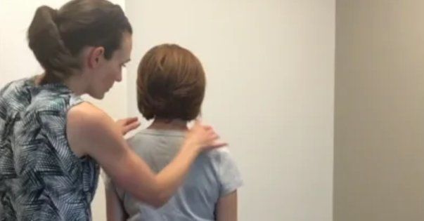
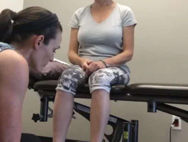
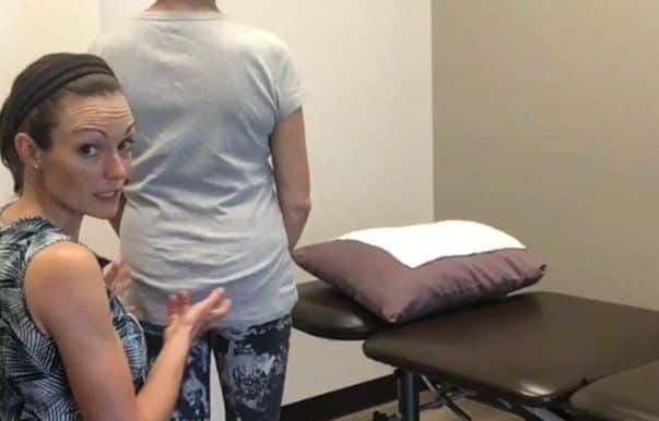
Case Study Author

Leigh Fortuna
Diane Lee & Associates
ISM Certified Practitioner
102-15303 31 Ave, Surrey, BC V3Z 6X2
Clinical Mentorship in the Integrated Systems Model
Join Diane, and her team of highly skilled assistants, on this mentorship journey and immerse yourself in a series of education opportunities that will improve your clinical efficacy for treating the whole person using the updated Integrated Systems Model.
{"first_class":"1","title":"Heading","show_title":"0","post_type":"course","taxonomy":"","term":"","post_ids":"3346","course_style":"recent","featured_style":"custom_block_1","masonry":"0","grid_columns":"clear1 col-md-12","column_width":"200","gutter":"30","grid_number":"1","infinite":"0","pagination":"0","grid_excerpt_length":"100","grid_lightbox":"0","grid_link":"0","css_class":"block-small-thumb","container_css":"","custom_css":""}
We will come together for 3 sessions of 4 (4.5) days over a period of 6-8 months with lots of practical/clinical time to focus on acquiring the skills and clinical reasoning to put the ISM model into practice. Hours of online lecture and reading material and 12 hours of in-person lecture are...
More Info