Story
Her physician referred Mrs. T for a physiotherapy assessment. She was assessed first time on 8th April 2016.
Ms. T is a 55-year-old nurse who has long history of recurrent thorax pain past 15- to 20-years. She remembers her symptoms have been recurrent since teenage, but after her second pregnancy symptoms have started to appear more frequently. She has given birth to two children, both with C-section. Before her recurrent issue of back pain in mid thorax area, she remembers having at least two lumbagos before her first pregnancy. When she had the LBP episodes, she also had right knee pain that aggravated with running.
When she goes running, after few kilometers she experiences upper extremity numbness on both arms, she feels pain and discomfort in her cervical-thoracic junction and pain in left shoulder as well as in the mid thorax between her shoulder blades. After flaring up, pain symptoms can go on for 3-5 days. Her upper extremities numbness gets better when she stops running and shakes her arms for a while.
She also reported right arm numbness when not sleeping in her own bed.
She has tried different treatments on her symptoms such as dry needling, physiotherapy and various manual therapy treatments. All treatments have provided temporary or none relief on her symptoms.
Medical history
She has taken painkillers for the pain, when pain is flared-up. Painkillers dulls the pain, yet won’t take it away completely.
She has been in a car accident 20-year-ago. She drove a car off the road ending upside down in a ditch. She had seatbelt on and she didn’t have any injuries due the accident based on the medical assessment after the accident.
There is a history of brain and heart diseases in her family. She has been in a heart assessment year 2010 where there were noted a small leak on aortic flap. She has had shorts episodes of chest pain, which concerns her.
She had a protrusion on L4 radiating symptoms on her right lower leg in 2006.
Her right knee was RTG-imagined in 2008 after period of pain and discomfort. In the assessment there was diagnosed knee hydrops, her knee was punctured at that time.
Meaningful Complaint
At the initial assessment, Mrs. T complained two areas of pain:
- Pain between shoulder blades, aggravated with running.
- Pain in the thoracic-cervical junction, aggravated with running.
She also reported numbness of her upper extremities while running and when sleeping somewhere else than in her own bed. Numbness starts from her arm going all the way to around her wrist. Her symptoms are clearly aggravated with activities and will go on for 3-5 days after flare-up.
Diane’s question
Given that Mrs. T has experienced chronic pain for 15 – 20 years, what findings from her story suggest that her pain is centrally mediated and what findings suggest there is also a peripheral mediation component?
Lasse’s answer
The fact that she has symptoms 3-5 days after a flare-up and relief of symptoms when shaking her hands after provocation suggests that there is a peripheral mediation component in her story.
Numbness of the right arm when not sleeping in her own bed, symptoms on both upper extremities, cervical-thoracic junction, left shoulder and mid thorax area after few kilometers of running, the fact that painkiller only dulls the pain, small leak on aortic flap and history of knee hydrops implicates centrally mediated component.
Cognitive Belief
In Mrs. T’s family women are prone to have same kind of symptoms as she does. Her mother and grandmother have had mid-thorax pain and upper extremity numbness, so she strongly feels that she is structurally prone to have back stiffness and other symptoms as noted before.
Diane’s question
Do you feel that this cognitive belief is a barrier to her recovery and if so, how would you address this barrier?
Lasse’s answer
I believe this cognitive belief is a barrier and it is crucial to her recovery that is addressed. I address this barrier by educating her how the body works, how different tissues react to different stimulus e.g. central nervous systems and brain plasticity can change it in short periods of time. Addressing this while changing experience how she moves are major components of the treatment.
Meaningful Tasks
Based on Mrs. T’s story, the meaningful asks were identified as follows:
- Running
The screening tasks that were chosen to further assess were chosen from the requirements to perform the above meaningful task – running. Screening task side was determined by her subjective experience which side was harder to perform in terms of balance, effort and ease. They included: Standing postural screen, seated trunk rotation to the right, one leg standing on the left side.
Diane’s question
Why did you choose to assess the one leg standing task only on the right side when running requires her to transfer load alternately through both legs? Why did you choose seated trunk rotation to screen as opposed to standing trunk rotation since standing thoracic rotation would simulate the requirements of running?
Lasse’s answer
One leg standing tasks was chosen by which side she felt harder to lift the leg and/or maintaining balance. I chose seated trunk rotation over standing trunk rotation because she felt clear difference between sides and had provocation of familiar symptoms in seated trunk rotation, but experienced no difference in standing rotation even though there was significant difference between sides. Screening tasks were chosen to promote change in the experience of the movement to help to change her cognitive beliefs.
Diane
Excellent answers! A key feature of the ISM approach is to bring awareness to different strategies. If the patient can’t feel a difference, you are not likely to change the brain map for that task.
Screening Tasks
Standing Postural Screen
This screen was chosen as a screening task to define starting position of other tasks, so that we can determine what changes in posture, alignment, control and biomechanics are relevant in other screening tasks.
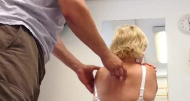
There were no reported symptoms during evaluation of the static standing postural screen. Mrs. T stood with a left intrapelvic torsion (IPT) (added information after questions below – accompanied with left transverse rotation (TPR)). Right hip is anterior relatively to the right acetabulum. The low thorax is rotated to the left. Specifically, 9th thoracic ring is in right translated/left rotated position in standing. In the mid thorax 4th thoracic ring is left translated/right-rotated position and 5th thoracic ring is in right translated/left rotated position. Her C4-6 were left translated/right rotated.
Diane’s questions
- Is there an associated transverse plane rotation (TPR) of her pelvis in standing?
- For all the regional rotations noted please list those that are congruent with the direction of the pelvis and those that are incongruent and why it is important to note this?
Answer
- Yes, there was a TPR left in the standing postural screen.
- In the regional findings congruent rotations in relation to the rotation of pelvis are: right hip anterior translation, 9th thoracic ring, 5th thoracic ring. Incongruent rotations are: 4th thoracic ring, C4-6. It is important to note this due the fact that some congruent rotations might be simply following other sites rotating the same way.
Diane’s further response
Yes, congruent regional findings suggest that the two regions are ‘following one another’. Incongruent regional findings often direct you to the location of the driver between regions.
Left one leg standing
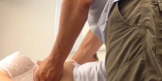
This screen determines the ability to transfer load in one leg standing and is crucial assessment for her meaningful task of running, which requires single leg loading. Side was determined by asking her which side is more effortful. When she lifts her right leg, her pelvic girdle feels unstable.
In this screening task her pelvic girdle feels unstable. When she performs the screening task her 9th thoracic ring translates right/rotates left. 4th and 5th thoracic rings maintain its positions, 4th thoracic ring keeps rotated to the right and translated to the left and 5th thoracic ring keeps rotated to the left and translated to the right. C4-6 rotates even more to the right. At right SI-joint Ilium fails to rotate posterior in relation to sacrum. Left hip rotates internally and adducts.
Manual correction of the left hip resulted better mobility of the right SI-joint and partial control of the 9th thoracic ring. 4th thoracic ring keeps rotated to the right, translated to the left and 5th thoracic ring keeps rotated to the left, translated to the right. Correcting the left hip and 9th thoracic ring resulted best experience as task felt stable and easy. With 9th thoracic ring correction her cervical spine and thorax stays better aligned except 4th and 5th thoracic rings, which stays in the same position during the task. With 9th thoracic ring correction her left hip feels, in her words, “wobbly”.
Correcting 4th and 5th thoracic ring was impossible due stiff end-feel on thoracic rings when correcting the thoracic rings. With a manual hip correction 5th thoracic ring could be corrected a little, 4th thoracic ring did not correct with any manual correction.
Primary driver for this task is left hip and secondary driver is 9th thoracic ring. It is still unclear what is driving the 4th and 5th thoracic rings, yet I found no correction to correct the site in initial assessment. Area is to be assessed further.
Seated trunk rotation to the right
Screen determines ability to perform normal biomechanics, control and alignment during her meaningful task of running, which requires trunk rotation on the opposite side of the loaded leg when performed optimally. As stated before, this task was performed seated due the experience of the movement.
In the screening task (seated trunk rotation to the right), she reports familiar stiffness in her mid thorax at T4-T5 area. When she rotates to the right C4-6 keeps rotated to the right, 4th thoracic ring maintains rotated right and translated left. 5th thoracic ring keeps rotated left and translated right.
Diane’s question
Given your findings noted above, which segments/thoracic rings are incongruent to the task and would therefore be corrected first to assess the impact of this correction on trunk rotation to the right? Why?
Lasse’s answer
From the finding above, 5th thoracic ring is incongruent to the task as it should rotate to the right and translate left. Assessing impact on correction of 5th thoracic ring to the other sites should be assessed first. As noted below also 9th thoracic ring is incongruent to the task and should be assessed.
9th thoracic ring rotates left and translates right even more when she rotates right. After 9th thoracic ring fails to transfer load optimally, she reports stiffness at 4th and 5th thoracic ring area. 9th thoracic ring correction results more range of motion and no symptoms in mid thorax area.
Diane’s question
What happened to the biomechanics of the 4th and 5th thoracic rings with the 9th thoracic rings correction and what happened to the 9th thoracic ring with the 4th and 5th thoracic ring correction. These tests are essential for confirming your hypothesis that the 9th thoracic ring is a primary driver for this task.
Lasse’s answer
4th and 5th thoracic rings couldn’t be corrected with or without 9th thoracic ring correction, even though her experience of the task improved with 9th thoracic ring correction. With 9th thoracic ring correction correcting 5th thoracic ring resulted stiff end-feel with no change on alignment of the rings. This area should be assessed further.
With 9th thoracic ring correction 4th and 5th thoracic rings corrected a bit, they didn’t feel as compressed as without correction. In the task, with 9th thoracic correction 4th and 5th thoracic ring starts to rotate to the right but fails to keep alignment as 5th thoracic ring get jammed (without better word to describe it) right after rotation and 4th and 5th thoracic ring get compressed again. (see Video 1seated_rotation.mov).
Diane's Question
The right hip correction should occur as she sat or at least be assessed in sitting especially since it was a driver in the one leg standing task and running does not occur in sitting. Was the femoral head still anterior in the seated position? Was the pelvis still in a left IPT? Please hypothesize what the impact of a rotated pelvis and asymmetric hip position could be on the 9th thoracic ring and one underlying mechanism for this possible relationship.
Lasse’s answer
Right hip couldn’t be manually corrected in this task. Ít was translated anteriorly in relation to acetabulum in the seated position.
Right femoral head is anterior was anterior in the seated position and I was unable to manually correct it.
Screening Task Summary
Findings suggest areas of the body where to start the treatment. The primary driver is an area of the body that results the best total response in the task. The secondary driver is an area of the body that results partial improvement in the body, not fully correcting other site of failed load transfer. This knowledge helps to coordinate what areas of the body need to address treatment.
In summary:
- Right OLS primary driver – Left hip secondary driver: 9th thoracic ring
- Seated trunk rotation: Primary driver – 9th thoracic ring
4th and 5th thoracic rings were glued together and couldn’t be corrected in any position. This site will be assessed in further appointments. 4th and 5th thoracic rings felt to be compressed, meaning no joint play and no movement of the thoracic rings when correcting the area. With 9th thoracic ring correction and in child pose 4th and 5th thoracic rings could be corrected partially but they moved together implicating that area should be assessed further.
Diane’s question
I’m curious as to why you would defer further assessment of the 4th/5th thoracic rings to a later appointment. Maybe releasing this region would result in your best correction? Even in an inverted position the 4th and 5th thoracic rings were not capable of independent motion. Tell me more about your clinical reasoning process to leave this compressed/stiff region that is so highly relevant to running until later?
Lasse’s answer
Correcting 4th and 5th thoracic rings gave a compressed feeling – no hard end-feel (joint restriction) or no particular vectors. Hip correction gave her better experience and quality of movement. If hip would be driving 4th and 5th thoracic ring dysfunction, releasing that area would have aggravated her symptoms. Also, her previous treatments included multiple manipulations at T4-5 level with short duration relief of symptoms. Due to these facts I decided not to assess 4-5th thoracic rings within the treatment sessions where time is limited.
If the area wouldn’t be the primary driver, I was going to make her symptoms worse or she had no change at all. The hip correction made a clear difference in her experience of the task in positive manner and so buying her in to my approach, not going to the same area as everyone else and having a short time effect.
Diane's response
good clinical reasoning
Her lower cervical spine correct itself with 9th thoracic ring correction suggesting it doesn’t need to be addressed at this time.
Diane’s question
What type of system impairment could make corrections at the 4th/5th thoracic rings impossible and what findings are there in her story that would support the possibility that this type of system impairment existed?
Lasse’s answer
An articular system impairment (specifically fibrosis) would make it impossible to correct the thoracic rings. Very short neural vectors could create glued rings also e.g. m. multifidus. In her story the history of car accident, consistent discomfort in that area supports the possibility of articular system impairment.
Diane's further comment
A history of trauma (car accident) is enough to be causal for stiff joints. Pain is not as relevant as the fact there was trauma.
Vector Analysis
Diane’s question
Please describe the quality (direction, length, speed) of the vector impacting the right hip as well as the 9th thoracic ring. Please describe for both regions, what you felt in your hands on both the correction and release of the correction that suggested the underlying system impairments (neural & visceral). Also describe the vectors you felt when attempting to correct the 4th and 5th thoracic rings.
Lasse’s answer
Vector analysis of the left hip: The left hip was assessed in supine in hip flexion where femur head started to lose centered position, left hip was first taken to position where femur head was centered, 60 degree of flexion and was then taken passively to extension until femur head started to lose its centered position at 35-40 degree. Vectors to the hip was felt coming from inner thigh, long vector from distal end of the medial side of the femur and shorter vector came after from the medial side for the femur.
Vector analysis indicated over-activation of right adductor muscles; more specifically the main vector was from the adductor brevis and adductor longus muscles.
Vector analysis of the 9th thoracic ring (see video 2Vectoranalysis_TR9): The 9th thoracic ring was assessed in supine lower extremities in flexion. Vectors came from the right side of the thoracic ring from anterior ventral aspect of the right rib. First vector was short sucking the rib medially fast and the next vector were felt pulling the rib slowly toward right side of her umblicus. Vector analysis indicated 9th thoracic ring vectors from visceral structures right side of her frontal low rib cage; more specifically it is coming from right liver/gallbladder.
Motor Control
Manually correcting the 9th thoracic ring made right hip to feel unstable during one leg standing on the left side and correcting the left hip partially corrected 9th thoracic ring. Correcting manually the left hip to the centered position and keeping it corrected with 9th thoracic ring partial correction made best result of experience in the initial assessment on the one leg standing on the left side. This suggested that ability to coordinate deep muscle system was to be tested.
Motor control was tested supine with lower extremities flexed.
We initially tested Mrs. T’s ability to recruit pelvic floor muscles and transversus abdominis muscle in supine with external palpation. Tests showed that she had poor recruitment response over her pelvic floor on her left side. The clinical palpation tests were confirmed via ultrasound imaging examination.
During lying in prone, legs flexed, she was able to recruit transversus abdominis muscle with a verbal cue, but she was unable to maintain contraction on her transversus abdominis muscle at the level of 9th thoracic ring while breathing normally. Optimally a person should be able to keep the contraction of transversus abdominis muscle while breathing normally. She had a proper recruitment response over transversus abdominis muscle with verbal cue, but she held her breath while doing so. When she was asked to maintain contraction and breath normally, she failed to do keep the contraction during a verbal cue.
Inability to control lower thorax may result in excessive tone in superficial muscles i.e. QL, EO, IO, IC and so could relate to pelvic control by creating a pull to the pelvic girdle and/or affect lower abdominal pressure.
With the pelvic floor, she had no recruitment response over her left ischiocavernosus muscle and left transverse perineal muscle.
After releasing 9th thoracic ring and left hip, we assessed her deep muscle system again with following findings:
- Transversus activation was still the same, but with 9th thoracic ring correction she had better contraction and she was able to breathe normally while maintaining the contraction.
- With the pelvic floor findings were the same as before, but with 9th thoracic ring correction she had better resting activation over her left ischiocavernosus muscle and left transverse perineal muscle, but recruitment response was still weak.
We tested active straight leg raise test resulting left leg to be harder to lift. With 9th thoracic ring correction and a verbal cue to connect with her pelvic floor lifting left leg was easier. Suggesting better recruitment patterns for the task.
Motor control is an important part of the physiotherapy, and it is crucial to assess deep muscle systems endurance and ability to contract or relax it with intention. Tests suggest that motor control should be included on treatment plan. Motor control should be assessed more after release.
Diane’s question
How do these findings help you determine if specific isolated muscle recruitment training is indicated? Is it? If not, why not?
Lasse’s response
Isolated muscle recruitment training is indicated since she can’t breathe normally while activating deep muscle system. Her ability to coordinate her pelvic floor muscles and transversus abdominis muscle may improve after release pieces of the puzzle, but it needs to be assessed and determine after all the drivers are found. She should be able to have a recruitment response over the deep muscle system muscles keeping the activation on for at least 10 seconds 10 times while breathing normally.
Diane’s further response
This sequence of results suggests that once all the vectors impairing her hip and 9th thoracic ring are released, she will have a better recruitment response of both her TrA and PFM and be able to breathe with no ring correction. In this case, no isolated muscle training would be required.
Hypothesis
Mrs. T’s primary driver is 9th thoracic ring with vector analysis suggesting visceral vector coming from anterior part of the right the thorax, more specifically liver and/or gallbladder. My hypothesis is that her body has had to compensate different stresses over time, suffering car accident and two pregnancies. For this reason she has learn thigh gripping strategy to maintain hip control. Pelvic floor contains muscles that control hip joint as well. If femur head loses centered position in the hip socket, it will change relations of the pelvic floor muscles.
Diane’s response: Above you state: “Correcting manually the left hip to the centered position and keeping it corrected with 9th thoracic ring partial correction made best result of experience in the initial assessment on the one leg standing on the left side.” Therefore this would be defined as a co-driver or secondary driver as you mention below. Depends if they are related 50/50 or if one has more precedence which it seems like the 9th thoracic ring does.
Diane’s question
How does abdominal cavity pressure (below the diaphragm) compress the shoulder girdle? You state that you couldn’t correct the 4th or 5th thoracic rings which could have been significantly impacted in her MVA 20 years ago with a resulting system impairment that has not been considered in this story to this point.
Lasse’s answer
Given that the diaphragm connects to the 6th thoracic ring and psoas connects to diaphragm, dysfunction of these structures have a capacity to compress 4th and 5th thoracic ring. Over activation of the diaphragm is capable to increase abdominal cavity pressure hence compress the shoulder girdle if abdominal and pelvic floor muscles fails to maintain the pressure. If the system cannot maintain abdominal pressure and deep system muscles fail to control the system, upper thorax will be compressed due pressure changes in the thorax and hyper-activation of global muscles trying to do deep systems work.
Diane’s summary
So if I understand this correctly…. Her inability to control her 9th thoracic ring and her left hip are secondary to dysynergistic recruitment patterns of TrA (not sure if left or right side or both) and the left side of her superficial pelvic floor (layer 1). She has compensated for these deficiencies by overactivating her left thigh adductors and while you mention generally ‘hyper-activation of global muscles’ the vector analysis of the 9th ring didn’t reveal this – it revealed a visceral vector from the liver/gall bladder. After releasing the visceral vectors did your 9th ring vector analysis reveal any specific global muscle over-activation? I’m assuming the 4th and 5th rings remained glued given your comment immediately below.
Lasse’s comment
Her dysynergistic recruitment patterns of TrA and the left Pelvic floor is not secondary to her inability to control her 9th thoracic ring and her left hip, but a result of dysfunctional thorax-pelvis junction. I feel that her visceral vectors (liver/gall bladder) are creating reactive response over the diaphragm, internal oblique muscles, external oblique muscles, extensor spinae muscles. After releasing 9th ring vector no specific global muscle vector were felt, as well as 4th and 5th rings still remained glued.
Thoracic-cervical junction and 4th and 5th thoracic rings should to be assessed later on. With no possibility to correct the area and no change in the areas alignment with other corrections suggest that there are still drivers to be found.
Treatment Plan
Initial treatment session (RACM)
Release
Primary driver (9th thoracic ring) (see video 3Release&align_TR9): Secondary driver (hip):
- Release adductor longus and adductor brevis muscles of the left hip, using positional release with awareness and trigger points.
- Release of 9th thoracic ring with ring stack and breathe, combining it with vector specific stretch. Correcting 9th thoracic ring alignment as much as possible, then rotating thorax to find the specific vector, in this case rotating thorax upward and lateral (see video 4Seated_rotation_re-test for post release improvement in seated right rotation). After local release, rotating lower extremities were used with 9th thoracic ring alignment to find vector pulling to the 9th thoracic ring.
Align
- 9th ring alignment corrected left hip alignment after release. Best cue: let your thoracic rings float. 9th thoracic ring and left hip was first released manually.
Control
- Pelvic floor contraction was evaluated with ultrasound initially and after release of the left hip and 9th thoracic ring
- Pelvic floor contraction was possible after hip release, but she was unable to maintain contraction. – This suggests an endurance deficit.
Relationship between the 9th thoracic ring alignment and hip alignment and impact on pelvic floor recruitment were assessed both manually and with ultrasound imaging. 9th thoracic ring alignment cue (let your thoracic rings float) clearly restored her hip alignment to neutral. This was felt manually (femoral head centered) and her pelvic floor ability to recruit and relax was seen via ultrasound imaging and felt manually externally.
Move
She was given three home exercises:
- Maintaining of left hip abductor range of motion moving lower extremity passively in supine using strap while maintaining 9th thoracic ring alignment. Breath 3-4 times at the end – Lateral costal breath, which gave the best sensation of release perform 2-3 times before other exercises.
- Supine 9th thoracic ring alignment combined with Pelvic floor activation three second hold 10 times a day 2-3 times a day to work on pelvic floor endurance.
- Left one leg standing 10 high quality repetitions, using mirror and/or her hands to ensure optimal thorax and optimal hip alignment, three times a day. Palpating thoracic rings and hip was taught to her step by step, finding out the easiest way for her to be sure she is maintaining optimal strategy for the task.
Exercises was to be done in given order concentrating on increasing pelvic floor endurance and control, maintaining hip range of motion and promoting brain plasticity to create better movement strategy for meaningful task.
Follow-Up treatment sessions
After the initial treatment session she reported ease of her mid-thorax symptoms. After two week after the first visit she was able to run half marathon without flaring up any symptoms. She still had some feeling in her cervical-thoracic junction and arm numbness while running, but symptoms didn’t stay for days. She felt she could run more effortless as before.
In the follow up treatment sessions left hip needed less of releasing and needed more concentration on control. 9th thoracic ring needed releasing on every session, but on third session we noted that 4th and 5th thoracic rings could be partial corrected manually. With 4th and 5th thoracic ring correction range of motion in thoracic rotation increases flaring up cervical-thoracic junction stiffness and pain. 4th and 5th thoracic ring relationship with shoulder girdle and cervical spine is to be assessed later on.
In the follow up treatment sessions home exercises were progressed to thoracic rotation in sitting and in lunge, one leg standing with partial one leg squat and hip rotation via trunk and pelvic.
Diane’s comment
This is a great case report that highlights the many ‘layers of the onion’ that our patients with long histories of multiple injuries present with. While I may have released the more resistant vectors of the 4th and 5th thoracic rings earlier than Lasse did, he chose to go with restoring function of her left hip and 9th thoracic ring. This treatment did not resolve her arm symptoms while running, she was able to perform at a higher level with less effort, so key pieces were resolved in her total story.
One last question Lasse
At this point it appears that the 4th and 5th thoracic rings are not the next driver to go after because of the flare up of her cervico-thoracic stiffness and pain. When you were finally able to correct these rings what happened to C4-6? If you didn’t assess this, can you hypothesize how you would have determined whether to further release the mid-thorax or move into an assessment/treatment of her neck/thorax junction?
Lasse’s comment
I haven’t seen her yet to cover that, but my believe is that after 4th and 5th thoracic ring release there could be vectors to the C4-6 area from breathing systems muscles and shoulder girdle muscles, such as scalene muscles, serratus anterior muscles, trapezius muscle. Shoulder girdle coordination will be one key element to be assessed and treated if there is something dysfunctional. Integrating shoulder girdle control to the core control in her meaningful task is progression after learning how to isolate cervical spine, shoulder blade and thorax from each other.


Case Study Author

Clinical Mentorship in the Integrated Systems Model
Join Diane, and her team of highly skilled assistants, on this mentorship journey and immerse yourself in a series of education opportunities that will improve your clinical efficacy for treating the whole person using the updated Integrated Systems Model.
{"first_class":"1","title":"Heading","show_title":"0","post_type":"course","taxonomy":"","term":"","post_ids":"3346","course_style":"recent","featured_style":"custom_block_1","masonry":"0","grid_columns":"clear1 col-md-12","column_width":"200","gutter":"30","grid_number":"1","infinite":"0","pagination":"0","grid_excerpt_length":"100","grid_lightbox":"0","grid_link":"0","css_class":"block-small-thumb","container_css":"","custom_css":""}
We will come together for 3 sessions of 4 (4.5) days over a period of 6-8 months with lots of practical/clinical time to focus on acquiring the skills and clinical reasoning to put the ISM model into practice. Hours of online lecture and reading material and 12 hours of in-person lecture are...
More Info