Story
Miss. A. was referred for assessment from another physical therapist. She was initially assessed on May 12, 2017.
Miss A. is a 16 year old competitive dancer with a goal of dancing professionally. She dances in multiple disciplines up to 25 hours per week, but ballet is reportedly her strongest discipline and that which she is most passionate about. Her primary reported complaint at the time of assessment was continued left foot pain after suffering an avulsion fracture of her navicular on December 6, 2016. After the injury, she was stabilized in an aircast for 7.5 weeks and then commenced physiotherapy treatments 1-3 times per week. As she would improve in her range of motion and strength she would return to dancing, and on multiple occasions would have to subsequently stop due to secondary foot/ankle “injuries”. She spent the dance season resting, then returning to full dance, then being forced to stop due to reinjury or pain. She reported being frustrated about this as she would return to dancing in the numbers she would be choreographed in, and then have an injury (rolling her ankle, for example), then be told she could not compete for that number. After several attempts she was “pulled” out of all numbers by her teachers as changing the choreography to bring Miss A in or out of the numbers was reportedly a challenge. This information is included in the story as Miss. A emphasized several times that this was incredibly frustrating as she believed she had the capability to perform and compete. X-ray imaging had shown the fracture to be healed and Miss. A stated she felt she should be able to dance given her injury had healed.
Diane’s question:
There is a cognitive belief that is inconsistent with this story. It may present as a barrier to her recovery if not addressed. What is it?
Kristine’s answer:
Miss A has a cognitive belief that her fracture was healed and that she has the capability to perform and compete despite having repetitive re-injuries indicating that she was not at a level of recovery required for her to perform. She was focused on her pain and symptoms especially during training yet prided herself in her ability to perform without letting them “stop” her.
Diane’s response:
Absolutely! And why could this present as a possible barrier for your rehab program and how would you address this barrier with her?
Kristine’s answer:
This could be a potential barrier to rehabilitation as during rehab sessions she would focus on the symptom of pain and not on the quality of movement of her foot or the rest of her body, and this could limit her ability to progress through rehabilitation exercises. This would then limit her function in dance as she would potentially not have the motor control capacity needed to perform the tasks she was performing, but would continue anyways, thus utilizing non optimal strategies and potentially causing exacerbations of injuries. There was also inconsistency as she would use the injury as a limitation in daily life and require numerous treatments to address her various complaints, yet she would not allow the same injury to stop her from doing her chosen activity which was dance. I could address this by setting solid expectations around restrictions in dance while participating in a rehabilitation process, which, if not met, would impact her ability to participate in rehab.
With further questioning Miss A shared that prior to the fracture she had had chronic pain in her left lateral knee and hip, left hamstring, and that she was being treated for chronic ITB syndrome as well. She reported being seen weekly before her fracture by a physiotherapist and the treatment focused on releasing her left ITB and hamstring. She also had mild neck pain and stiffness that she attributed to dancing and posture. This was reportedly never assessed or treated. When asked whether she had prior challenges with her dance technique, or if she had any corrections that were being continually made by her teachers, she did not have any insight. Her mother did say however, that Miss A would wear through her pointe shoes at a rate that seemed much higher than other dancers (one pair for every 30 minutes on pointe) and that she always found this odd.
Diane’s question:
What part of the navicular was avulsed and by what structure? Did her mother say ‘how’ she would wear through her pointe shoe i.e. what there a side, or part, that broke down first? How would this information help you understand that relationship between her prior proximal complaints and her foot injury?
Kristine’s answer:
The navicular was avulsed at the tuberosity at the most lateral aspect of the tibialis posterior insertion site. By looking at her old pointe shoes it was evident that the platform was wearing out quickly – and faster on the left more-so than the right. The shank of the left shoe broke down first and even had a palpable break at the level of the midfoot. I interpreted this wear pattern to potentially be that she was loading more heavily on her left foot compared to the right, and also that she had excessive mid foot flexion that was not controlled at end range (so she was gaining extrinsic stability from the shoe itself). Upon questioning she also stated that she tends to switch her shoes occasionally (the left and right foot) as it seems to help them last longer.
Diane’s comment:
Good hypothesis. It appears that something extrinsic to the foot is altering her base of support to be more to the left foot and something intrinsic to the foot is overloading the midfoot region.
Kristine’s response:
Your words describe my thoughts perfectly!
Other pertinent history included a left ulnar fracture in 2008 and physical urticaria and dermatographism suggesting a heightened immunological response to food and environmental allergens. This limits treatment techniques as she has had allergic reactions to any proprioceptive tape she has tried.
Prior to starting dance at age 11 Miss A had trained as a gymnast. She reported having multiple small injuries but nothing that “stood out”. She did however report having a significant fall on the high beam at age 9/10 where she injured her tailbone and had to stop training for a “week or so” while it healed. It was never x-rayed and there was no post injury rehabilitation.
When she was initially assessed on May 12th she had a goal of completing her grade 8 ballet exams May 30th, having several weeks off dance in the spring, then returning to summer intensives and full dance by mid summer.
Meaningful Complaint
Miss A complained of pain “over her left navicular bone” with any dance, and occasional pain “over her cuboid” with more prolonged dance (greater than 2 hours) or pointe work (over 10 minutes). She described the pain as sharp with dancing and achy afterwards. She reported occasional achy pain with walking as well. On questioning Miss A reported that she was weaker on her left ankle, but the pain was her primary complaint.
Cognitive Belief
Miss. A reported that she believed the majority of her symptoms were caused by the fracture in December 2016, but that she knew there was a connection to her other symptoms and that there “must be something else going on or it would be better by now”. She was tearful during the subjective assessment and she reported being fearful that she wouldn’t be able to dance again. She also reported a belief that she needed to complete her ballet exam as if she didn’t it would prevent her from moving forward with her class and would impact her long term success as a dancer. She also reported that it had been very difficult to refrain from dancing “full out” upon her trialed returns to dance as it was competition season and thus there was a strong focus on performance. She reportedly did not see a prolonged departure from dance as an option as she had already taken time off as the fracture healed.
Diane’s comment:
She definitely doesn’t have ‘fear avoidance’ but her inability to pace for healing is problematic.
Meaningful Task
For Miss A. the meaningful task was a full return to full time dance training (>25 hours per week) and performance.
Screening Tasks
Based on Miss A’s expressed goals and challenges, the meaningful tasks identified at the time of the assessment included the following.
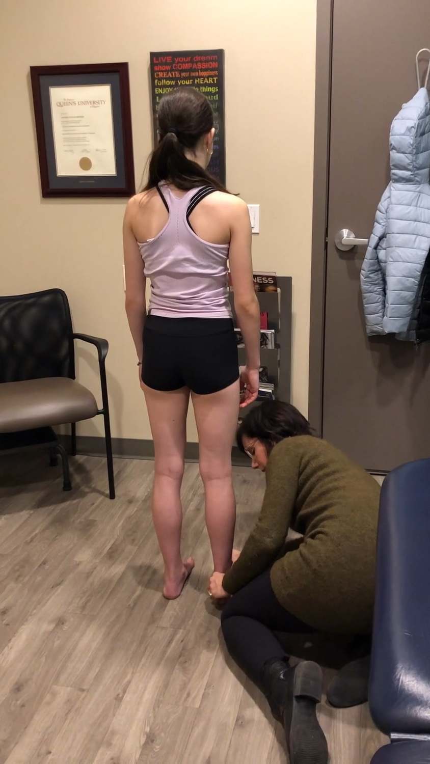
1) Standing postural screen
2) Relevé – in first position
3) Demi-plié – in first position
For both tasks the upper extremities were left in neutral first position. These 2 tasks were chosen as they are the basis for any jump or turn in ballet and would allow us to assess the alignment, biomechanics and control of the left foot while doing a bilateral standing task.
Diane’s comment:
These are good choices since there wasn’t one particular movement in dance identified as being more problematic than another. Dance in general was chosen, which can make it difficult to choose screening tasks.
Kristine’s comment:
It was difficult to determine screening tasks as Miss A could not decipher what movement(s) made her symptoms worsen. I struggled with whether to assess in turnout or neutral foot position, as she dances in multiple disciplines. I chose turnout in ballet first position as ballet is her primary area of study and where she has the highest goals set for herself.
Standing Postural Screen
Even though Miss A’s meaningful task was ballet dancing it was important to assess her standing posture to determine existing postural alignment and control within and between regions while weight bearing through her feet, especially given that she also experienced some mild pain with walking. The standing postural screen also allowed for assessment of the starting positions for both the relevé and demi-plié tasks.
Optimal alignment in standing includes a neutral pelvic girdle with no rotations or torsions, and both SIJs controlled, femoral heads centred in the acetabulum with the knees in neutral and the talus centred in the mortice, with weight bearing through the four corners of the foot, and the foot in neutral. It also includes a neutral head and neck on a stacked thorax, with no rotations within the thorax or cervical spine and no torsions within the cranium.
In standing there were no reported symptoms.
Kristine’s original presentation of her findings:
On assessment of Miss A’s standing posture there was an intracranial torsion to the left and the sphenoid was rotated to the left. The mandible was deviated right and compressed on the left. There were multiple Cspine shifts including C2,3 and 7 to the right and C1,4,5,6 to the left. She had a mild intra shoulder girdle torsion to the right and her left sternoclavicular joint was compressed. The upper thorax was compressed but there was no overall thoracic or pelvic rotations. The hips were both centred equally, the left ankle was compressed at the distal tib-fib joint and the hind foot was in slight inversion.
Diane’s comment:
There has been an update since your last submission and course in how we organize findings in ISM. This came from our trying to help clinicians expedite their findings and to avoid a ‘laundry list’ of findings and getting way off track with corrections. We are now encouraging people to find drivers in what we’ve called ‘functional units’ first and then play each driver off one another to determine the ultimate drivers. For your standing screen findings, this new way of organizing your knowledge would look like this:
Functional Unit #1 (3rd thoracic ring to hips) (FU#1):
Pelvis: Neutral and controlled
Hips: Centered
Thorax: Upper thorax compressed, no significant thoracic rings translated/rotated
Driver: No driver found for FU#1 for since this was a starting screen for subsequent tasks.Functional Unit #2 (2nd thoracic ring to cranium) (FU#2):
Shoulder girdles: mild torsion to the right, left sternoclavicular joint (SCJ) compressed
Cervical: C2, 3, 7 right translated/left rotated, C1, 4, 5, 6 left translated/right rotated
Cranium: Intracranial torsion left with a congruent left rotation of the sphenoid
Driver: No driver found for FU#2 for since this was a starting screen.Functional Unit #3
Foot: left tib-fib joint compressed, left calcaneus slightly inverted to the lower leg
No driver found for FU#3 for since this was a starting screen.I took this information from the information you provided above and I think you’ll agree it is much easier to read/follow when organized like this rather than in a narrative sentence.
Kristine’s response:
SO MUCH MORE CLEAR!!!!
And I think in a typical assessment in the clinic this is more how I’ve always organized my thoughts. I was trying to include every single thing for a case study but can see how getting too involved in unnecessary assessment is time consuming, often unnecessary, and can end up just flaring up a client and aggravating an already sensitive nervous system. (and I love the FU)
Diane’s comments & questions:
Reflecting on how findings are in relationship with one another expedites finding drivers. Often the first place to implement a correction is between regions where there is incongruence.
Please note if the following findings are congruent or incongruent to the others
FU#2:
- LICT and left rotated sphenoid: congruent (<- Kristine’s response)
- LICT and right deviation of mandible: incongruent (<- Kristine’s response)
Diane’s comment:
Kristine – this was left as congruent from your last draft of this report. Can you explain to me the osteokinematics that make a right deviation of the mandible congruent with a LICT?
Kristine’s response:
Nope I can’t I was completely off the mark. They are incongruent.
Diane:
Whew!!
Diane's question (cont'd):
- LICT and C2, 3, 7 right translated/left rotation: direction of rotation is congruent
- LICT and C1, 4, 5, 6 left translated/right rotated: direction of rotation is incongruent
Diane’s follow-up question:
In functional unit #2, given the above findings and their congruence relationship, IF you were going to find a driver for the standing task, where would your first correction be and why?
Kristine’s response:
For the standing task I would begin by correcting the cranium-atlas-axis (cranial region) as there is an incongruence between the cranium and C1, with the cranium and mandible and the cranium and middle cervical segments (C4, 5, 6).
Diane's question (cont'd):
- Fill in the blanks. In a right intra-shoulder girdle torsion the left clavicle should be rotated___ anteriorly___ and the right clavicle should be rotated ____ posteriorly___
- What joint do you mean when you say the ‘left ankle was compressed’?
Kristine’s response:
In screening the ankle in standing posture there was reduced play at the distal fibula relative to the right leg. The soft tissues at the lateral ankle/foot had restricted mobility and the distal fibula had a palpable feeling of being compressed towards the tibia, with the talus and calcaneus being compressed towards the fibula.
Diane’s clarification:
So, I think you mean that both the distal tib-fib joint and the lateral talocrural and lateral subtalar joints were compressed?
Kristine’s answer:
Yes
Diane's question (cont'd):
- What joints are involved in ‘hindfoot inversion’?
Kristine’s answer:
The subtalar 3 joint complex
Diane's question (cont'd):
- What are the resultant arthrokinematic glides at the relevant joints during hindfoot inversion?
Kristine’s answer:
During hindfoot inversion or pronation the anterior calculus rotates laterally and the talus plantar flexes and adducts relative to the calcaneus.
Diane’s comment:
Foot terminology is so confusing! In 2018 during the Denver Series, Cathy Rogers and I worked hard to come to an understanding of the osteo and arthrokinematics of the hindfoot joints because we found people were confusing inversion (which can only occur in open chain unless the talus tilts in the mortice – and this is sub-optimal – always). Hindfoot inversion is a turning of the entire foot such that the sole of the foot faces medially. Since the ground blocks the lateral aspect of the calcaneus from descending when standing, inversion forces the medial aspect of the talus superiorly i.e. tilts the talus in the mortice. Pronation, on the other hand cannot occur in open chain and requires ground reaction forces to produce the following biomechanics. During pronation (which occurs in midstance or during a squat) the talus plantar flexes and adducts relative to the calcaneus and the calcaneus laterally rotates (as you said) relative to the talus. Lateral rotation of the calcaneus is totally different than inversion, or tilting of the calcaneus. Lateral rotation of the calcaneus requires the anterior aspect of the calcaneus to glide laterally and the posterior aspect to glide posteromedially (follows the plane of the posterior ST joint). There is no tilting of either the talus or the calcaneus in pronation of the hindfoot.
Diane's question (cont'd):
- What is the impact on the talus in the mortice when the hindfoot is held in inversion?
Kristine’s answer:
The talus remains in dorsiflexion/abduction and external rotation.
Diane’s comment:
This is true in the open chain position. When the foot is on the ground and the hindfoot is inverted, the talus tilts medially in the mortice.
Diane's question (cont'd):
- What is the impact on the navicular at its related joints if the hindfoot cannot move out of inversion for the screening tasks you have chosen?
Kristine’s answer:
For demi-plié if the hindfoot cannot move out of inversion then the calcaneus cannot laterally rotate beneath the talus, and there is the potential for compensatory increased external rotation of the navicular relative to the talus to allow for the pronation that is required to demi-plié. For relevé the foot remains in supination so the navicular will not be as affected by the limited hind foot mobility
Because of ballet being her meaningful task a standing postural screen was also completed in ballet first position. In this position with the hips externally rotated the head, neck thorax and foot/ankle remained in the same relative position. The pelvis posture did change however and there was now a transverse plane rotation to the right, with the sacrum counternutated on the left. There was compression at the sacrococcygeal joint and a compressed left proximal tib fib joint.
Diane’s questions:
To clarify, when Miss A. was standing in ballet first position, the pelvis as a ring was rotated to the right in the transverse plane and the left SIJ was ‘unlocked’ (sacrum was counter-nutated). How did you determine that the sacrum was in this position?
Kristine’s answer:
When Miss A stood in ballet first position the pelvis was rotated to the right and the sacrum was in a position of counter-nutation on the left. This was assessed purely by palpating the sacrum positioning relative to the innominate bones.
Diane’s comment:
Several inter-tester reliability studies have shown that we are not very reliable with ‘pure positional evaluation of bone points’. I have therefore emphasized in teaching that we use motion to confirm the positional relationships between bones. To understand where the sacrum is in relationship to the innominate in standing, I now teach therapists to have the patient offload the side of interest by shifting their weight to the opposite leg (in this case the left SIJ therefore start by shifting weight to the right leg), and then while monitoring the ILA of the sacrum with one hand and the entire innominate with the other, have the patient begin to take load on the left leg just until they come back to standing (i.e. 50% body weight). Nothing should happen between your hands. If the innominate is felt to anteriorly rotate relative to the sacrum (sacrum counternutate) prior to 50% of body weight load, then we can say that the sacrum is truly counternutated relative to the innominate in standing.
Diane’s question:
Can you clarify what you mean, and how you determined, that the left sacrococcygeal joint was compressed and that the left proximal tib fib was compressed? What tests determined this?
Kristine’s response:
What I meant was that upon palpation of the sacrococcygeal joint there was more palpable tension on the left side than there was on the right, and that in standing when I provided a gentle “wiggle” palpation to the left and right sides of the coccyx there was a “stickier” feeling on the left side. The coccyx seemed to be deviated to the left and had good mobility to the left compared to the right (again this was palpated in standing only).
For the left proximal tib-fib joint this was again noted on the scan that the left proximal fibula was compressed superiorly and anteriorly relative to the right.
Diane’s questions:
Did you do any further tests to determine drivers in either standing position? If not, why do you think this wasn’t necessary?
Kristine’s response:
In other sessions (not her initial assessment) I did look at breath in standing to determine if there were other drivers (or changes, or different drivers) that I was missing that could have been adding to her non-optimal alignment in standing alone, both in neutral and in turnout. I rationalized focusing more on the drivers in her tasks as I was finding that no matter what was happening in her body she would never stop moving (even in our waiting room she is standing moving her foot in and out of tendu’s and practicing her releve. I honestly went straight into movement tasks as I found that if she wasn’t going to ever stand still assessing her in standing wouldn’t be meaningful to her…and if I could find some cueing that she could work on throughout the day it would hopefully assist her motor learning as she could incorporate the cues all day long.
Diane’s response:
Exactly – standing was not her meaningful task, dancing was and the standing evaluation was just to give you a reference point for movement analysis changes during the movement screening tasks. No need to find drivers in standing for this case.
Relevé
Assessing Relevé in first position is an opportunity to see how weight is transferred and how motor control is maintained while rising onto the forefoot bilaterally. For a dancer this is an essential movement as it forms the basis of many postures and actions in dance. Optimal strategy for a first position relevé includes maintaining the body in neutral alignment as described in the standing postural screen while flexing at the talocrural joint and mid foot while extending through the MTPs. The foot intrinsics should maintain foot control with no over activation of the tibialis posterior and flexor hallucis longus musculature.
Diane's question:
Relevé requires a controlled foot. What position (supination or pronation) provides more form closure and thus passive control for the foot?
Kristine’s response:
supination
Diane's question:
Can you describe the specific osteokinematic position of the subtalar joints, talonavicular, calcaneocuboid, cuneiforms and metatarsals beyond the sagittal plane motion that you have noted, that are required for optimal control in releve?
Kristine’s response:
For optimal control in releve the foot is maintained in supination. Osteokinematically, at the subtalar joint the anterior calcaneus rotates medially and the talus dorsiflexes and abducts relative to the calcaneus. The navicular is rotated internally relative to the talus and the cuboid is in external rotation relative to the calcaneus. The medial cuneiform internally rotates relative to the middle cuneiform, and the lateral cuneiform laterally rotates relative to the middle cuneiform. They are all drawn dorsally in space. The first metatarsal is in a position of adduction, internal rotation and plantar flexion and the second is in a neutral position ((of rotation) and plantar flexes. The remaining metatarsals are in a position of external rotation, adduction and plantar flexion.
Diane's question:
Which muscle from the lower leg attaches to the navicular and would be of interest to assess in this task given her story?
Kristine’s response:
Tibialis posterior
Kristine’s Original Presentation of Findings for Relevé
During Miss A’s relevé the left ICT increased very early in the movement and the right TPR of the pelvis increased early as well. The left SIJ remained unlocked.
She lost intrinsic foot control (as observed by her foot sickling in) at the same time as the ICT increased. All other sites of impairment remained the same during this task.
Correcting the left ICT made the right TPR of the pelvis worse.
Diane’s question
What happened to the left SIJ control i.e. nutated sacrum. I’m assuming this cranial correction did not restore control to the left SIJ?
Kristine’s response:
no there was no change
Correcting the sacrococcygeal joint improved the cranium, pelvis, and also improved the foot control as she was able to complete a relevé while maintaining her foot position
At the end of the range of motion of the relevé she did lose foot control and went into excessive mid foot flexion and MT1 and 2 rotation. This was not seen with the other assessed corrections as she was not able to perform the full relevé task due to the early loss of control through the foot.
Diane’s original questions:
- Is the direction of rotation of the cranium and pelvis congruent or incongruent?
Kristine’s response:
Congruent
Diane’s response:
it’s actually incongruent. The cranium rotated left (left ICT) and the pelvis rotated to the right.
Kristine:
ummmm yep brain fart.
Diane:
LOL!
Diane’s original questions:
- Define ‘foot sickling in’.
Kristine’s response:
In relevé if there is a loss of mid foot control, or supination, and it falls into a pronated position with increased weight through the medial forefoot, dancers refer to this as a sickled foot.
Diane’s response:
In ISM, we reserve the terms ‘pronation and supination’ for the optimal osteokinematic motion that occurs in the hindfoot, midfoot and forefoot during a squat or midstance of gait (pronation) and heel rise or push off of gait (supination). A sickled foot (shape of a C or sickle) is neither pronation or supination. Rather it is often a collapsed shortening of the medial longitudinal arch often associated with external rotation of the first ray. You are right that it is absolutely a result of loss of intrinsic muscle control and excessive superficial muscle dominance.
Kristine:
Thank you for the clarification – to be sure I understand – is the naming different because there is no ground contact of the hind foot? Diane’s response: not really, a ‘sickle-shaped’ or C-shaped midfoot as you describe is often an adduction collapse associated with either excessive internal or external rotation of the navicular – neither of these motions occur in pronation or supination. It is a buckle of the foot.
Diane’s original questions:
- Two immediate biologically plausible mechanisms for these findings to be related come to mind:
- the loss of intrinsic control of the foot may be causing adverse neural tension that cannot be adapted to within the rest of the system (local primary musculoskeletal problem)
- the nervous system does not have enough length going into releve and the foot may be attempting to “shorten” the distance from coccyx to foot to prevent further tension in the nervous system (primary nervous system problem)
Clue: the fall on the tailbone in gymnastics is a clue here. The foot ‘sickling’ may be an attempt to “shorten” (or approximate?) the nervous system as there may not be enough mobility and/or ability to permit elongation of the nerves passing through the foot.
- Two immediate biologically plausible mechanisms for these findings to be related come to mind:
Diane:
The New Process of Finding the Driver(s) for the Releve Task
To update to the new way of expediting finding drivers I have reorganized your findings below. See what you think.
Findings in FU#1:
Pelvis: Right TPR increased early, left SIJ remained unlocked
Hips: no comment
Thorax: no comment
Driver for FU#1: pelvis – since only one site of impairment is mentionedFindings in FU#2:
Cranial: left ICT increased very early in the task
Cervical: no mention
Upper thorax: no mention
Driver for FU#2: Cranial – since only one site of impairment is mentionedFindings in FU#3:
Foot: Left foot lost alignment and control
Knee: no mention
Driver for FU#3: left foot – since only one site of impairment is mentionedRelationship between drivers of the functional units:
Correcting the cranium (FU#2) worsened the alignment of the pelvis and did not provide pelvic control
Correcting the pelvis (FU#1 – sacrococcygeal joint) improved the alignment of the cranium (and thus the neck) and also improved control of the left foot as she was able to complete a releve and maintain optimal foot position until the end of the range of motion of the releve. At this point, her foot excessively flexed at the mid-foot.
Drivers for releve task:
Primary driver: pelvis
We don’t say the sacrococcygeal joint is the driver or the SIJ – both joints are part of the pelvis, thus the pelvis is the driver.
Secondary driver: left foot
Diane’s question:
Which biologically plausible mechanisms for adverse neurodynamics (a or b) does this finding support – a primary MSK problem or primary neural/dural problem?
Kristine’s answer:
This supports a primary neural/dural driver for the SIJ, cranium and foot with a secondary local muscleskeletal driver for the intrinsic control
Diane’s response:
Actually, these findings support a primary musculoskeletal cause for the adverse neuromuscular tension since correcting the alignment of the pelvis (sacro-coccygeal joint) resulted in improved position, biomechanics and control of multiple others sites of impairment. When an inability to permit elongation of the nervous system is the underlying impairment, correcting one site of impairment always worsens something else. There is no MSK correction, or combination of corrections in the body that results in improvement of others and the task.
Kristine’s response:
gotcha. I was interpreting it as since the pelvis corrected the cranium/neck and the vector was dural that it was a neural system impariment. But yes I understand that if the neural system does not have the ability to elongate that no single correction would make improvements in other body regions)
Demi-Plié
Assessing demi-plié in first position provides an opportunity to assess how the foot responds to a squatting task with the hips in a turnout position, as used in ballet. Optimal strategy for a first position plié is similar to that of a squat, in that there should be no increase in rotations or torsions within the pelvis, the femoral head position should be maintained centred in the acetabulum, the knee should not change in compression and should stay aligned along the second metatarsal.
Diane's question;
Please describe the optimal foot and ankle biomechanics required for squat/demi-plie.
Kristine’s response:
For a demi plie the foot and ankle remain on the ground through the entire movement and the foot and the leg is in a turned out position from the hip. In moving into squat/demi-plie the foot needs to move out of supination and into pronation, and then return back into supination when rising up from the squat/demi-plie.
The optimal biomechanics for pronation are as follows (the biomechanics for supination were already explained above): At the subtalar 3 joint complex the anterior calcaneus rotates laterally and the talus plantarflexes and adducts. The navicular moves into external rotation relative to the cuboid and talus. The cuboid internally rotates relative to the calcaneus and navicular. The medial cuneiform externally rotates and drops plantarly in space as do the medial and lateral cuneiforms, while the lateral cuneiform internally rotates relative to the middle cuneiform. In the metatarsals, the first abducts, dorsiflexes and externally rotates relative to the second, while the remaining 3 internally rotate, abduct and dorsiflex relative to the second. The phalanges extend.
Kristine’s Original Presentation of Findings for the Demi-Plie Task
During Miss. A’s demi-plié she had a very early increase in left lateral translation at C1 followed by her left foot shifting (observed shift included shift in COM laterally, increased activation of tibialis posterior and flexor hallucis longus, and increased MT 1 external rotation)
Diane's question:
What happened to the ICT?
Kristine’s answer:
It did not change.
She also had altered mechanics at the knee whereby at terminal extension on return to standing there was a reduced palpable external rotation of the tibia relative to the femur. This movement pattern increased with increased repetitions of the task.
With correction of CI the pelvis control improved fully, knee control and mobility partially improved, and foot control improved partially. With correcting the left foot position and manually correcting C1 there was the best correction of the entire task.
Diane's question:
What happened to the cranium (LICT) with the correction of C1?
Kristine’s answer:
partial correction
Diane's question:
What happened to the sacrococcygeal joint with correction of C1?
Kristine’s answer:
no change
Diane's comment:
So far it seems that you haven’t found the driver(s) if things are only partially correcting. Play your drivers off one another to determine the relationship between drivers for different tasks. Please list drivers for this task: you have two sites of impairment (knee and foot) that only partially improved with correction of C1. You note that a co-correction of the left foot and C1 was the best correction but don’t mention the cranium – missing piece here especially given the relationship between an ICT and the atlas.
Kristine’s response:
Correcting C1 partially corrected the cranium and correcting the cranium partially corrected C1. So I am hypothesizing that they are co drivers. This was a case where I didn’t have enough hands to do all the manual corrections properly. I should have/could have corrected C1 and the cranium together then cue the correction of the cranium until she had a good correction (>50%) then manually corrected C1 and the foot together to see what effect that had.
Diane's response:
The cranial region consists of the cranium, C1 and C2 so a complete correction of the cranial region would include co-correction of the cranium (temporal bones, occiput and sphenoid), OA and AA joints. You had an amazing response to the coccyx correction, but no mention of the coccyx in this task. Given the connection between the coccyx and C1 through the dura – this may have been playing a key role here. In the releve task, the cranium completely corrected as did the pelvis and for the most part the foot, when the coccyx was corrected.
Kristine’s response:
I assessed C1 together with the foot primarily because I could get a better manual correction given the positioning. Also, the position of the pelvis did not change with the demi plie as it did with releve. I found it crazy and fascinating that with demi plie there was such a large change in the cranium and C1, but with releve there was change in the pelvis. At the time it was so apparent and curious I believe I may have gotten caught up in the fascination of it all and didn’t properly assess all the driver possibilities.
Diane's question:
When you corrected the foot did you get any correction in C1?
Kristine’s answer:
Only partially and not consistently
Diane's clarification:
So for this task (demi-plie) you feel that C1 and the left foot were co-drivers, yes?
Kristine’s response:
yes
Diane's question:
So for the two tasks you have 3 drivers however, the relationship of the coccyx in the demi-plie task was not tested.
Kristine’s response:
No. It wasn’t fully according to my notes. In hindsight I believe I must have screened this but did not accurately chart the findings. (as I am typically pretty thorough in my ISM assessment – I’m assuming retrospectively that I had checked this but with no change did not record it as meaningful).
Diane's comment:
This is a great example of why we now teach finding drivers in functional units (this concept is new for Kristine and therefore is not expected for this report). ISM certification is all about updating and fine-tuning ISM skills so here we go – let’s organize this demi-plie task as we did for the standing screen and releve and see what is missing, where the tangents occurred and how the assessment of this task could have been improved.
Using Functional Units – demi-plié in first position
Functional Unit #1
Pelvis: maintained the same starting position as first position standing (RTPR/LSIJ unlocked) – no change in task
Hips: no mention of any aberrant findings
Thorax: no mention of any change from starting position
Driver FU#1: only one site of impairment therefore at this time the pelvis is considered the driver for this unit
Functional Unit #2
Cranium: no change in LICT
Neck: early left lateral translation C1, no mention of any change in any other cervical segments or upper thorax (TR 1 or 2)
Driver FU#2: Co-driver of both cranium and C1 – only a partial correction occurred with correction of either. A co-correction was not tested on the task of relevé
Functional Unit #3
Left foot: loss of center
Left knee: you note that there was a loss of external rotation of the tibia when coming out of the demi-plie – this finding is more relevant to releve than demi-plie. I would want to know if the tibia internally rotated at the initiation of the demi-plie – that would be more relevant to this task.
Driver FU#3: only one site of impairment for demi-plie – left foot
Relationship between drivers of the functional units:
Pelvic correction (sacro-coccygeal joint): the relationship of this driver was not tested on the others
Cranial region co-correction of cranium and C1 restored pelvic control – no mention if the alignment of the pelvis improved, partially improved the control of the left foot
Left foot correction: inconsistent partial correction of C1
A co-correction of the left foot and C1 resulted in best performance of the task but the pelvis was not tested.
Conclusion: Co-driver cranial region and left foot BUT the pelvis is not known in this task.
Screening task summary
Findings from the above assessment helped guide further assessment and treatment. In ISM, a driver is that area or areas of the body which, when corrected, results in the best total body response and best performance of the screening task. If one sole correction does not create a full change in the alignment, biomechanics and control of the task, there may be a secondary, tertiary, or co-drivers whereby more than one area of the body needs to be corrected in order to have the best perceived experience in the body and create the opportunity for best alignment, biomechanics and control.
To summarize the findings for Miss A’s postural screen and meaningful task assessment:
1) standing postural screen: multiple areas of impairment, worsening when the feet are in a ballet first turned out position
2) releve: pelvis primary driver with a secondary left foot driver at end range of the task
3) demi-plie: Cranial primary driver with secondary left foot driver
Diane's comment:
Deciding drivers is sometimes done on hypothesis when joints are stiff or a complete correction of a body region is not possible without treatment. The test finding here that confirms the foot is a secondary driver and not a co-driver is that correcting the foot resulted in ‘less’ of a correction of the C1 than correcting C1 had on the foot. C1 is primary, foot is secondary but I still wonder what role the coccyx played in all this!
Kristine’s comment:
The story that made sense to me was that the coccyx injury created the dural tension that caused the cranial/C1 adaptation that then drove the neural tension that caused the navicular fracture…….and then at the time of the assessment there were the 2 different tasks that biomechanically had different drivers…but that there was an overwhelming emotional component to it all that added to the stress on the nervous system thus increasing the impact the cranium had on it all, making the cranial driver more powerful than the initial coccyx driver. Does that make sense?
Diane's response:
Absolutely – I think you are bang on!
Drivers & Motor Control
In all three screening tasks there was non optimal alignment, biomechanics and control of the left mid foot.
With performing a relevé there was still loss of end range motor control even with the primary driver (the left coccyx/pelvis region) corrected. With correction of the primary driver and the secondary foot driver she was able to perform the task but she did not have the motor control to perform this task independently without correction and constant verbal cueing.
Diane's comment:
However, the motor control changed with correction of the drivers so specific muscle training may not be necessary here. The problem may be about capacity i.e. strength and endurance.
With performing a plié there was loss of motor control of the foot in both directions that improved with correction of CI and the foot.
Assessment of the motor control of the foot was completed after the vector analysis.
As her function improves through the course of treatment, higher loads, increased repetitions and more challenging screening tasks will be used to further assess the biomechanics, alignment and control of the left foot mechanics.
Further assessment of the drivers to determine underlying system impairments
Once a body region has been determined as a potential driver we utilize further tests to determine which system, or systems (articular, neural, visceral, myofascial) is/are impairing function of that driver.
A listening approach was used during tests of active mobility, and passive mobility to determine which system(s) impairments were causing the sub-optimal alignment, biomechanics and control in the different drivers. Listening involves correcting the driver to the point of first resistance (R1). Once the ground forces have been neutralized (by having the patient lift each foot off the ground) and the body has been allowed to process the correction, the correction is released and the clinician passively listens (feels through the hands) the direction, depth, length and quality of “stop”) of the first vector that pulls the driver back to its non optimal position or alignment.
Diane's comment:
Is it ground forces or is it the compensatory position of the foot that we are allowing to change (if it can) by resetting the foot? The ground reaction forces hold the foot in a particular position, then when we correct something proximal to the foot, lifting the foot while maintaining the correction allows the open chain foot to change with the correction (neural and myofascial vectors release) and setting the foot on the ground creates different ground reaction forces.
Diane's comment:
What follows is an excerpt from the new Thorax book which was written in conjunction with Gail Wetzler updating listening terminology. Gail teaches the ‘listening’ courses for Barral and we have agreed on this terminology.
‘Active and passive listening’ techniques are used during analysis of alignment, biomechanics and control, correction and release, to understand more about the vectors that are impacting function of the driver. According to Gail Wetzler, physiotherapist and director of the Barral Institute of Advanced Manual Therapy, the term ‘listening’ was coined by osteopath Dr. Rollin Becker (2000). Dr. Jean-Pierre Barral (2007) introduced the concept of Becker’s listening protocol to manual therapy through his novel ideas for assessing and treating the visceral system. In this approach, both general listening of the entire body, and local listening of a specific structure within the body, are done passively; the patient is not asked to do anything actively. Dr. Gail Wetzler developed the ‘Listening Courses’ for the Barral Institute and introduced the concept of ‘active and passive’ listening. We had the opportunity in 2015 to discuss and integrate the visceral listening protocols to those of the Integrated Systems Model and came to the following agreement for use of the terms active and passive listening (Wetzler and Lee 2015):
Active listening occurs when manually analyzing the biomechanics and control of a body region as the patient is moving.
There are two forms of passive listening:
First, when correcting alignment and/or facilitating the biomechanics of a body region during a screening task, attention is paid to the location (left/right, front/back, inside/outside etc.), length (long/short) and strength (strong, moderate, weak) of the first vector resisting correction.
Second, as the correction is released, the hands remain in contact with the body region and attention is focused to determine:
Which bone moved first? If two drivers (body regions) are being palpated at the same time, and both regions move simultaneously, then there is often a vector between the two drivers.
What direction did the vector pull the bone to and did the force come from inside the thorax/abdomen/pelvis/cranium (visceral system) or outside (neural, myofascial, articular)?
How long and strong is the vector (long, short, strong, weak)?
These are both passive listening techniques (Wetzler and Lee 2015).
Kristine’s response:
Love it! I have taken VM1-4 and NM 1-4 and Gail’s LT1 through Barral and found they have helped my hands tremendously in palpating the connections and vectors between structures. I know my strength is knowing and trusting what I’m feeling with my hands, and my weakness is articulating in biomechanical terms what my hands feel is happening. (Why this process is SO challenging for me but so necessary!)
Diane's response:
This is what ISM certification is all about. Helping clinicians articulate what they feel with language that is logical, biologically and biomechanically plausible and well clinically reasoned.
Primary Driver – pelvis (coccyx) for releve
In the sacrococcygeal region the initial listening during a passive correction exposed a neural vector. The left sacrococcygeal joint was found to be compressed. When the coccyx was corrected, and the correction released, passive listening revealed a vector that was initially short in an inferolateral direction from the coccyx suggesting increased tension in the sacrotuberous ligament, then a second tension in the sacrospinous ligament.
Diane's comment:
In ISM ligaments are part of the articular system, not the neural system. Can you clarify why you have the system impairment listed as neural when the listening took you to these ligaments?
Kristine’s response:
My understanding from the neural courses with the Barral Institute is that there are dural connections from the spinal dura and cauda equina into the sacrum and these ligamentous structures.
Diane's comment:
The perineurium is the continuity of the dura peripherally so if I understand correctly you were picking up a listening that was from the nervous system within the ligaments. I haven’t a clue how to differentiate nerves within ligaments to be honest! It likely doesn’t matter as long as treatment is precise and results in a positive clinical change. We may never completely understand all the mechanisms and anatomy of the human body! Do you have a way of differentiating the connective tissue of the ligament from neural tissue within it?
Kristine’s response:
This is where my language is terrible. The nerves feel like nerves. They are tiny, hairlike, and when irritated have a wirier feel to them.
Diane's response:
So it’s more the impression you get when palpating the ligament and its contained structures that helps you differentiate the structure – I get it.
As is possible in clients with longstanding motor control issues, when a dominant vector is released other vectors may be exposed. With Miss A’s initial assessment, once these short vectors were released using a neural manipulation technique (Barral Institute), over activation was then noted in the left obturator internus and left bicep femoris (especially where the sciatic nerve branches into the tibial and common perineal nerves) muscles.
Diane's question:
What test was used to determine over-activation of the left obturator internus and left bicep femoris? In an ISM approach, vectors next to be treated are usually determined by correcting and releasing the driver. In this case, you note that the coccyx was the driver. I have never picked up a biceps femoris ‘listening’ from a coccyx correction but then again I don’t do many coccyx corrections! If you determined this from direct palpation of the muscle itself – how did you determine that these muscles were not merely reacting to something else?
Kristine’s response:
A passive listening technique was utilized to assess the vectors that were impacting the compression on the sacrococcygeal joint. One the initial vector was released the coccyx was gently compressed and as it released the listening was followed first fairly deep towards the small portion of obturator internus that was palpable externally, then (using the same listening technique at the coccyx) the next listening was directed down along the path of sciatic nerve where the biceps femoris was also found to have several trigger points. I did not believe the muscles themselves to be the issue, but rather the increased tension in the neural components as palpated in the nerve “bud”.
Primary Driver – Cranial region (C1) for demi-plie
In the standing screen C1 was non-optimally translated (Diane – we don’t use the word shift anymore remember…) to the left and this worsened with the demi-plié. When Miss A was put in a supine position the C1 and cranium alignment were consistent with her standing posture. Vector analysis after release of the correction again showed a neural system impairment with overactivity in Rectus capitus posterior minor (RCPM).
Diane's question:
What did you correct C1 or the ICT to reveal this vector?
Kristine’s response:
Both correcting C1 or the ICT exposed the same vector – rectus capitus posterior minor.
Once RCPM was released using a release with awareness technique a very long dural vector was noted into the spinal dura.
Secondary Driver – left foot for demi-plie
In the left foot, passive listening was used during correction of the subtalar, talocrural and midfoot joints and again a long, deep neural vector suggestive of the tibial nerve was noted. Correcting the cranium/C1 (cranial region) facilitated correction of the foot suggesting that the twists in the musculoskeletal system were taking up all the potential of the nervous system to permit elongation.
Diane's question:
I reorganized what you were saying in the paragraph above – have I changed the context of what you intended to say?
Kristine’s response:
Not at all. perfect.
Once the tibial nerve was released using a neural manipulation technique (Barral Institute) the second vector was from over-activation of the tibialus posterior muscle (neural system impairment, the muscle was not tight but rather excessively activated at rest). This was released using a release with awareness technique and followed by a dry needling technique. Once these longer vectors were released a more detailed articular assessment was possible and there was joint stiffness at the calcaneocubiod and cubonavicular joints.
Diane's question:
Can you be more specific as to the direction of the joint restrictions and how they relate to the position of the midfoot you found? Was there any underlying stiffness of the subtalar joints? Can you add a sentence about how you treated this stiffness.
Kristine’s response:
There was a decreased dorsal glide of the cuboid on the calcaneus and decreased glides between the cuboid and navicular. They were treated with grade 4 joint glides of the 2 joints. The subtalar joint was generally stiff and so it was treated using grade 4 traction glides.
Hypothesis
It is hypothesized that Miss A’s story began when she was 9/10 years old and sustained in the fall where she “injured her tailbone”. As she never had any rehabilitation after this injury she developed compensatory strategies for this injury which led to an increase in dural tension. With her quick transition at age 11 to competitive dance and pointe work (and with her already pre-existing neural tension) she developed strategies for ballet technique that were non optimal, leading to the avulsion injury of her left navicular in December 2016.
Because her initial post-fracture rehabilitation focused solely on the foot range of motion and strength, and did not pick up the neural impairments and non optimal motor control strategies that were pre-existing, she continued to utilize the same non-optimal strategies she had pre-fracture and thus failed to progress in her rehabilitation.
On assessment, Miss A also did not have the proper alignment, biomechanics, and both craniovertebral and intrinsic foot control to do one (never mind multiple) repetition of a left unilateral releve, and was instead compensating for this by over activation of her flexor hallucis longus and tibialus posterior muscles, thereby adding to the local stress on the recently healed navicular bone.
In addition to this, because she was returning to a high level of dance in both number of hours and challenge of task, she was now showing signs and symptoms of pure overuse, and her unwillingness to stop dancing posed a serious challenge to her rehabilitation potential.
Diane's comment:
Fabulous hypothesis – I totally agree.
Treatment Plan
Based on the principles of the Integrated Systems Model (ISM), the treatment plan was developed to first release and align the different drivers for the different screening tasks, then teach a new strategy to control, and then incorporate this into movements as identified by the patient’s meaningful tasks and goals. The initial treatment plan was as follows:
R: Primary drivers:
- Coccyx for relevé: sacrotuberous and sacrospinous ligaments, sciatic nerve at junction to tibial nerve
- C1 for demi-plié: release RCPM, spinal dura at C1,
Secondary Driver:
- Foot for demi-plié: tibial nerve, tibialis posterior, flexor hallucis longus, joint mobilization of calcaneocuboid and cubonavicular joints
A: Align CO-C1 – best cue was “imagine there is a fishhook between your head and your neck and someone is gently pulling on a line towards the sky”, while aligning the foot with “imagine the four corners of your foot connected to the ground, keeping your calf relaxed. For the tailbone the best cue was “imagine you have a fish weight attached to your tailbone and it is being pulled gently down towards the floor, making sure it is untucked from between your legs; and if it was a tail – that it would have the freedom to wag just a little bit”
C: The connect cue that was used in the foot to help engage the intrinsic muscles as opposed to over-activating tibialus posterior and flexor hallucis longus was “Engage the muscles in your foot as if you were wearing a slipper that was slightly too small, but do not let your calf get ‘grippy-grippy’”.
Diane's question:
Not sure if you have looked at your ISM Series support lately. I recently (this year) updated Part 3 with tons of lovely videos while teaching in Japan on assessment and foot training developed by Rachael Corbett. Given your interest in the foot – these videos would really help consolidate your ISM language and assessment of the knee and foot – have a look!
Kristine’s response:
THANK YOU!!! I’ve now watched them about 17 times.
M: Incorporate the above align and connect cues into the following exercises:
- Commence with demi-plié descending only as far as can control, doing 10 repetitions 5 times daily. Plié depth to be increased as control improves
- Commence with bilateral relevé in first position at the bar, 10 repetitions x10 daily, progressing to different ballet positions as control improves
Miss A progressed quickly through these exercises and we were able to initially progress her exercises to more challenging tasks as her foot control increased, taking her through all ballet positions and adding on more challenging arm and neck positions as well.
Followup treatment sessions
Miss A continued to dance full time throughout her treatment despite recommendations to stop or at least decrease her hours of training, which greatly complicated treatment.
Diane's comment:
This loops us right back to the first question/comment!!!
To remind you only – hopefully you’ve already answered this above.
Diane's question:
There is a cognitive belief that is inconsistent with this story. It may present as a barrier to her recovery if not addressed. What is it?
Kristine’s answer:
Miss A has a cognitive belief that her fracture was healed and that she has the capability to perform and compete despite having repetitive re-injuries indicating that she was not at a level of recovery required for her to perform. She was focused on her pain and symptoms especially during training yet prided herself in her ability to perform without letting them “stop” her. This conflict is conceptualized within the avoidance-endurance and goal pursuit models of pain, whereby she would protect her foot and focus on the physical symptoms and pain in her foot, yet would continue to push though these symptoms in order to protect and pursue her dance-related goals.
She was performing tasks much more complex than her motor control allowed for up to 20 hours per week. In August 2017, she had much improved alignment and control through her primary drivers but her foot alignment and control was still non optimal at end range and with multiple repetitions and she continued to report pain in her foot with dance. She was referred back to her physician with a request for further imaging, and an MRI showed continued effusion at the fracture site and also at her liz Frank ligament. Thankfully this imaging aided in changing her belief and helped her understand that she did in fact need to rest more than she had been and focus more on her rehabilitative exercises and less on intensive dance training. She is now being seen once weekly for physiotherapy treatment and has reduced her hours of dance to one third, with many restrictions in place. Treatment consists of releasing and aligning the overactive muscles and improving dural mobility, and progressing the connect and movement tasks as appropriate. As her activity tolerance increases the frequency of physiotherapy treatments will decrease when her newly acquired movement patterns become automatic. It is anticipated that as she returns to more intense dance again the frequency will have to once again be increased to weekly so that any over activation patterns can be addressed before any re-injury occurs.
Diane's comment:
Excellent report Kristine – well done!
Skills Demonstration
Synopsis of Video Case Study:
This synopsis is for the following videos which are not the same person as the report.
Story
Miss A. is a 13 year old competitive dancer. She reported having tightness in her right hamstring that developed after a jumping turn. There was initially back pain which had resolved with local treatment of her lumbar spine. She reported the tightness was worst with “full stretch” of her right leg and that it felt like there was a knot that just wouldn’t let her stretch any further.. Her history was otherwise clear for any injuries or medical concerns.
Meaningful complaint
Miss A’s meaningful complaint was tightness in the back of her right thigh that increased when she had her leg in full stretch. She reported it was worst with her leg straight and her trunk folded over her leg.
Cognitive belief
Miss A believed that she had a knot in her right hamstring but was concerned as it wasn’t working out with the foam rolling and stretching she typically does as self care.
Meaningful task
The meaningful task chosen to assess the hamstring “tightness” was a standing forward fold with the legs in neutral as this reproduced the feeling of tightness when she was assessed in the clinic
Screening tasks
The screening tasks chosen for Miss A were a standing postural screen and a standing forward fold.
Standing Postural screen:
- Functional Unit #1 (3rd thoracic ring to hips)
generally compressed mid thorax with multiple translations and rotations:
R 6 (especially), 4 – translated right/rotated left
R 5, 3 translated left/rotated right
pelvis – TPR and IPT right
Hips – right femoral head slightly anterior
Driver: No driver found as this was a screening task - Functional Unit #2: (2nd thoracic ring to cranium)
small ICT Left
sphenoid left rotated
mandible compressed on left
C1 translated right
Driver: No driver found as this was a screening task - Functional Unit #3 (Knee-ankle-foot)
knee – compression right proximal tib fib joint
foot – Right talus adducted and medially rotated
Driver: No driver found as this was a screening task
Forward Fold:
- Functional Unit #1: (3rd thoracic ring to hips)
thoracic rings – no change
pelvis – no change
hip – no change -seats well
NO drivers found in FU #1 for this task - Functional Unit #2: (2nd thoracic ring to cranium)
*** NOT SHOWN ON VIDEO (as there was no change in the pre-video screen my mind forgot to demonstrate this on the video)
in the pre-video assessment there was no change in the cranium or upper thoracic rings during the standing forward fold screen
NO drivers found in FU #2 for this task - Functional Unit #3 (Knee-ankle-foot)
knee – increased compression and non optimal position
foot – increased adduction and medial rotation of talus
2 potential drivers found in FU #3 – knee and foot
Screening Task/ Driver Summary
FU #1: no drivers identified
FU #2: no drivers identified
FU #3:
Correcting the foot worsened the alignment and compression of the knee, partially corrected the cranium/C1 and fully corrected the pelvis and thorax, somewhat improved the client’s experience of the meaningful task
Correcting the knee gave a partial correction of the cranium/C1, gave a full correction of the pelvis and thorax ( this was not well shown on the video), and increased the compression of the knee, and somewhat improved the client’s experience of the meaningful task
Correcting the foot and knee together gave a full correction of the pelvis, thorax and cranium/C1 and significantly changed the experience in the client’s body for the meaningful complaint
Correcting the cranium/C1 could have been assessed next to see what impact that correction had on the knee and foot together for the postural screen and the forward fold task. Because there was no change in the cranium/C1 with the forward fold, it was not identified as a potential driver for this task…..however I would still be interested to see the connection for other tasks and for the standing postural screen
For the meaningful task of forward fold the knee and foot are co-drivers.
Further Assessment of the Knee and Foot including Vector Analysis of Both
Vector Analysis of the foot – the distal tib fib was not able to fold (dorsiflex) over the talus. The first vector limiting this motion was from tibialis posterior. Once this was released the second vector was from flexor hallucis longus. Both were released using release with awareness techniques. This was a neural system impairment
Vector Analysis of the knee exposed a trigger point and over activation of the popliteus muscle. Again this was released using a release with awareness technique.
On the video it is apparent once Miss A is standing after the vector released the knee and foot that there is still more vectors to be assessed and released in the foot, but this was not captured in this session or in this video.
Hypothesis
It is hypothesized that when Miss A completed the jump she had a technical error in the landing that caused loss of motor control of her knee and foot that then caused the increased activation of tibialis posterior, flexor hallucis longus and popliteus. This was now causing altered biomechanics at the right knee that, with a forward fold, did not allow for the eccentric lengthening of biceps femoris muscle and caused the tight feeling that was Miss A’s meaningful complaint.
Follow up treatment sessions
In follow up treatment sessions (and in the continuation of this session) the right foot and knee mechanics would be re-assessed and more vector analysis would be done. The motor control of the right foot and knee would be assessed further to determine the potential cause for the increased activation of the knee and foot muscles, and specific exercises would be prescribed to assist with increasing the capacity of the intrinsic muscles of the foot.
Case Study Author

KristinePrice
Revive Wholebody Health
ISM Certified Practitioner
206, 150 Bellerose Drive, St Albert, AB T8N 7H7
Clinical Mentorship in the Integrated Systems Model
Join Diane, and her team of highly skilled assistants, on this mentorship journey and immerse yourself in a series of education opportunities that will improve your clinical efficacy for treating the whole person using the updated Integrated Systems Model.
{"first_class":"1","title":"Heading","show_title":"0","post_type":"course","taxonomy":"","term":"","post_ids":"3346","course_style":"recent","featured_style":"custom_block_1","masonry":"0","grid_columns":"clear1 col-md-12","column_width":"200","gutter":"30","grid_number":"1","infinite":"0","pagination":"0","grid_excerpt_length":"100","grid_lightbox":"0","grid_link":"0","css_class":"block-small-thumb","container_css":"","custom_css":""}
We will come together for 3 sessions of 4 (4.5) days over a period of 6-8 months with lots of practical/clinical time to focus on acquiring the skills and clinical reasoning to put the ISM model into practice. Hours of online lecture and reading material and 12 hours of in-person lecture are...
More Info