Story
Ms. Z is a 14 year old female, complaining of left foot medial arch pull and bilateral calf pain while performing sports activities. She experiences pain when she is doing “too much” exercises, especially lunges and push-ups. It is not comfortable for her to stand in ski boots and she experiences a sharp stabbing pain in the bottom of the left foot when she is skiing downhill. Ms. Z started skiing when she was 3 years old. Her pain started developing when she was 10 -11 years old. At this point she stopped enjoying skiing and her yearly skiing trips with her family. She experiences the same kind of pain during ice skating.
Diane’s question:
The IASP recently updated the classification of pain into three phenotypes: nociceptive, neuropathic and nociplastic. Please provide a definition/description of each phenotype and then from your initial story classify, with reasoning, Ms Z’s pain.
Kate’s response:
The IASP provided following explanations to the pain phenotypes.
- Nociceptive pain is the one “that arises from actual or threatened damage to non-neural tissue and is due to the activation of nociceptors.”
- Neuropatic pain is “caused by a lesion or disease of the somatosensory nervous system.”
- Nociplastic pain “arises from altered nociception despite no clear evidence of actual or threatened tissue damage causing the activation of peripheral nociceptors or evidence for disease or lesion of the somatosensory system”. In order for the pain to be classified as nociplastic it needs to last over 3 months, have regional distribution, cannot be fully explainable by nociceptive or neuropathic mechanisms, and needs to show signs of hypersensitivity.
Ms. Z meets all four requirements for her pain to be classified as nociplastic. She complains of pain for over 3 years. She perceives it in the foot and in the calf region, there is no tissue or neurological damage evident at this time. It looks like in Ms. Z’s case she has hypersensitivity for movement. Her pain comes every time she uses dorsiflexion of her foot for a long time. Her pain could have started as a nociceptive type when she fell off her bike, her right foot did not sustain evident trauma at the time of the fall but it could have developed dysfunction as a form of compensation for left foot non-optimal mechanics.
Diane’s comment:
If we only use time for phenotyping pain, then persistent nociception in combination with sensitization may be missed. I think the fact that her pain is clearly identifiable from her as to what makes it worse, questions the phenotyping as being only nociplastic.
She fell off her bike when she was 9 year old and fractured her left foot (fifth phalanx). She was treated with buddy taping for 6 weeks. She had no complications afterwards.
She fractured her left hand 3rd metacarpal bone during gymnastics at 10 years old. She was treated with a cast, and was left with a small displacement of bones.
Diane’s question:
Does this further information suggest a specific functional unit that will require assessment? Why?
Kate’s response:
Functional unit that needs to be assessed is FU#3. This is the unit that Ms. Z feels her pain in and this is the unit that sustained trauma.
Meaningful Complaint
Pain in both calves and at the bottom of left foot during skiing and standing in ski boots.
Cognitive and Emotional Beliefs
Until recently, Ms. Z believed it was normal that legs and feet hurt during skiing. She had a talk with her mother and learned that normally legs should not hurt during skiing. Her current belief is she has too short muscles in her calves. When she is massaging the bottoms of her feet she can hear crackling. She believes that it is stretching the bottom of her feet.
Diane’s question:
What tests could you include in your assessment to confirm/negate her cognitive beliefs?
Kate’s response:
She could have done the Gastrocnemius -soleus length test. Ms. Z would stand in a lunge position facing the wall with the front foot 5 cm away from the wall and rear foot 1 foot length away from the heel of the front foot. Both feet and pelvis should face forward and hands have support on the wall. Ms. Z would do anterior lunge, maintaining good position of the feet and leaving heels touching ground. If she was able to touch the wall with her front leg’s knee cap, while holding her heel down, it would mean that her front leg soleus muscle has normal length, if her posterior leg’s heel would stay down touching the floor, that would mean that her back leg gastrocnemius muscle has proper length.
Diane’s response:
Did you do this test to gain more information about her cognitive beliefs and if so, what were the findings?
Kate’s response:
During the test Ms. Z was able to reach the wall with the front knee cap on both legs. Her knees were tracking correctly and she did not experience pain in the calf. That would indicate that her L and R soleus length is not a problem. During testing for the gastrocnemius length Ms. Z was able to keep her heels down but felt severe calf burn in left and right leg. She tried to compensate with rotation of her pelvis toward the posterior leg, but after queuing to keep her pelvis in the frontal plane, she was still able to keep her posterior heel on the floor but her pain would increase. The above testing shows that Ms. Z has enough length of soleus muscles. The stretch of the gastrocnemius muscle gives her burning in her calves but does not provoke her pain under the left medial arch.
Diane’s question:
Given your definition of the three phenotypes of pain, does burning then fit with pain from the gastrocnemius? Support your answer.
Kate’s response:
Burning pain is most often associated with neuropathic pain. Sometimes it can also arise from nociception originating in the deep structures like viscera or muscles, but in those cases it tends to be diffuse and difficult to localize. Since Ms. Z’s burning calf pain is localized; it is probably not caused by gastrocnemius muscle, but by a chronic neuropathic component.
Diane’s comment:
Absolutely, and the musculoskeletal system will then try to align itself so as to avoid any stretch on the posterior nerves in the calf.
Meaningful Tasks
Based on Ms. Z’s subjective history and her goals, the meaningful tasks identified included half squat and trunk rotation in half squat. Standing bare foot was used as a postural screen.
Diane’s question:
I’m not sure from what you have written that I understand what her goals are? What aggravates her pain may not necessarily be her functional goals. Can you clarify please.
Kate's response:
Ms. Z’s examination was done in the winter season prior to her yearly skiing trips with family and friends. Her primary goal was to enjoy skiing. Due to pain while wearing her ski boots and during skiing she was not able to have fun. Her secondary goal for the future sessions is to be able to work-out without calf/foot pain.
Screening Tasks
Standing Postural Screen, Half Squat, Trunk Rotation in ½ squat position
Standing Postural Screen Related to the Screening Task
Diane’s question:
What is the purpose of identifying findings of alignment, biomechanics and control in a start screen test?
Kate’s response:
Every movement has its starting position and the most optimal alignment of the structures participating in the movement. This alignment allows for the most efficient, most effortless and the safest movement for the body. We screen for the non-optimal alignment and then assess if it affects the movement in a negative way. Finding positional incongruencies in the standing postural screen helps to set the most efficient assessment path.
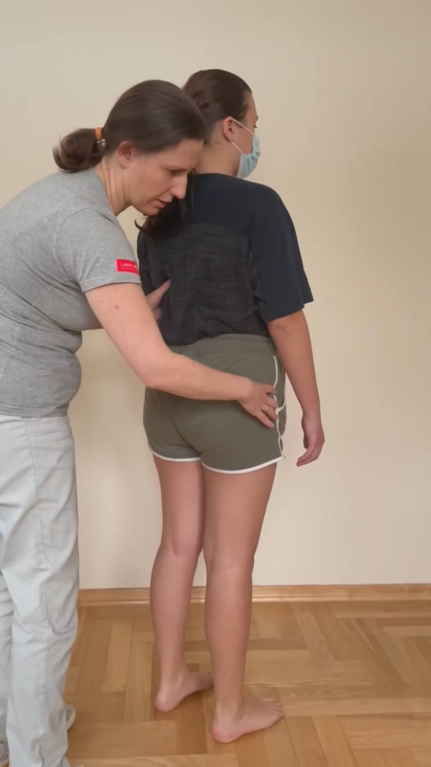
Findings for the Standing Screen
Functional Unit #1 (Thoracic Ring (TR) 3 to the hips)
Pelvis: In standing, her pelvis was rotated to the left in the transverse plane (TPR) with left SI joint unlocked (right innominate rotated anteriorly relative to the left innominate, sacrum counternutated relative to left innominate).
Diane’s question:
To determine if the sacrum is nutated relative to the ipsilateral innominate in standing (locked) versus counter-nutated (unlocked) a standing weight shift test is used. If the SIJ gives way (unlocks) before 50% of the total load from the trunk is taken through the tested leg, we say that the SIJ is unlocked in standing. At which approximate percentage of load did Ms Z’s left SIJ unlock?
Kate’s response:
In Ms. Z’s case left SIJ unlocked when she reached about 50% load from the trunk.
Hips: Right femoral head was forward compared to the left one. Right femoral head placed anteriorly in relation to the acetabulum, an incongruent finding for the pelvis.
Diane’s comment:
To clarify – the right and left femoral heads are congruent to the pelvis TPR; however, as you note the right femoral head is incongruent to its acetabulum. Can you explain why we say this latter point is incongruent and the significance of the finding to the right hip joint?
Kate’s response:
Starting position for the standing task is for the both femoral heads to be centered in their acetabulums. Right femoral head seating anteriorly in the acetabulum is incongruent with the requirement of standing task.
In optimal conditions in standing body weight should be transferred equally within the pelvis and to the femoral heads and necks. Non-optimal strategies, postural alignments or muscle imbalances, can lead to anterior translation of the femoral head in an acetabulum followed by uneven distribution of loads. Ms. Z’s R femoral head is anterior relative to the R ilium, if a situation like that lasts for a long time it can lead to impairments of an articular system.
Diane’s comment:
Almost! When the pelvis rotates to the left in the transverse plane, it is expected that the right hip will be anterior to the left in space (this is a congruent finding). It should never be anteriorly translated relative to its acetabulum – this is an articular shear lesion/impairment.
Thorax: The lower thorax in left rotation, which was congruent to the positional finding of the pelvis (L TPR). The TR6 was noted to be left translated/right rotated, which was incongruent to the positional finding of the pelvis and the lower thorax. TR9 was congruent to the regional rotation of the lower thorax (right translated/left rotated).
Diane’s question:
In ISM, there are two definitions of incongruence. One is that the direction of rotation between segments or body regions (regardless of the plane of motion) is opposite to each other and therefore incongruent. What is the second definition?
Kate’s response:
We talk about the second type of incongruence when the body region is in a position that is not congruent with requirements of the task being assessed. This is why it can possibly interfere with optimal biomechanics of the task.
Taking into consideration above definitions of incongruence first two statements below are the example of the incongruence of the body segment in relation to the task (in this case it was standing), and last statement below would be the example of incongruence between two body
Diane’s response:
Excellent answer!
Summary of incongruencies in FU#1
- Counternutated position of the sacrum in relation to the left innominate.
- The right hip was anteriorly translated in the acetabulum.
- The 6th thoracic ring was incongruent with regional rotation of lower thorax.
Functional Unit #2 (second thoracic ring to the cranium)
Upper Thorax: The 2nd thoracic ring and manubrium were noted to be “floating” between left translated/right rotated and right translated/left rotated.
Diane’s question:
In ISM, we call this an infinity sign. In cranial osteopathy, it is called a lateral fluctuation sign. What is the hypothesis that drives these biomechanics and what implication does it have for further assessment?
Kate’s response:
I am not sure If I understand the hypothesis correctly because I cannot find a lot of information on that subject. In ISM we talked that as the central nervous system, dura mater and its extension into the peripheral nervous system has its limited extensibility. As the systems are subjected to different pulls from non-optimal alignments and increased pressures, they can reach their expandability limits. Intracranial dural tension can also be influenced by altered intracranial venous sinuses drainage. It can be compromised by suboptimal alignment of the cranium, neck, clavicle, manubrium and upper thoracic rings. If the mobility of the nervous system is altered, we can feel fluctuating changes in alignment of the bones as the system is adjusting it to its needs. More specific testing is needed to establish if loss of nervous system mobility causes suboptimal alignment or on the contrary if suboptimal alignment is the reason for loss of mobility of the nervous system. It tells us which system should be prioritized during treatment. If the underlying cause will not be addressed in the first place, any change that will be made during treatment will be most likely temporary.
Researching lateral fluctuations in cranial osteopathy, took me to Dr. Sutherland theory. He stated that lateral fluctuations are happening in a fluid body. They are manifestations of the fluid lesion and can be felt when the system tries to balance itself. These fluctuations happen on the transverse axis from one side of the fulcrum to the other – like an infinity sign.
Diane’s response:
I believe the latter part of your answer to be correct from my investigation of this sign. It is a ‘fluid lesion’ and in osteopathy thought to be driven by the flow of cerebrospinal fluid when the expansion and contraction phases on the left and right sides of the body are not synchronized. For us in using ISM, it means that positional findings of the cranium, TR2 and the pelvis will not be consistent nor valid. The first treatment is to restore the fluid flow in the CSF and this will stop this finding. I too admit that I have lots of questions about this finding!
Cervical Spine: C1, C2, and C7 are translated to the right and rotated to the left. The alignment of the cervical spine was congruent to the positional finding of the upper thorax.
Diane’s question:
I thought the TR2 was ‘floating’ and therefore alternating in its direction of rotation? Wouldn’t this therefore make both the position, and congruence, of the upper thorax therefore variable?
Diane’s note and question:
C1 doesn’t translate relative to C2 as long as the transverse ligament is intact so can you please clarify what is meant when you say ‘C1 is translated right rotated left’?
Kate’s response:
Whole cervical spine as a unit was translated right and rotated left. Since TR2 was changing directions of rotation, I could not assess congruence between cervical spine and TR2.
Cranium: Cranium was in left ICT (left temporal bone was posteriorly rotated and the right temporal bone was anteriorly rotated). The ICT and sphenoid bone were congruent in relation to each other and to the positional finding of the cervical spine (left rotation).
Kate’s summary: The rotations in the transverse plane between the hips, pelvis, lower thorax are congruent to the left (TR6 is segmentally incongruent to this), the neck and cranium are congruently rotated to the left and TR2 has the infinity sign – alternating between left and right rotation.
Diane’s question:
Where was the starting position of the first thoracic ring?
Kate’s response:
I did not check the position of the TR1 at the initial assessment, but I did on the follow up session and it was rotated right. It means it was incongruent in relation to the cervical spine but congruent with shoulder girdle rotation right.
Shoulder Girdle: Shoulder girdle was rotated to the right, which was incongruent with the rotation of the cervical spine. Bilateral downward rotation of the shoulder blades. Right clavicle depression and posterior rotation, which was congruent with right rotation of the shoulder girdle but incongruent with the left rotation of the cervical spine.
Summary of incongruencies in FU#2
- Right rotation of the shoulder girdle was incongruent with rotation of the cervical region and cranium.
- The TR2 and manubrium were changing from left translated/right rotated and right translated/left rotated.
- The TR1 was evaluated on the follow up visit and it was in R rotation. It was incongruent with the L rotation of the cervical region, cranium and lower thorax.
Summary of incongruencies in the start screen: TR6, TR1, Shoulder girdle are incongruent to pelvis, lower thorax, neck and cranium.
Functional Unit #3 (lower extremity)
Knee: Right tibia in internal rotation.
Diane’s question:
This finding is often referred to as a ‘reverse screw home mechanism’. What is the normal screw home mechanism (alignment of the tibia relative to the femur) in knee extension and what are some of your hypotheses of why this mechanism can be reversed?
Kate’s response:
In normal screw home mechanism tibia rotates externally at the last 15 degrees of knee extension. It means that optimally in standing tibia should be in external rotation in relation to the femur. In Ms. Z’s case her right tibia is rotated internally in relation to the femur.
In Ms. Z’s case I suspect an involvement of either hamstrings (semimembranosus or semitendinosus muscle) or gracilis muscle in preventing medial tibial condyle from anterior glide and holding tibia in internal rotation. At the same time the hamstring group could be responsible for holding the right femoral head in anterior position in the acetabulum. My second hypothesis is that since Ms. Z’s right foot is pronated in standing, tibia and fibula could adjust to its position and stay in internal rotation in standing.
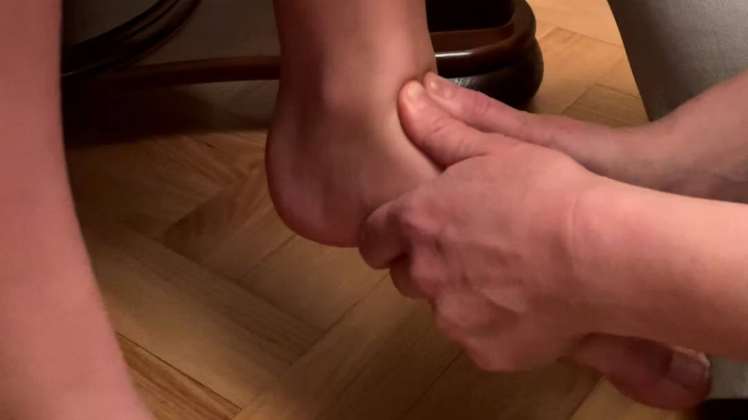
Feet:
- right calcaneus in the neutral position in relation to the ground, Achilles tendon bisects calcaneus, right talus plantar flexed and adducted in relation to the calcaneus, which means right foot was pronated and was in congruent position with internal tibial rotation.
- left calcaneus and talus tilted laterally, which means left hindfoot was laterally tilted as a unit, relative to the mortice.
Diane’s comment:
both postures are often interpreted as a pronated foot, yet they are clearly different. The talus of the right foot is not tilted relative to the mortice, the plantar flexion/adduction is relative to the calcaneus – this is pronation as you note. The left talus and calcaneus are both tilted relative to the mortice.
Summary of incongruencies in FU#3
- Right tibial internal rotation and right foot pronation are not optimal for standing task (intra-regional incongruence for the task and the position of the tibia and foot are congruent to each other).
- Left hind foot lateral tilt shows inter-regional incongruence between hind foot and tibial position and also intra-regional incongruence for standing task.
Screening task analysis 1/2 squat
I chose half squat for the screening task since standing in ski boots is similar to standing in a small squat position. Skiing itself also requires constant action of half squat.
Findings for the 1/2 squat
Functional Unit #1 (TR3 to the hips)
Pelvis: Ms Z started with the left sacroiliac joint already unlocked. It stayed that way through the squat task. Her pelvis changed from left transverse rotation on standing to no rotation in squat and returned to left rotation on coming up from squat to standing. It suggests non optimal biomechanics on returning from a squat to standing.
Hips: Left femoral head started centered in the acetabulum in standing and stayed centered through the squat. Right femoral head started anteriorly in the acetabulum and translated back on the squat but did not center fully. On return from squat to standing right femoral head translated forward first and then was followed by pelvic rotation to the left. So, some of the pelvic L TPR is in relationship to the biomechanics of the right hip but not all.
Diane’s question:
If the right hip did not center completely going down into the ½ squat and yet you note that there was no rotation of the pelvis at this point, then the hip is not the only thing responsible for the TPR of the pelvis. Why? What else do you hypothesize from your start screen findings could result in the right hip still being forward, yet the pelvis in neutral in this ½ squat position?
Kate’s response:
Internal tibial rotation and right foot pronation could have caused functional R leg shortening and put pelvis in the L TPR. Since in the ½ squat position the left foot should go into pronation and L tibia should internally rotate that could neutralize position of the pelvis in ½ squat.
Diane’s response:
Great hypothesis!
Thorax: The lower thorax started in a non-optimal position in rotation to the left, it corrected during squat. The 6th thoracic ring started in left translation/right rotation and did not change its position throughout the task.
Diane’s question:
If I understand correctly, both the pelvis and the lower thorax fully corrected to a neutral position of rotation in the ½ squat; however, the right hip remained anterior and the TR6 remained left translated/right rotated. How do these findings facilitate your decision for the next step of your ISM assessment?
Kate’s response:
The elements that have not corrected during the task will be my primary suspects and should be the first ones to be analyzed.
Summary of incongruencies in FU#1
- Counternutated position of the sacrum in relation to the left innominate (unlocked left SIJ).
- The right hip did not fully center in an acetabulum and stayed partially translated forward in the acetabulum.
- TR6 stayed incongruent with regional rotation of lower thorax throughout the task (intraregional incongruence). TR6 being in right rotation is also in inter-regional incongruity with right hip anterior position.
Analysis of the drivers for FU#1:
- Correction of the hip in relation to acetabulum, partially corrected TR6 rotation, but did not help with left SIJ control.
- Pelvic rotation correction did not help right hip position, TR6 or left SIJ control.
Diane’s comment:
A complete pelvic correction would correct the TPR and provide control (force closure) for the left SIJ. This may then change the strategy that is impacting TR6. Without a complete pelvic correction, you don’t know the impact of correcting the pelvis on TR6.
- TR6 correction helped to control left SIJ in the squat (joint unlocked later in the task). Since it was possible to correct the ring fully, we can suspect neuromuscular system impairment, not an articular one. It did partially correct the hip in the socket but did not correct the rotation of the pelvis.
- Co-correction of the TR6 and R hip gave the most of the correction in the unit 1
Driver for FU#1: the right hip was the primary driver and the TR6 was the secondary driver. The persistent unlocking of the left SIJ is still something to consider and is therefore also a driver.
Diane’s comment/question:
I’m not sure of this based on your findings? You haven’t mentioned
anything about patient’s experience with any of these corrections – can you add that piece please.My analysis:
Hip Correction: n/c in pelvic control and partial correction TR6
Partial Pelvic Correction (just the TPR: n/c hip, n/c TR6, n/c in SIJ – therefore you weren’t completely correcting the pelvis because a complete correction of the pelvis provides control)
TR6 correction: partial improvement in pelvic control and rotation, partial correction of right hip
I think you are getting a bigger ‘bang for buck’ from the TR6 correction as opposed to the right hip from just findings – if the co-correction of TR6 and the right hip improved her experience the best then you have a co-driver. The next decision about drivers must involve patient experience.Kate’s response:
Looking at the findings I agree that correcting TR6 seems to give better results than hip correction. As to the patient’s experience, when performing a ½ squat lasting for a short time only, Ms. Z did not feel much difference with any of the above corrections. Staying in the ½ squat position for a longer time, would give her feeling of the feet getting tired. With no corrections feet would get tired in 28 seconds of being in a ½ squat position, correcting only TR6 would prolong that time to 33 seconds, correction of the right hip to 39 seconds and co-correction of TR6 and right hip would give her best result of 45 seconds before feeling tired in her feet. That would indicate that TR6 and right hip correction gives the best experience to the patient.
As to the pelvis correction in my first assessment I just corrected TPR without providing compression to the pelvis. After your reminder, I reassessed using pelvic alignment and compression component. While I was holding the corrected and compressed pelvis, the patient was not able to give me feedback on the TR6 or R hip position so I corrected the pelvis manually and then used a mobilization belt to apply compression. I confirmed that L SIJ was not unlocking anymore in the squat and then checked position of R hip and TR6 in the ½ squat – both were correcting with pelvic compression but patient’s experience got worse and time for her feet to get tired in the squat shortened in comparison with the one with no corrections at all.
Diane’s comment:
Nice! Now you know for sure that the pelvis is not a primary driver; however, because no correction yet has provided full control in the squat it is definitely still a secondary driver for the ½ squat task.
Final drivers FU#1 given this discussion: Thorax (TR6) and right hip – either primary/secondary or co-driver and the pelvis.
Functional Unit#2 (TR2 ring to the cranium)
Upper thorax: The TR2 and manubrium started by “floating” between left translated/right rotated and right translated/left rotated and were still “floating” in half squat position.
Cervical spine: C1 to C7 all started in suboptimal position of left rotation but all corrected going down to the squat.
Cranium: Cranium started in suboptimal position in left ICT and corrected going down to the squat.
Shoulder girdle: Started in the right rotation and corrected going down to the squat position. Bilateral shoulder blades downward rotation was also corrected during the squat. Shoulder biomechanics going down was correct but non-optimal going from squat to stand.
Summary of incongruencies in FU#2
- The TR2 and manubrium alternating from left translated/right rotated to right translated/left rotated did not change with squat task.
- Right shoulder biomechanics was optimal going down to the squat and non-optimal going back up.
Analysis of the drivers for FU#2: no driver analysis was done since all incongruencies were corrected going down into the squat. The second thoracic ring could not be assessed as the driver due to changes in its position.
Diane’s comment:
Excellent reasoning: Going down into the ½ squat is the task being analyzed, not coming up and all sites of impairment in FU#2 completely corrected going down. You may have quickly avoided doing all this if you used a FU#2 quick screen after finding your FU#1 drivers. What is a FU#2 quick screen? With no correction, check head and neck rotation range of motion and experience in a ½ squat. Compare the range and experience in the ½ squat with the FU#1 drivers corrected. If the experience worsens, or the range of motion reduces, then you have made something worse in FU#2 with your FU#1 correction and you have to go into FU#2. If there is no change in performance or experience with the FU#1 drivers corrected, then you can avoid assessing FU#2.
Driver for FU#2: no driver
Functional Unit #3 (lower extremities)
Knees and Feet: During the normal foot biomechanics going down in to the squat task: tibia should internally rotate in the relation to the femur, talus should dorsiflex in a mortise and plantarflex and adduct in relation to the calcaneus, calcaneus should go into external rotation under the talus. Medial arch should lengthen. Mid foot and forefoot should fan around the axis of the second ray. Fanning would involve navicular bone external rotation in relation to the talus and cuboid, cuboid internal rotation in relation to the calcaneus and navicular, medial cuneiform external rotation and lateral cuneiform internal rotation in relation to the navicular and middle cuneiform, middle cuneiform remaining in neutral as to respect to the lateral and medial cuneiform, 1st metatarsal bone external rotation, abduction and dorsiflexion, and 3rd, 4th and 5th metatarsals internal rotation, abduction away from 2nd metatarsal and dorsiflexion).
- Right foot started in the suboptimal position of pronation. During squat talus decreased its pronation 50%, mid foot folded in instead of fanning out and foot shortened instead of elongating. Since the right tibia already started in internal rotation it did not further internally rotate during the squat.
- Left foot and knee also started in suboptimal position of hind foot lateral tilt and left tibial internal rotation. Hind foot did not change its position during the squat. Mid and forefoot folded in instead of fanning out. The foot shortened and the tibia did not internally rotate relative to the femur.
Diane’s comment:
Excellent description of expected biomechanics compared to patient’s findings.
Summary of incongruencies in FU#3
- Right and left foot suboptimal biomechanics during squat. During squat talar abduction and dorsiflexion for the right foot, no change in the position of the hind foot in the left foot, folding action for the mid foot for both feet.
Analysis of the drivers for FU#3:
- For the right foot correction, I corrected the talus alignment in relation to the calcaneus and mortise. It allowed for the mid foot and fore foot to fan during squat and for the calcaneus to move gently into lateral rotation under the talus; it did correct the starting position of the tibia but did not help with the tibial ability to rotate internally during squat. Correcting the position of the talus did not help it to adduct and plantar flex during a squat (it felt like the change of the relation between talus and calcaneus came from calcaneal movement only).
- Left hind foot tilt correction (setting calcaneus in neutral in relation to the ground and stacking talus over the calcaneus) allowed for tibial internal rotation during squat, it did not help with midfoot and forefoot fanning. It did not help calcaneus to move under talus and talus move into adduction.
- Right tibial derotation corrected partially talar tilt but did not allow for forefoot and mid foot fanning, or the calcaneus to move under talus.
- Correcting either foot alignment did not affect the other foot.
Diane’s question:
Were you able to physically rotate the left calcaneus relative to the talus? This presentation ‘sounds like’ an articular system impairment of the left subtalar joint.
Kate’s response:
Setting the calcaneus in neutral in relation to the ground would set talus in neutral in the talocrural joint. Rotation of the calcaneus in relation to the talus would create a lot of resistance, I agree that it looks like an articular restriction.
Driver for FU#3:
Right and left foot were considered as codrivers for FU#3.
Relationship between drivers of all Functional Units in the ½ squat task
- Correction of FU#3 (both feet) did not restore control of left SIJ (pelvis), and partially corrected TR6 and fully corrected the right hip.
- Correction of FU#1 drivers did not visually correct the position or mobility of the feet.
- Co-correction of both FU#3 and FU#1 drivers (both feet and TR6) allowed the patient to stay in the squat position for 55 seconds before her feet “got tired”, but it did not fully restore SIJ control.
Diane’s comment:
Final drivers for the ½ squat task: Primary: Both feet and TR6, secondary pelvis.
Screening task body twist in half squat
Half body twist is important in skiing. During turns the lower body turns one way and the upper body stays facing down the slope. Overall lower extremities and pelvis turn the opposite way than the thorax, cervical spine and cranium. Since I did not have the ability to test lower body rotation in standing, the patient executed the thorax rotation one way while standing with a gentle knee bend to mimic position in a ski boots. Ms Z did not feel any difference between the left and right body twist but to complete the right body twist she shifted her body to the left, starting at 45 degrees of the twist.
For the purpose of the “skiing twist” head and neck stays in neutral in relation to the thorax, thorax and shoulder girdle rotates one way, pelvis rotates opposite way, one leg rotates internally and foot pronates, and opposite leg rotates externally and foot supinates.
Findings for body twist in half squat
Functional Unit #1 (TR3 to the hips)
Pelvis: pelvis started in a good position and had optimal mechanics on left and right twists. In the left body twist right innominate rotated anteriorly and right one posteriorly. Opposite was true for the right body twist. Her sacroiliac joints remained locked.
Diane’s comment/question:
But her start screen standing findings and ½ squat findings were:
“Ms Z started with the left sacroiliac joint already unlocked. It stayed that way through the squat task. Her pelvis changed from left transverse rotation on standing to no rotation in squat”
Are you saying that when you asked her to ½ squat and then twist her left SIJ was now not unlocked?
Kate’s response:
She started with unlocked L SIJ and during body twist the relation between L ilium and sacrum did not change so the joint did remain unlocked but did not unlock any further. Her R SIJ started in a locked position and the relation between R ilium and sacrum did not change during the task, so R SIJ remained locked.
Diane’s comment:
Therefore her pelvis is still a site of impairment here in that the left SIJ is unlocked. Once it unlocks, there is ‘no further unlocking’.
Kate’s response:
It makes sense. I will add full pelvic correction component to my assessment of the FU#1 in this task and make sure to check pelvic control influence on other incongruencies in that unit. Unlocked pelvis can create a number of compensations in order to complete the task
Hips: Right hip started in anterior position in relation to acetabulum. During the left and right body twist it still stayed anteriorly in the right and left body twist although it did partially center with the right body twist.
Thorax: Thorax started in a good position with an exception of the TR6 that was in the left translation/right rotation. On the left body twist all thoracic rings did translate right and rotate left. On the right body twist all thoracic rings translated left and rotated right but from 45 degrees of the body twist, TR6 got worse.
Diane’s comment:
If I understand this correctly, the TR6 was able to left rotate congruent with the other thoracic rings during left rotation of the thorax; however, during right rotation it translated left/rotated right FURTHER than the rings above and below it? If so, this is a control problem, not one of restriction of this ring OR it is compensating for something else.
Kate’s confirmation:
I have rechecked it and I can confirm that TR6 was able to rotate left together with other TRs, and during right rotation starting in about 45 degrees it would translate left/rotate right further than the other TRs (this is the same moment Ms.Z starts the shift of the trunk to continue the twist).
Diane’s comment:
So this is truly an inter-ring control problem.
Kate’s response:
Inter-thoracic ring control problem is present in the absence of intra-ring impairment and when the strategy for the alignment, biomechanics or control of any of TR 1-10 is suboptimal. It can be caused by trauma or by habitual bad posture or incorrect movement patterns. I believe that in Ms. Z’s case, TR6 can compensate for something else as you also mentioned as a possibility. My further assessment showed that if the pelvis is controlled and L SIJ is not unlocked during the body twist task, TR6 position is fully controlled.
Summary of significant findings in FU#1
- The right hip remained partially anteriorly translated in the acetabulum in both body twists.
- The TR6 left translation/right rotation got worse with the right body twist.
- L SIJ started in an unlocked position and remained unlocked throughout the task.
Analysis of the drivers for FU#1:
- Right hip correction partially corrected TR6 during right body twist, (it delayed but did not eliminate body shift to the left), it did not change control in L SIJ- it was unlocked through the task.
- TR6 correction, centered the right hip in the acetabulum in the right and left body twist and eliminated the body shift during the right body rotation, it did not change control in L SIJ- it was unlocked through the task.
- Compression to the pelvis with a mobilization belt allowed for L SIJ start and stay locked through the task, it corrected R hip and TR6 alignment, allowed TR6 to stay properly aligned during body twist and eliminated body shift during the R body twist. Ms. Z also noted that the right twist was the easiest to perform when the compression belt was on her pelvis.
Driver for FU#1: The pelvis (control of the L SIJ) was considered the driver for FU#1 in the right body twist screening task.
Final drivers FU#1 given this discussion: Thorax (TR6) and right hip – either primary/secondary or co-driver and the pelvis.
Functional Unit#2 (TR2 to the cranium)
Upper thorax: TR2 continued to present with an alternating rotation (infinity) sign.
Cervical spine: cervical spine C1-C7 started in neutral position in the ½ squat and there was no change in position of the neck during the right and left body twist (patient was asked not to rotate her neck and head during body twist since during skiing head stays facing down the slope).
Cranium: started in neutral and there was no change in its position during the right and left body rotation.
Shoulder Girdle: started in the correct position and stayed in a proper position throughout the left and right body twist.
Summary of incongruencies in FU#2
- The TR2 remains alternating in rotation through the screening task.
Analysis of the drivers for FU#2:
- no driver analysis needed for FU#2
Diane’s comment:
Again, a quick screen of FU#2 would eliminate the need to assess specific segments in this task IF correcting the pelvis did not worsen head and neck rotation.
Kate’s response:
That is very good advice. Thank you.
Functional Unit #3 (lower extremities)
Knee: During the body twist to the right, the left tibia rotated internally relative to the femur and the fibula rotated internally relative to the tibia. During the body twist to the left right tibia rotated internally relative to the femur and the right fibula rotated internally relative to the tibia. Superior tibiofibular joint had optimal mechanics in this task.
Right foot: started in the pronated position, supinated and folded with the right body twist but it did not fully pronate (it returned to starting position) and did not fan with the left body twist.
Left foot: started in a non-optimal position of lateral tilt of hind foot. Hind foot corrected tilt and mid and forefoot folded with the left body twist but instead of pronation on the right body twist the hind foot laterally tilted as a whole and mid and forefoot did not fan.
Summary of incongruencies in FU#3
Sites that showed suboptimal mechanics on the right body twist were
- Left hind foot tilted as a whole
- Left mid and forefoot did not fan
Sites that showed suboptimal mechanics on the left body twist were
- Right foot did not fully pronate
- Right mid and forefoot did not fan
Relationship between drivers of all Functional Units for Left Body Twist Task
The driver in FU#1 for this task is the pelvis (left SIJ control)
- Correction of the pelvis:
- corrected the initial position of the right foot but it did not improve on the right foot pronation and fanning during left body twist.
- did not improve the initial position of the left foot, did not improve its pronation or fanning.
- Ms. Z reported more ease in body twist.
In FU#3
- Correction of right talus to be in neutral and left hind foot tilt allowed for the full control of the L SIJ. Ms. Z reported the most ease during body twist.
Both feet were considered co-divers for body twist in small squat position screening task.
Screening Task & Drivers:
- ½ squat final drivers: Primary both feet, secondary thorax (TR6) and pelvis (L SIJ control)
- Left body twist drivers: Primary both feet (co-driver)
Further Assessment of Drivers (both feet, thorax (TR6) and pelvis (LSIJ control)
Feet
Further assessment of both feet was done to determine the underlying system impairments using active and passive mobility tests with active and passive listening during both the correction and release of the correction.
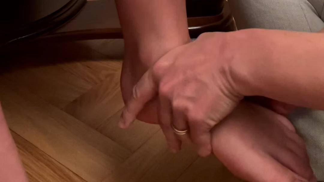
Assessment of the right foot:
- Active mobility assessment: calcaneus went from being laterally rotated in relation to talus in closed chain position (standing), to medially rotated in relation to the talus in open chain position (sitting) and the talus neutralized in relation to the mortise. During plantar flexion the talus was gliding anteriorly, and with dorsiflexion it glided posteriorly but only at initial phase of dorsiflexion, then it stopped. In standing the starting position of this foot was in slight pronation and then full pronation was limited in the left body twist. The hindfoot could supinate in the right body twist.
- Passive mobility assessment and vector analysis: Dorsiflexion/Posterior glide of the talus relative to the mortice was limited with muscular type of end feel at approximately 50% of expected ROM, the anterior glide was normal. Passive listening to the vector is leading to a long posterior vector. Manual vector analysis showed primarily tibialis posterior problem. Passive mobility testing of the subtalar joint showed decreased medial rotation of the calcaneus in relation to the talar bone.
Diane’s question:
This is inconsistent with your active mobility findings. In the body twist you report that supination occurred in the right body twist and that pronation was limited in the left. Medial rotation of the calcaneus relative to the talus occurs in supination and lateral rotation in pronation. How do you reconcile this inconsistency between active and passive mobility of the right subtalar joint?
Kate’s response:
Taking into consideration finding from the further passive listening, restrictions from calcaneo-navicular ligament, it is possible that since during active mobility testing all joints in the foot are moving and navicular bone folding would allow for medial rotation of calcaneus under the talus. During passive mobility testing the navicular bone is being immobilized by the tester’s grip and will not fold and loosen the ligament and calcaneus bone will not be able to rotate medially under the talus. Another reason for the difference could be due to the position of the testing. Active testing of the subtalar joint is done in standing (closed chain position) and passive testing in sitting (open chain position).
Diane’s comment:
You note that in sitting the calcaneus is medially rotated in relation to the talus and perhaps this starting position has taken up some of the available range for more medial rotation. In other words, if the starting position of the joint is already in medial rotation, it is easy to interpret that medial rotation is limited when tested passively if the start position isn’t considered.
- Joint distraction of all four joint aspects one by one showed restrictions in the antero-medial part. Then distraction to the first resistance and release was used for passive listening. It revealed a short medio-anterior vector suggesting calcaneo-navicular ligament.
Diane’s question:
The tibialis posterior vector can also compress this joint. How did you differentiate the calcaneo-navicular ligament from Tib post?
Kate’s response:
Compression from the tibialis posterior would create a long pull in a cranial direction. To confirm that vector is coming from tibialis posterior distraction of an antero-medial aspect of subtalar joint would be held and the different points of tibialis posterior would be palpated. During the palpation of the part that needs release, compression of the distracted aspect would be felt. In the case of the calcaneo-navicular ligament vector, the pull in antero-medial aspect of the subtalar joint is short and in the direction compatible with the course of the calcaneo-navicular ligament.
Diane’s comment:
Nicely reasoned!
- Passive mobility of the mid foot showed the navicular and medial cuneiform bones not folding. Passive listening suggested involvement of flexor hallucis longus.
- Active control assessment showed loss of some active control during squat and OLS- foot pronated. It should be reassessed after all foot vectors are released.
- Passive control tests were not done.
Assessment of the left foot:
- Active mobility assessment: in open chain position (sitting) talar tilt in talocrural joint corrected partially, calcaneus did not change its position in relation to the talus. Talus did glide anteriorly with plantar flexion, but not posteriorly with the dorsiflexion. During body twist the whole hind foot tilted medially more instead of pronation in the right body twist, and returned from the tilt to the neutral instead of supination in the left body twist, there was no midfoot and forefoot fanning or folding.
- Passive mobility assessment and vector analysis: in the talocrural joint, talus glided anteriorly but did not glide posteriorly, it stopped with muscular end feel. Passive listening pointed toward soleus muscle problem. Passive assessment of the subtalar joint showed calcaneus being compressed posteriorly on both sides but lateral more than medial side. Passive listening using distraction showed soleus muscle involvement. Passive assessment of the midfoot showed decreased navicular fanning and no restrictions in the mobility of cuboid and cuneiform bones.
Diane’s question:
Did the compression of the subtalar joint restrict medial or lateral rotation of the posterior subtalar joint? These findings are highly suggestive of an articular impairment of the posterior subtalar joint. Which arthrokinematic glide (anterolateral or posteromedial) would be restricted with pronation and which with supination. What were your arthrokinematics findings?
Kate’s response:
Looking at posterior subtalar joint, if anterolateral glide of calcaneal bone in relation to the talus is restricted, supination of the hindfoot will be limited, if posteromedial glide of calcaneal bone in relation to the talus is restricted, pronation will be limited.
During Ms Z’s examination the posterior lateral aspect of subtalar joint was most compressed limiting anterolateral calcaneal glide, and therefore medial rotation of the calcaneus, but also the posterior medial aspect was compressed partially limiting posteromedial calcaneal glide, and therefore lateral rotation of the calcaneus (all relative to the talus).
- Active control assessment: Patient was not able to sustain proper foot alignment in the squat and OLS. It should be reassessed after all foot vectors are released.
- Passive control tests were not done
Assessment of the Pelvis
- Active mobility test showed symmetrical movement on the left and the right side. When flexing the right hip, the right innominate was minimally rotating posteriorly in relation to the sacrum, when flexing the left hip, the left innominate was minimally rotating posteriorly in relation to the sacrum. It is a normal finding.
- Passive mobility test showed restrictions to the left upper part of the sacroiliac joint with a short vector toward multifidus muscle. Lumbar multifidus palpation showed decreased muscle bulk on the L4-L5 level.
- Active control test showed loss of a control in L SIJ at 50% of weight load in standing and in ½ squat task. Correction of both feet allowed for the full control in the L SIJ in the left body twist.
Diane’s question:
To reach a diagnosis of the pelvis for the ½ body squat (no driver brought back left SIJ control) can you now clinically reason the test findings from
- Active mobility
- Passive mobility
- Active control
Is this an articular, neural, visceral or myofascial system impairment of the LSIJ based on your clinical reasoning?
Kate’s response:
Based on the above findings pelvic control is not considered as an issue in the left body twist since it can be restored with a bilateral foot correction. Since the active mobility test is normal, it means that we can rule out an articular system impairment. In case of suspected articular system impairment of stiff joint, both active and passive mobility tests would show a decreased mobility of the joint on one side in comparison to the other side. In case of suspected articular system impairment of joint laxity, active mobility test could be restricted due to bracing strategy but passive mobility test would show increased mobility of one joint in comparison to the opposite side. None of the above options are true for Ms. Z. Based on Ms. Z history – no surgical history, no pelvic trauma, no gastroenterologic problems etc. and above testing the most probable system impairment is the neural one. She is losing L SIJ control with 50% of weight load in the ½ squat task and the joint feels restricted in passive mobility in the upper part of the left SIJ.
Assessment of the Thorax (TR6)
- Active mobility assessment-TR6 was a driver in ½ squat task. During a properly done 1/2 squat with the hip hinge, thoracic rings should not flex, extend or rotate. Since TR6 starts in left translated/right rotation position, left trunk rotation was used to assess if TR6 can actively get to the neutral position. It was possible for TR6 to get actively to a neutral position.
- Passive mobility assessment and vector analysis – during correction attempts of TR6, it was possible to correct it fully with use of minor force. This finding rules out an articular system impairment and suggests a possible visceral or neural vector. Passive listening, on release of the correction, revealed a vector from the anterior left rib to midline, which moved into the abdominal cavity (half deep to the pyloric sphincter region). This finding suggests a visceral vector. Significant tenderness of that region was reported during palpation.
Hypothesis
Ms. Z first pain complaints started about 1.5 years after her L foot injury on a bike. Even though she had fractured only her fifth phalange, her foot probably sustained more injury. She had never had any post injury physical therapy treatment or functional evaluation. With time the foot developed tightness and lack of mobility in the talocrural joint. Her mother reported she did have moderate flat feet prior to injury. This, in conjunction with growth spurt (fast elongation of the bones and need of muscle to catch up with bone growth, changes in balance distribution and body weight) could have led to development of two different non optimal biomechanics in a right and left foot and lead to body compensation in higher regions.
Treatment
Treatment – First Two Sessions
Release
Right foot: Primary vector for the talocrural joint, posterior tibialis muscle, was released with a neural technique called ‘release with awareness’. Secondary vector, soleus muscle, was released in the same manner. Primary vector for subtalar joint anterior /medial part was released – calcaneo-navicular ligament with cross friction massage technique. Primary vector, for navicular and medial cuneiform bones, flexor hallucis longus was released using ‘release with awareness’ technique.
Diane’s question:
Can you describe the principles of the ‘release with awareness technique’ and why this technique is considered a ‘neural release’ and not a technique for an articular system impairment.
Kate’s response:
Release with awareness technique is a neural technique lowering the tone of the muscle. It consists of few steps:
- Therapist looks for the part of the muscle that is the most tense.
- Therapist monitors tension of the muscle with gentle palpation.
- Therapist reduces the stretch of the muscle spindle by shortening the muscle either by approximation of its fascicles or by changing position of the joint.
- After the muscle tone lowers down a little, the therapist is searching for the best cue for the patient to soften the muscle. When the muscle softens, verbal reinforcement is given to the patient.
- After full release of the fascicles is achieved muscle and surrounding tissues are being taken into the stretch.
The technique is neural technique since it is influencing the nervous system. By changing the amount of input from muscle to the spinal cord it changes the amount of output to the muscle and lowers its tone. By using verbal cues for the patient to lower muscle tone, we allow for their brain to learn that it is ok to let go of this specific area and that there is no need to guard it with increased tone. Patient also learns to recognize tension vs. relaxation in this specific region. Nowadays many of us are disconnected from our bodies and are not able to distinguish when our body tenses up. This technique would not work on articular impairments since such impairments require vector specific, precise manual mobilization. Verbal cues to ‘let go’ have been found to be less effective with these impairments.
Left foot: Primary vector for talocrural joint, soleus muscle, was first attempted to be released with a “release with awareness” technique. Since it was not giving expected results, a different release technique was used. Muscle was directly stacked in all 3 planes, dorsi and plantar flexion of the foot was used to stimulate Golgi receptors. Soleus muscle was only partially released. Due to time restrictions of the first session, inhibition taping with KinesioTape was done to the soleus muscle and no further manual treatment was done to the left foot during this session.
In the follow up session, reassessment showed that all of the release techniques and patient’s home exercises for the soleus muscle partially decreased tension in the muscle, partially restored talar posterior glide but did not restore mobility of subtalar joint. It means that soleus muscle was not a primary vector for a subtalar joint. Passive listening to subtalar vectors was repeated and articular restrictions showed as the primary vector. Grade III mobilizations, joint distraction and cross friction massage of soft tissues around the joint were used.
Diane’s question:
Can you describe specifically which direction of glide, and at which joint (anterior or posterior) the Grade III mobilization was applied?
Kate’s response:
Antolateral glide of the calcaneus in relation to the talus in the posterior subtalar joint was applied.
There were a lot of soft tissue restrictions at the attachment of the Achilles tendon. After the release of the articular restrictions passive listening of the subtalar joint was repeated and soleus muscle, then tibialis posterior and short anterior calcaneal ligaments showed up as vectors. Muscles were treated with “release with awareness” technique and calcaneal ligaments (tibiocalcaneal, posterior tibiocalcaneal and calcaneofibular) with cross friction massage.
Reassesment showed good subtalar joint mobility, and some remaining restrictions in the talocrural joint. Talar posterior glide had hard end feel but only at the medial third of the joint, with short posterior vector. Distal interosseous membrane was treated with grade III distal tibiofibular joint mobilizations into anterior and posterior fibular glide relative to tibia. Talar mobility was restored.
Navicular bone listening, at the talonavicular joint, showed short articular vectors – navicular bone was treated with Grade III and IV talonavicular joint mobilizations. Navicular bone was mobilized into external rotation in the relation to the talus in order to restore its ability to fan. General forefoot, midfoot fanning, folding mobility was done.
Diane’s question:
When was the visceral vector impacting TR6 treated?
Kate’s response:
This vector was treated on the Treatment no. 3. I think that if it was treated earlier it could have given faster results for the patient.
Align
Right foot: After the releases, the right calcaneus was aligned properly both in sitting and standing. During standing the talus was also properly aligned but during the squat it was not adducting (medially rotating relative to the calcaneus). To facilitate proper calcaneal position taping was done.
Diane’s question:
What is your hypothesis as to why, if all the joints were now free to move, the hindfoot was not able to properly pronate? Can you describe how you taped this joint?
Kate’s response:
There could be few reasons why at this point calcaneus was not adducting. First of all for the half squat there were more drivers along with right foot (Left foot, TR6 and pelvic control). If all of the drivers are treated and the calcaneus still is not pronating during the squat, it will point toward motor control issue.
I used a taping technique to distract of the medial part of the subtalar joint. First I anchored 2” wide cover roll tape on the anterior medial part of calcaneal bone, distracted calcaneus caudally away from talus and sustained it while laying down the tape under calcaneal bone, along its lateral side and up the distal third of lateral side of fibula. I laid down 1.5” wide Leukotape in the same manner on the top of the cover roll tape. It is important not to pull the calcaneus laterally during the taping so the talus can remain in neutral position. The role of the taping was to change proprioceptive input from subtalar joint.
Left foot: After first treatment releases alignment of the left calcaneus was also corrected in standing but talus remained adducted and navicular bone and first ray were internally rotated. Due to time constraints nothing further was done to left foot alignment at the first session.
Diane’s question:
The left foot initially presented with a hindfoot tilt – I need more information about the specific impairment of the posterior ST joint to understand this post treatment finding. Sounds like the talus is now pronating with respect to the calcaneus in standing but I’m unclear how the reduction of the hindfoot tilt was achieved.
Kate’s response:
During the first treatment short release was done with cross friction massage to the subtalar joint it did cause at the end of this session that talus was pronating with respect to the calcaneus. By the second treatment the left foot was presenting again with hindfoot tilt not a pronation. Second treatment was done as described above.
After the second treatment even though left foot mobility was restored it did not show proper biomechanics during meaningful tasks. Muscular control training is needed to regain proper function during the tasks. For the squat task also remaining drivers need to be addressed.
Connect
Right foot: Cues to allow for the foot to pronate during the squat and supinate during getting back from the squat were used to train right foot mechanics during this task.
Left foot: No connect cues were used after first treatment. The cues for the foot pronation were used after the second treatment.
Move
Ms. Z was given a home practice:
- Trigger Point releases with small ball for the right tibialis posterior and right soleus muscles.
- Trigger point releases with a small ball for the left flexor hallucis longus, and left soleus muscles.
- Practicing gentle right foot pronation during squat and body twist in small squat.
Treatment 3
After restoring foot mobility and control TR6 was still right rotated and left translated, and pelvis was still unlocking during ½ squat task.
Diane’s question:
What from your ISM assessment could have predicted this response?
Kate’s response:
The part of assessment that predicted this outcome was an analysis of relationships between drivers for ½ squat task. It showed that correcting feet position is helping to restore TR6 position only partially and does not impact pelvic control, which is consistent with findings above.
TR6 vector was treated with induction of pylorus technique, stomach mobility technique and surrounding tissue indirect stack to release technique. All of the above techniques were taught to me by Ramona Horton PT, during the Mobilization of Visceral Fascia: The Gastrointestinal System Seminar. After releases, TR6 remained corrected throughout the ½ squat. To see if connect cues will be needed for TR6 to stay in alignment load will be added to the squat task in future sessions. Cobra stretches and self tissue mobility techniques were added for the home program.
For the pelvis and the loss of L SIJ control, the patient was given several connect cues to activate her deep sacral multifidus muscle. The best and only working cue was to lift the hair on her low back, like her dog when it gets angry. Activation was confirmed by the palpation of the muscle. When activation was sustained throughout ½ squat the L SIJ stayed locked. Multifidus activation awareness was done during treatment and activation exercises given as home program.
Diane’s question:
So, correcting Ms Z’s feet resulted in L SIJ control in the left body twist but not in a ½ squat task. How do you explain this? Why is motor control training required in one task and not the other?
Kate’s response:
I am not exactly sure but it could be that our brain develops different movement patterns for each type of movement. It takes a while for a brain to create a new “substitution pattern”; it is possible that loss of pelvic control in squat started to happen earlier than the one in a body twist. Correcting Ms Z’s feet was enough for the muscle to remember how to control the pelvis in a body twist but not in the ½ squat. It will require muscle retraining and changing activation pattern to return to pelvis control in ½ squat task.
Reassessment of the ½ squat task after TR6 vector release and with multifidus activation showed that if Ms. Z puts her feet in the correct starting position, L foot has proper pronation and supination mechanics including fanning and folding, but right foot still failed to pronate and fan.
Follow up treatment plan
In the follow up treatments we will concentrate on retraining her R foot biomechanics for the ½ squat. If this training does not successfully restore pronation and supination of the right foot – further assessment will be done for remote drivers. Both feet training for strengthening and control will be implemented for both tasks ½ squat and body twist.
To gain L SIJ control in ½ squat, deep multifidus activation exercises will be implemented. When a patient will be able to activate the muscle with ease, exercises will progress to activation of dMF during squat movement, activation in a squat with added arm movements, and activation with added weight/resistance.
To ensure TR6 control during squat different exercises with gradually increasing load in the squat task while TR6 position is monitored will be added.
Training will be concluded with integration of the control of all drivers for ½ squat task.
A lot of children and adults currently lead very sedentary lifestyles. It is getting harder to motivate children to engage in physical activity. In Ms. Z’s case, she tries to enjoy skiing and her work-outs but it gives her low grade pain in her legs. Her pain was not significant enough for her to believe that it is abnormal to have it during skiing, but on the other hand it would stop her from being active. She is young and her giving up on physical activity can influence her activity habits for the future. In her case the ISM assessment showed multiple drivers for a chosen task. Addressing all of them might be a key that will allow her to get back to enjoying skiing.
What I find the most frustrating is that it is difficult to come by a specialist who can thoroughly assess and address foot problems. In a lot of cases problems are ignored in children for many years and if treated, the orthotics are recommended as an only treatment solution. I hope that learning about foot assessment and treatment will help me spread awareness that there are other more effective options to treat them.
Further Follow-up Sessions
Mrs. B’s weekly treatment gradually transitioned from an emphasis on releasing the cranium to controlling her new alignment as we added sensorimotor retraining exercises common to post concussive rehabilitation. We also worked on incorporating cranial alignment into various yoga positions during her teaching.
Her progress was not strictly linear; she had good and bad days depending on how much she pushed herself or what her work schedule was like. She also suffered a shoulder injury during a yoga practice which changed our focus for a few sessions. Her dizziness symptoms gradually reduced in both intensity and frequency as her new strategy of cranial control (cranium and OA and AA joints) became more automatic and her sensorimotor systems became more robust.
Her initial significant change in symptom presentation was likely due to draining the dural venous system inside her cranium. As I mentioned, the cranial nerve supply to the eyes has intra- and interdural segments which are tensioned by the venous backflow in the sinuses which pulls on the cerebellar tentorium. Her lingering symptoms of frontal bone headaches, “yucky” feeling and recall issues gradually improved as she worked to create a new habitual strategy to control her cranium, keeping the jugular foramen patent and prevent the backflow of pressure from reoccurring.
Diane’s comment:
Excellent ISM Certification Case Report Kate!
Case Study Author

Katarzyna Niebrzydowska
KTS Physical Therapy
ISM Series Graduate ISM Certified Practitioner
69 Guyon Avenue, Staten Island, NY, USA
Clinical Mentorship in the Integrated Systems Model
Join Diane, and her team of highly skilled assistants, on this mentorship journey and immerse yourself in a series of education opportunities that will improve your clinical efficacy for treating the whole person using the updated Integrated Systems Model.
{"first_class":"1","title":"Heading","show_title":"0","post_type":"course","taxonomy":"","term":"","post_ids":"3346","course_style":"recent","featured_style":"custom_block_1","masonry":"0","grid_columns":"clear1 col-md-12","column_width":"200","gutter":"30","grid_number":"1","infinite":"0","pagination":"0","grid_excerpt_length":"100","grid_lightbox":"0","grid_link":"0","css_class":"block-small-thumb","container_css":"","custom_css":""}
We will come together for 3 sessions of 4 (4.5) days over a period of 6-8 months with lots of practical/clinical time to focus on acquiring the skills and clinical reasoning to put the ISM model into practice. Hours of online lecture and reading material and 12 hours of in-person lecture are...
More Info