Story
Ms. A is a 45-year-old female. She is self-employed and has a varied week that is split between studying for a PhD, lecturing and working with clients as a bio-mechanist. She enjoys walking her dog and spends a lot of time working on her nutritional and general health. She first presented being concerned about her back. She reported that it was a ‘daily issue’ at present, and recently, whilst on a course, was told that she had a spondylolisthesis of her L5 vertebra on S1. She was significantly concerned about this and decided to explore treatment options before her condition deteriorated.
Diane’s question
This opening paragraph suggests that her complaints are mainly cognitive (concerned about ‘her back’, concerned about a spondylolisthesis at L5-S1). Can you explain more in this paragraph exactly what her concerns were (i.e. sensorial (pain limiting function), cognitive (beliefs about what a spondylolisthesis is) and what specifically the ‘daily issue’ was?
Jen’s response
Ms. A was primarily concerned about pain when walking. She had put on weight over recent years as her activity levels had dropped. She wanted to lose weight, but was nervous of making her back pain worse. Often exercise would increase the pain to the point that she was unable to undertake activities of daily living unaided. She had not been able to find any form of pain-free exercise at the time and/or afterwards, which was very bothersome. Her cognitive beliefs were that she had a mechanical weakness in her lumbar spine that would only get worse the more she pushed her activity levels. She had not been able to find any treatment or self-correction that improved her pain sufficiently to increase her activity levels. She was worried that if her Lumbar spine was ‘unstable’ that she may suffer further neurological symptoms and that she may even suffer irreversible damage to her spinal cord. Her long-term goal was to be able to run/jog, but she felt that she needed to be able to walk without pain first, so, in that sense her goals were realistic. Her daily issue was right-sided lower lumbar pain (that was ‘fairly’ diffuse) at constant low level, increasing with activity, and right groin pain that increased variably, but generally after a few minutes of walking her dog. She had numerous other aches and pains, but first she wished to focus on her low back pain experienced whilst walking and running.
Past medical history
In the past Ms. A had been a personal trainer and was incredibly fit and active. She was working out daily and pushing her body to the limit, with high-intensity, weights and endurance training. She also enjoyed martial arts and had had many injuries with this over the years. In 2006 she awoke with paresthesia in both legs and was diagnosed (on MRI) as having prolapsed her L4/L5 and L5/S1 vertebral discs. She was referred to a rheumatologist (the reason for this is unclear, but is apparently the norm for where she lives) and medicated. She also received acupuncture for her pain and over time the pain reduced. She has had some form of low back pain (LBP) ever since and has had a long history of analgesic medication plus various therapies.
Other relevant medical history includes: Chronic Fatigue Syndrome (CFS); Fibromyalgia; Post-Traumatic Stress Disorder (PTSD); Deep Vein Thrombosis (DVT) and Pulmonary Embolism (PE); Left shoulder adhesive capsulitis; shortened ulna on the right possibly from an injury during martial arts (she is unsure); bony spurs in her left ankle; multiple whiplash injuries and concussions.
Over the past three months she has been working with a company specializing in orthodontic work to see if orthodontic procedures improve her pain and function. She had already agreed to work with them as a case study. She has now had braces fitted on her teeth and wears a mouth guard day and night. She reports that she had poor alignment of her canines, and that the braces were fitted to improve this. I believe that use of the mouth guard is to try and reduce tension in her temporo-mandibular joint. We discussed that the concurrent dental treatment may make it difficult to fully assess our progress or identify the need to alter our treatment. She was unable to opt-out of the orthodontic treatment and wanted to persist with her physiotherapy, so we agreed to note it as an external ‘correction’ with which we cannot presently interfere.
Meaningful Complaint & Task
Ms. A’s meaningful complaint is right-sided low lumbar back pain and a fear of spondylolisthesis. Her meaningful task is to be able to run again without low back pain and with confidence that she is not making any of her back pathology any worse.
Diane’s question
Did you get any more detail about her ‘fear of spondylolisthesis’? What specifically was she afraid that it would do? Create more pain? Reduce her function? Paralyse her?
Jen’s response
Ms. A had a definite fear of worsening pain and function. Her concern was that if left untreated it could cause permanent neurological damage and/or may require surgery. She is a care giver for family members and is self-employed and has a strong desire and need to stay as active as possible. She also wishes to increase her activity levels to improve her fitness and lose weight. She was frustrated as she had not been able to increase her activity levels without an immediate and lingering increase in lumbar pain.
Cognitive Belief
Her cognitive beliefs are that she has an old physical injury to her back which affected her vertebral discs in her lumbar spine. She is very concerned about having a spondylolisthesis and making her back worse. She feels de-conditioned in general and unable to exercise in the way that she would like. Her belief is that there is a physical reason for her pain. She is open to the fact that her past life-trauma may be a contributing factor.
Diane’s question
Given the length of time Ms. A has been experiencing pain, do you suspect any of her pain to be centrally mediated or mediated by her cognitive beliefs? What features from her story guide your clinical reasoning process?
Jen’s response
Ms. A has had low back pain for many years and the information that we have from pain science would suggest a strong likelihood of central sensitisation. Central sensitisation can occur when the nervous system misinterprets normal stimuli as a threat. The sympathetic nervous system (fight/flight mechanism) can become more dominant and stress-hormone levels rise. Sleep can be affected which in turn will lessen the body’s ability to heal. Ms. A has reported PTSD and fibromyalgia in her past medical history which suggests a change in the way that her body is managing and coping with stress. One hypothesis is that if the nervous system is already altered due to stress it is more likely to have a difficulty with regulating pain information. Fear avoidance can also impact how the nervous system perceives pain and a chronic cycle may then become embedded.
Diane’s further question
How can you address these realistic hormonal and nervous system changes with the ISM approach?
Jennifer’s answer
The ISM approach was developed to help clinicians to organise their knowledge and personal scope of practice to quickly and effectively treat the ‘whole person’, together focusing on the patient’s functional goal. The model incorporates the definition of ‘health’ as being a state of complete physical, mental and social well-being and not merely the absence of disease or infirmity’ (WHO Constitution, The Pelvic Girdle 4th ED). The ‘whole person’ approach includes input to the ‘body-self neuromatrix’ (Melzack, 2001, 2005, The Pelvic Girdle 4th Ed.) and related outputs. The inputs include, for example: cognitive-related brain areas (memories, past experiences); sensory signaling systems (musculoskeletal, cutaneous); emotion-related brain areas (Limbic system, hypothalamic-pituitary-adrenal axis). The outputs to brain areas producing, for example: pain perception, such as action programs (involuntary and voluntary action patterns) and stress regulation programs (hormonal). By understanding that a balance is required for all the aforementioned systems, the clinician can incorporate the knowledge that they have to maximise the effectiveness of their treatment ensuring that each system has been addressed.
Once a ‘driver’ has been identified and a ‘likely and lovely’ hypothesis created, the therapist can discuss with the patient how their impairment may have occurred. At this point past fears or concerns can be more easily alleviated, as an alternative theory has been offered. The hypothesis is reinforced through the patient experiencing how it ‘feels’ to move in the most optimal way. Commonly we describe this as a ‘wow’ moment, a catalyst for change. Having the patient ‘on board’ is one of the most fundamentally important parts of the treatment program. We know that to retrain a new movement pattern or strategy, we need to optimise neuroplasticity. The most effective way of doing so is to have focused attention and awareness; to make the task/exercise specific to the functional goal and to have a high level of repetition (quality over quantity). Positive feedback (reward) is also considered to be of importance as is sensory input (tactile cuing). By disproving unnecessary fears (“I may be paralysed”), creating a safe and goal-specific treatment program, and the patient experiencing the change within their own body (with positive and tactile reinforcement) they can begin to retrain optimal movement strategies, that in turn will change the input and output of the neuromatrix.
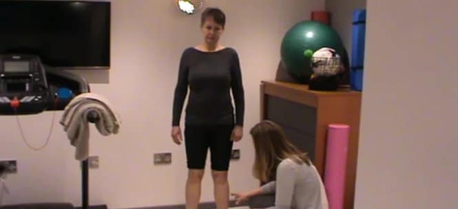
Screening Tasks
Standing postural screen
The reason for choosing the standing screen is to establish the starting point of Ms. A’s body; how she organises herself in an upright position. Multiple torsions are likely to be felt in most people’s bodies, and they may change frequently, however having a baseline enables the therapist to determine likely sites of sub-optimal performance that can then be tested further within the screening tests. It is useful to start with a standing screen before looking at specific tests for running as any sites that demonstrate sub-optimal alignment, biomechanics and/or control are likely to be relevant and be used to determine the overall ‘driver’. It will also give an idea of any habitual posture.
Diane’s comment
Excellent reason for doing a standing postural screen.
Standing screen results
Pelvis
Her pelvis was rotated to the left in the transverse plane (TPR (L)) and she also had an intra-pelvic torsion (IPT (L)) to the left which means that the right innominate was anteriorly rotated, left innominate posteriorly rotated. The sacrum was rotated to the left and nutated. A rotation of the pelvis is a physiological movement; however, it is non-optimal for quiet standing and is therefore charted as non-optimal.
Diane’s question
How did you determine that the left and right sides of the sacrum were nutated relative to their respective innominate (and therefore the physiological term you used was an IPT) vs one side or the other being counter-nutated and the SIJ therefore being unlocked? Is there any difference in the TPR or position of the left and right innominate in either an IPT or when there is an unlocked SIJ?
Jen’s response
The position of the sacrum is palpated in relation to the innominate on either side. It is the relative position that will determine if the SIJs are in a ‘locked’ or ‘unlocked position’. For a TPR to the left the right innominate is anteriorly rotated and the left innominate posteriorly rotated. The sacrum should be nutated more so on the right (moving with the right innominate to rotate to the right) than the left. In an unlocked position the sacrum would be counter nutated in relation to the innominate on that side.
Hips
Her right head of femur was anterior within the acetabulum compared with the left. It is possible that the right hip is therefore causing the pelvis to rotate to the left, but we cannot determine this without further testing, so again, although physiological we note the finding as non-optimal.
Diane’s question
I think when you use the term physiological you are implying that the position of the bones ‘make sense’ or are congruent with what should happen when the pelvis rotates to the left, am I correct in this assumption? If so, tell me what the right femoral head position would be that would suggest it’s position was not congruent with a left TPR/left IPT?
Jen’s response
With a left TPR/ left IPT the right femoral head will be taken more anteriorly relative to the left as the direction of rotation is away from the right hip. The femoral head should always stay in a neutral position within the acetabulum to allow a full range of movement without becoming impinged. If within the TPR/IPT the right hip was positionally too anterior, or held posteriorly and excessively internally or externally rotated it would be non-optimal for that position. The hip should always remain ‘centred’ to be able to spin within the joint.
Diane’s response and further question
Correct. So if the pelvis was rotated to the left in the transverse plane and the left hip was anterior relative to the right, would this be a congruent or incongruent finding and what is the significance of this finding with respect to the finding of drivers?
Jen’s response
In this case the finding would be incongruent. Where there is an incongruence it is suggested that another vector is acting on it as it is ‘going against’ the rest of the structures it that area. Where this occurs it should be assessed as a potential driver. If this is the case If by correcting the left hip, the pelvis neutralised (improved) and it had the best over-all effect on the other sub-optimally performing sites (including the task), it could be considered the driver. If by correcting the hip, the other sites including the pelvis got worse, it could be suggested that it is reacting to the pelvic TPR, to brace against it, and would therefore not be the driver.
Diane’s response
Both scenarios you list above are correct. It also could suggest that there are at least 2 drivers, one which is causing the hip to translate anterior (e.g. the hip itself) and another that is causing the pelvic ring to rotate (e.g. the thorax).
Lumbar spine
On palpation of the lumbar spine all vertebrae were rotated to the left which in standing is congruent with the pelvis and right hip for left rotation, but non-optimal for quiet standing.
Thorax
Multiple upper thoracic ring shifts were found on either side, in the mid-axillary line.
- TR (thoracic ring) 3 – translated to the right and rotated to the left (congruent with pelvis/hip/lumbar spine)
- TR4- translated to the left and rotated to the right (incongruent with TR3, pelvis, hip, lumbar spine)
- TR5- translated to the right and rotated to the left (congruent with TR3, lumbar spine, pelvis, hip
Cervical
Her right eye was deeper than the left eye (which suggests that her sphenoid bone is rotated to the right). The right temporal bone was anteriorly rotated and the left temporal bone posteriorly rotated which suggests an ICT (intra-cranial torsion) to the left with an incongruent sphenoid.
Her mandible was deviated to the right. On palpation her temporal bones felt as though they were ‘walking’. It is agreed between cranial therapists that the bones of the cranium should move rhythmically and symmetrically adducting and abducting, any asymmetrical movement is again noted as non-optimal.
Diane’s question
The position of her mandible is incongruent to the position of her temporal bones. Describe the possible resultant position of the left and right condyle of the mandible relative to the disc/articular eminence with the incongruent relationship between the temporal bones and mandible. This is likely a sore jaw! This is a question not covered on the ISM Series to date and requires an understanding of the arthrokinematics of the TMJ so let me know if this is new stuff for you.
Jen’s response
The incongruent position of her mandible and temporal bones would be likely to cause an asymmetrical jaw movement. In normal arthrokinematics of the jaw, the first 20mm of movement is rotational (at the inferior joint), after this (to open the mouth wider) the condyle and the disc glide anteroinferior along the articular eminence (superior joint motion). In optimal cases the disc will translate with the condyle of the mandible. Commonly, the disc can become displaced anteriorly, causing retro-discal tissue to become trapped and sore (the central part of the disc is not vascularised or innervated and so doesn’t cause pain). I could hypothesise in this case that the right condyle of the mandible would be posterior (putting compression onto the posterior articular surfaces and traction onto the anterior ones) to the articular eminence and the left condyle would be more anterior (creating the opposite effect), positionally.
Diane’s response
Great hypothesis! Some fine-tuning of the arthrokinematics in your answer. In the initial stage of opening the mouth the condyle should rotate (as you note) at the inferior joint (i.e. between the condyle and the disc). Subsequently, the condyle and the disc glide anteroinferior along the articular eminence (superior joint motion). This is very similar to the arthrokinematics of the sternoclavicular joint. When the mandible and temporal bones are incongruent, there is likely two vectors of force acting on the jaw. The normal resting position of the discs and the joint surfaces will be torqued and, as you note, the right condyle would be positioned more posteriorly thus putting compression on the posterior articular structures and traction on the anterior ones and visa versa for the left side.
Shoulder girdle/humerus
Her right clavicle was anteriorly rotated and left clavicle was posteriorly rotated (causing a torsion to the left) which is congruent with her pelvis, right hip and lumbar spine. Her right humerus was sitting anteriorly in the glenoid more than that of the left side. Using Shirley Sahrmann’s one third: two third theory for the shoulder it was therefore deemed as non-optimal. Her temporal bones (ICT) are congruent with her clavicles for the task of standing. Her sphenoid, C5 and TR4 are incongruent.
Information added after Diane’s question below: The best over-all correction for standing was TRs 3 and 4.
Diane’s question
There is no ‘driver’ listed here for this screening task. Later you do report on the driver. When is it essential to determine the driver in standing and when is it less relevant? Is it necessary for this case and meaningful task?
Jen’s response
It is essential to determine the driver for standing if the task is related to standing. It is less important for other tasks e.g. swimming that are not related to standing. For a task such as sitting it is important to understand how the patient has moved from standing to sitting (as sites can fail to transfer load effectively (i.e. lose optimal alignment, biomechanics and/or control), before they have reached their final position) and for other tasks it is useful to see how the body has re-organised itself (if indeed it has). For example, the driver for standing may change once the person starts to run, which is why it is important to assess the patient using more than one screening task. The idea is to identify the driver which makes the overall best change and improvement for function, alignment, biomechanics and control for the whole task.
Diane’s response
Excellent!
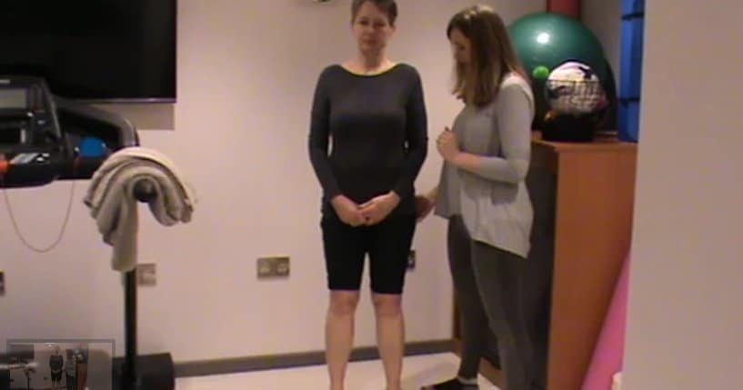
One leg stand (OLS)
On testing for control of the sacroiliac Joints (SIJs) in standing they were found to lose control bilaterally. This is determined by placing one hand on the spine of the sacrum and the other hand on one innominate, holding as much of the bone as possible to maximise the potential for feeling motion away from the axis of movement. As the patient starts to move, the therapist monitors for any movement between the bones. A ‘stable’ or well controlled joint would not have any relative motion between the sacrum and the innominate. Loss of control can occur at the initiation of movement, or late into the task. The timing is useful to note as it can help to determine which area of the body fails first. It was noted that both SIJs ‘unlock’ or move early in the task.
Diane’s question
Above, you note that Ms Abbot stood with her pelvis in a left TPR and left IPT. If I am understanding what you have written in the OLS findings above the moment she shifted weight from two legs onto one the innominate moved in a non-optimal manner relative to that side of the sacrum. Is this correct?
Jen's response
Yes
All the areas noted in the standing screen remained shifted. The best correction for this task was noted as the upper thoracic rings (3 and 4). TR4 is the ring not congruent with the pelvis, hip and lumbar spine. As TR 4 was corrected, 3 started to shift more on the opposite side. The rings were therefore ‘glued’ meaning that a vector needed to be released between them enabling TR4 to be fully corrected. The combination of correcting both TR3 and TR4 together improved the other sites over-all and the task felt easy, light and connected for the patient. I was not able to fully correct the 4th thoracic ring, but its correction made the best over-all improvement, indicating that there were likely to be other secondary drivers to consider as the treatment continued.
Diane’s question
When two thoracic rings are not able to move independently of one another, we refer to them as being glued. Can you list the possible neuromuscular vectors and articular system impairments that can ‘glue’ two rings together. Then, can you describe how you would differentiate a neural system ‘glued’ ring from an articular system ‘glued’ ring.
Jen's response
Neuromuscular vectors could include: the Intercostal muscles (internal and external); Articular system vectors for thoracic rings could include: fractures; joint subluxations/dislocations; ankylosis; intervertebral disc strains/tears/herniations and osteochondrosis of the relevant costotransverse joints and zygapophyseal joints.
If the vector is neuromuscular the barrier or end feel would have a stretch to it and there would be some play before the vector prevents movement. It is often possible to partially, if not fully, correct a neuromuscular vector during the assessment process. A glued ring has a very short vector of pull and the release after correction produces a very short inter-ring ‘listening’ or pull.
An articular system vector would have a stiffer and more rigid end feel and would not have play. In most cases it is not possible to correct an articular vector during the screening process, it will require treatment. It is often necessary to remove neuromuscular vectors before determining that there is an underlying articular vector. Any articular treatment can then be directed locally to the joint plane that you believe to be affected.
Seated trunk rotation (STR)
The task of STR was chosen as thoracic rotation is an important component of running. I wanted to look at available range of motion bilaterally and I find it easier for the patient to gain a sense of restriction points when sitting, rather than the standing ‘full body twist’. I was also considering its use for homework as it is easily reproducible and measurable for the patient.
In sitting, the pelvis was rotated to the right (TPR(R)) a change from when the patient was standing. She sat on an ‘unlocked’ pelvis which means that relative motion was palpated between the sacrum and innominate bones, indicating that the pelvis was not transferring loads appropriately prior to/into the task of sitting.
Diane’s question
Originally Jen had said that her pelvis was in a right TPR and a right IPT. It is not possible for her to be sitting in an IPTR and an unlocked pelvis. Why? This was my initial question to which Jen responded.
Jen's response
An intra-pelvic torsion requires that the sacrum be nutated bilaterally relative to both innominates. An unlocked pelvis is when the innominate is anteriorly rotated more in relation to the sacrum or the sacrum has counter nutated relative to the innominate on that side. Therefore, Ms. A was actually sitting with her pelvis rotated to the right in the transverse plane (right TPR) and the sacrum was counernutated on the right, and thus unlocked on this side. On palpation this occurred early during the task of stand to sit.
She had more restricted motion in right STR than left. The best correction for this task was the TR3 and TR4. In sitting TR3- translated to the right and rotated to the left. TR4- translated to the left and rotated to the right. TRs 3 and 4 were ‘glued’. TR5- translated to the right and rotated to the left. This was positionally the same as in standing. As the patient rotated to the right, the 3rd and 4th rings remained shifted. By partially posteriorly rotating the left rib of the 4th ring, waiting, monitoring and de-rotating the 3rd ring in response on the right, the rings were held in a more neutral position. Because they were ‘glued’ correcting one ring would impact the other, making it worse. The correction was performed in sitting and the range was assessed (to be better). The correction was then performed again in standing, and held as the patient sat again. More rotation was obtained by correcting the rings prior to sitting as the correction improved the pelvis, hip and lumbar spine position before the STR task was started.
Correcting the upper thorax revealed a very strong vector visceral vector (pulling towards the heart). A vector can be deep, or superficial, short or long and the nature of the pull can help us to determine what structure is likely to be causing it. In this case the pull was felt to be originating deep into the chest on the left side.
Correction of the cranium did not improve the cervical spine or clavicles. Correction of the cervical spine partially improved the clavicles, but did not improve the task. Correction of the humerus did not improve any other sites of sub-optimal performance. (I did not note and can not recall any specific impacts on the cranium on this occasion).
Diane’s question
A strong visceral intra-thoracic pull was noted during your correction of thoracic rings 3 & 4. Was the same vector noted when you released the correction?
Jen's response
The correction of TRs 3 and 4 was difficult to obtain due to local immediate vectors. As the correction was initiated a sense of pulling was felt upon both rings towards the left lower thorax in general and a sense of the whole left thorax being pulled down was felt as the correction was released (passive listening). I didn’t feel that a full correction was obtained at this point, but it was still the best over-all correction, as for standing. However, in hindsight the vector may have been more powerful/more easily palpated with the patient sitting as I had not been so aware of it when she was stood.
Diane’s subsequent question
I’m still very curious about her intra-cranial relationships. Her sphenoid position is incongruent with her ICT (temporal bones and occiput) and her ICT is incongruent with her mandible. Did you note if the TR3/4 correction had any impact on any of the findings in the cranium?
Jen's response
The TR3/4 correction partially improved the position of her temporal bones (I did not note sphenoid relative position at this point), corrections of the mandible/temporal bones did not improve the TRs. Given her on-going orthodontic work, my limited cranial work and the over-all improvement with the thorax (that didn’t seem to worsen the cranium), we had focused on the TRs as the primary driver.
Vector Analysis of the drivers (Thoracic rings 3 & 4)
A technique called ‘listening’ was used to establish a possible vector acting on the upper thorax.
Diane’s question
This term ‘listening’ comes from the Barral Institute osteopathic visceral approach and can be passive or active. Can you please define the difference between ‘active listening’ and ‘passive listening’ and which one you used to determine the noted visceral thoracic vector.
Jen's response
Active listening is a technique that involves using your hands to determine a vector type. For example, you would correct the thoracic ring to the point of first resistance (R1), and as you are correcting you are bringing your awareness to any resistance that you feel. As you gently and slowly let go of the correction you would focus your attention, through your hands, to the ‘feel’ and direction of the pull, thus ‘passively’ listening. This can help you to establish, for example, if the vector of pull is short or long. This is used to identify the first vector that is acting on a region that needs to be released (as you only perform vector analysis on the driver).
Diane’s response
Almost! Active listening is when the patient is ‘moving actively’ and the therapist feels (listens) to what is happening. Are the biomechanics consistent with the task? do you feel any ‘resistance’ to the movement? Passive listening is when the therapist passively moves the patient and feels for resistance during this correction of either alignment, biomechanics and/or control. Passive listening is also when the therapist lets go of this passive correction and feels for the location, quality (direction, strength, length) of the vector of pull. So, vector listening informs us of the primary side, direction, length and possibly the system of the underlying impairment.
The best corrections for quiet standing, STR and OLS were found to be thoracic rings 3 and 4. The ‘correction’ is applied by gently distracting the rings and ‘creating space’ in between them, the anteriorly rotated rib is then posteriorly rotated whilst monitoring the other rib of the ring and the rings above and below on the opposite (and same side) of the thorax. A full correction is achieved when optimal alignment is palpated and the task is improved in terms of ease, quality of movement, pain and range of motion. When the correction is slowly released the therapist can monitor through their hands for where the vector of pull is originating (passive listening).
Diane’s question
You mention that you monitor the opposite rib of the ring being corrected as well as the one above and below. What are you monitoring for? What are the possible things that could happen and what do those possibilities suggest?
Jen's response
When correcting a thoracic ring the first part of the correction requires space to be created between the rings to allow for the corrected movement to take place. It is possible for the rings to become compressed together making the correction difficult as the rings feel like one unit and are difficult to separate. Compressed rings are commonly found when the patient has a habitual long term change in their posture, such as Mums that carry babies on their hips and actively compress one side of their thorax to help to stabilise themselves as the baby gets heavier. Often the compression is caused by the intercostal muscles locally to the ring in question. Another possibility is that two rings are ‘glued together’. In this instance if you start to correct one ring by posteriorly rotating one side of the ring, the ring above or below that it is glued to will rotate more on the opposite side (worsen). With a glued ring you need to slightly correct both rings together to gain the best correction, and prevent worsening of the other ring. Glued rings often occur as the body tries to automatically correct a segment that is not being controlled well, for example after an injury that isn’t rehabilitated properly. This was the finding with Ms Abbot. Her TR3 and 4 were ‘glued’ together.
Diane’s addition
Two rings rotated in opposite directions can be compressed or glued. Compression is usually caused by long vectors that don’t glue the rings together such that when you correct one of the rings, the other automatically corrects. A glued ring is one that cannot move independently of one either above or below and is usually caused by vectors between them – the intercostals is one example of a neuromuscular vector that can cause two rings to be unable to move independently.
The primary and immediate vector palpated was visceral and was thought to be originating in the heart and left lower lung/diaphragm.
Diane’s question
What would the difference in ‘personality’ of the vector be if it was originating in:
- Iliocostalis (and for this to be the primary vector which way should the ring be translated – to the same or opposite side of the iliocostalis incriminated by vector analysis)
- Pectoralis minor
- Intercostals between thoracic rings 3 and 4
Jen's response
- If iliocostalis was the primary vector it would be felt posteriorly and the pull would be longer. The translation of the ring would be to the opposite side as iliocostalis pulls the posterior segment of the ring caudally therefore posteriorly rotating the same side of the ring which in turn creates a translation to the opposite side of the thorax.
- Pectoralis minor would prevent posterior rotation and would be felt quickly but not as immediately as an articular fibrosis or an intercostal muscle.
- Intercostals appear immediately on palpation, they can prevent separation of the rings initially as you start to correct and have a compressive nature.
Diane’s response
To avoid confusion, I would suggest reserving the use of the word compression or compressive to reflect those rings that can move independently and glueing for those that can’t. Intercostals most often glue, not compress, rings in my experience. How does a pectoralis minor vector prevent posterior rotation of a rib given its attachment to the coracoid process?
Jen's response
If Pectoralis Minor was the vector the rib would already be held in posterior rotation (preventing any further posterior rotation) and therefore resisting anterior rotation of the rib on that side. It would create a contralateral shift on the opposite side of the ring. The direction of pull would be superior in nature.
Diane's response
Pec Minor is interesting in that it can posteriorly rotate the rib OR the rib can be anteriorly rotated – it really depends on what the scapular stabilizers do in response to the ring shift so be prepared to find both!
It was difficult to correct the rings fully, however they produced the best over-all correction of the other sites of sub-optimal performance. It was decided that the first line of treatment would be to treat that vector. Anatomically the fibrous pericardium is attached to the cartilages of the 3rd to the 7th ribs on the left and to the central tendon and muscular fibres of the diaphragm. The patient has a history of physical trauma to her left lung (lung collapse and pulmonary embolus) and emotional trauma (P.T.S.D.). In Chinese medicine the lungs are the organ of grief and in cranial-sacral therapy the respiratory diaphragm is said to be potentially affected by physical and emotional trauma, with that in mind I felt there was sound clinical reasoning to initiate treatment. The correction (passively at this point) of TRs 3 and 4 improved her pelvic control and corrected her right hip. With the rings corrected passively (anteriorly by the patient) she transferred weight onto her right leg without her right SIJ unlocking, pain-free and with perceived ease of control by the patient.
Diane’s question
When there is a loss of SIJ control it is often assumed that there is an underlying motor control deficit of the postural pelvic stabilizers (TrA, pelvic floor and/or deep multifidus at the sacral level). In this case, why is it not necessary to assess the motor recruitment strategy for pelvic control?
Jen's response
In this instance, the correction of the driver ‘automatically’ restored optimal motor control for the pelvic stabilisers. When TR3/4 were corrected the SIJ remained stable. If this was not the case, if the driver was in this region, or if the control was poor, it would then be important to specifically re-train the motor control.
Treatment
Release component
The vector connecting the left diaphragm, heart and third and fourth thoracic rings (fibrous pericardium) was released with the patient in supine lying using release with awareness (RWA), fascial release techniques and directional breathing. We used a verbal ‘cue’ visualising and creating a sense of deep joy. The patient can use whatever imagery helps them to create and focus on the joyful feeling. It is not necessary for the therapist to know which image they choose. This patient easily identified an image, others may need examples and may take longer. We then progressed to ‘ring, stack and breathe’ (RSAB) a technique developed by Dr. Linda-Joy Lee. The driver rings are corrected and held (in this case by the therapist) whilst the patient takes six deep breaths. Each deep breath is inter-spaced by relaxed breathing. The patient is asked to direct the breath to the relevant section of the thorax, in this case the anterior upper thorax. RSAB can release multiple vectors connecting to the thoracic rings and is an efficient way of ensuring optimal release prior to further treatment. This treatment improved, but didn’t resolve the glued rings. The 4th ring was perceived as the primary driver with the 3rd ring compensating for it.
Diane’s question
What clinical finding led you to this perception that the 4th ring was the primary driver if the rings were glued?
Jen's response
Indeed, if two rings are ‘glued’ then they would jointly be perceived to be the driver. If, once the vector is removed between the two, and correcting one ring subsequently improves the other ring that was previously glued to it, then that ring would become the driver.
At this point the patient still needed to passively control TRs 3 and 4 externally, however they became easier to correct. Even with some release of the deep visceral vector, it was not totally released which meant that the patient needed to practice some release techniques at home to try and maximise the impact of the initial treatment.
Align component
The patient was taught how to manually self-correct the third and fourth thoracic rings anteriorly. Initially it was advised to do so in supine lying to optimise relaxation and reduce some of the compressive effects of standing. They were given the home-practice to try and eventually gain the correction using imagery only, if possible.
Connect component
The connect component relies on the use of the deep system. In this instance the patient was using a deep cue to connect and engage her intrinsic muscles (thoracic/segmental multifidus, levator costarum) locally to the 3rd and 4th thoracic rings. We used palpation into the segmental multifidus laterally at T3/4 specifically to help her brain to recognise exactly where to engage. Pain can cause a delay in the anticipatory contraction of the deep muscles. If this muscle contraction delay/absence is not corrected, the dysfunction can remain. You cannot strengthen a muscle that your nervous system isn’t using, therefore it is important to ‘connect’ to this system prior to or along-side re-training movement (Diane Lee, Diane Lee and Associates).
Movement component
The initial movement regime was separated into progressions. I anticipated a likely need for significant release over subsequent treatments with the primary vector being visceral. And additionally there were likely to be neural/dural vectors. My treatment approach was therefore very gentle.
Diane’s question
What findings from the story led you to believe there would be neural/dural vectors underlying the primary visceral one?
Jen's response
Ms A had reported multiple whiplash injuries and concussions in her past medical history. She had recalled some intermittent paresthesia in her lower limbs at the time of her initial injury and afterwards. She was also being concurrently treated with dental braces and a mouth guard. Given the direct link through the dural system from the cranium and cervical spine to the pelvis I could hypothesise that with injury, fixation and adaptive shortening it would be possible that the dural system had been impaired and would need time to adjust to the new input and positions that were being asked of it.
To ‘capture’ the dural system we placed a small rolled towel under the sacrum. Feet were to be placed on a wall to prevent supination. The patient then slowly and gently flexed the head and neck to create a gentle dural stretch. The progressions were to add the use of her upper limbs by attempting to control the 3rd and 4th thoracic rings whilst raising her upper limbs to 90 degrees. She could then add knee rolls (all the time controlling the rings and using breath to release any vectors) subsequently taking the correction into sitting, and then standing.
Diane’s comment
The cranium (including the cerebral falx and cerebellar tentorium and incongruent sphenoid techniques) have not been taught in every ISM Series since 2007. This piece has been added extensively since 2014 and continues to develop. While it is not necessary to have this ‘piece’ for ISM certification, I am looking forward to having you assist on the ISM Cranium, Neck and Upper Thorax course in Dublin in August 2017 so that your tool box is complete Jen!
Followup treatment sessions
Ms. A is not local to the clinic, we had therefore agreed to liaise over the internet to monitor progress and discuss any issues that may arise. We spoke just over a week after the initial assessment and treatment. She reported that her low back pain had improved a lot, but that she was getting some coccyx/pelvic floor pain and some left plantar fascia pain with walking.
Diane’s response
Not an unexpected response given the home program given. Why? How could you have ensured that she had enough posterior tibial nerve mobility and sacrococcygeal mobility to handle the home exercise program you gave her to avoid this over-dose?
Jen's response
Firstly, I should have tested passive sacroiliac joint (SIJ) mobility. In crook-lying one hand monitors for response posteriorly over the SIJ and using the other hand to distract the joint along the three parts/poles of the joint. If ischiococcygeus was over-active it would compress the inferior part of the joint, preventing a parallel glide between the innominate and the sacrum. Secondly, I should have tested the dynamic mobility of the posterior tibial nerve. One way of testing for compression or tethering is to have the patient in supine and perform a passive straight leg raise (PSLR) with a bias of pronation at the foot. I could also have given the home-practice of a ‘Tai-Chi Kick’ which should help with balance re-training, SIJ control and neural mobility as we progress towards running.
Diane’s further comment
More to come in Dublin – August 2017! Long dural/neural vectors can create incongruent rotations from the cranium all the way to the subtalar joint. With a story such as this, it is critical to ensure that the nervous system can tolerate the elongation that you are creating with the release of the skeleton. Often, when the dural/neural system is the underlying system impairment, multiple sites of FLT are merely because the skeleton is trying to shorten itself to give the nervous system more mobility. It may have been another reason for the inability to completely correct TR4.
I had omitted to assess the position of her coccyx at her first assessment, so I asked her to do this herself. She felt that it was deviated to the left. I hypothesised that her coccygeus muscle may be tight in response to us not monitoring it during the dural stretches. I asked the patient to gently correct the coccyx whilst thinking of her cues for her thoracic rings, then to step forwards. She reported that the foot pain was resolved with the coccyx and thoracic correction. We know that the third and 4th thoracic rings were poorly controlled and ‘glued’. Iliocostalis attaches posteriorly to the ribs and ilium, and if acting or reacting can compress the SIJ and cause dysfunction. In this case Ms. A’s pelvis was rotated to the left ((L) TPR) which worsened with weight transfer onto the right lower limb. We could hypothesise that when walking, increased tension and torque would occur at the knee, which could irritate the posterior tibial nerve. We could also hypothesise that the left coccygeus had increased in tone as a reaction to the pelvic TPR/IPT which was why on palpation it felt tight and sore.
We agreed on the following revised treatment plan:
- Release: Left coccygeus (manually first and then with imagery)
- Align: Thoracic rings 3 and 4 and coccyx.
- Connect: Cue to relax the pelvic floor and to connect to the ‘deep system’ at thoracic rings 3 and 4.
- Move: Step forwards (if easy and pain free continue for 30 reps, if possible).
Ms. A continued to have intermittent ‘flares’ of her pain for a few weeks, although the pain had improved to the point that she had been able to reduce a lot of her regular analgesic medication. We are continuing to work on the control of her thorax, and have also been working on coccygeus cues as her pelvic floor reacts quickly if she either pushes the dural release too hard or sits on a hard chair/surface for too long. The plantar-fascia pain has not returned. We are still taking note of any changes that occur with her ability to perform the exercises and her pain levels when her orthodontic braces are adjusted. Ms. A has also had to spend some time managing her stress levels and coping with the variation in her pain. We discussed not panicking if her pain levels increase, as in the past she has always tended to rush to have manual treatment straight away. She now has the ability to self-correct more easily which will enable us to add ‘layers’ to her treatment each time she visits the clinic.
Diane’s final words
Awesome Jen! If you see this patient again, and if she is still undergoing the dental work, it would be good to understand the relationship between the sphenoid, ICT and coccyx to further clarify the role of adverse neural tension here and then down to the lower extremities/feet.
Case Study Author

Jennifer Cardew
Cardew Physiotherapy and Performance
ISM Certified Practitioner ISM Series Graduate
Sea echo Seaton Park Torpoint Cornwall England PL11 3JF
Clinical Mentorship in the Integrated Systems Model
Join Diane, and her team of highly skilled assistants, on this mentorship journey and immerse yourself in a series of education opportunities that will improve your clinical efficacy for treating the whole person using the updated Integrated Systems Model.
{"first_class":"1","title":"Heading","show_title":"0","post_type":"course","taxonomy":"","term":"","post_ids":"3346","course_style":"recent","featured_style":"custom_block_1","masonry":"0","grid_columns":"clear1 col-md-12","column_width":"200","gutter":"30","grid_number":"1","infinite":"0","pagination":"0","grid_excerpt_length":"100","grid_lightbox":"0","grid_link":"0","css_class":"block-small-thumb","container_css":"","custom_css":""}
We will come together for 3 sessions of 4 (4.5) days over a period of 6-8 months with lots of practical/clinical time to focus on acquiring the skills and clinical reasoning to put the ISM model into practice. Hours of online lecture and reading material and 12 hours of in-person lecture are...
More Info