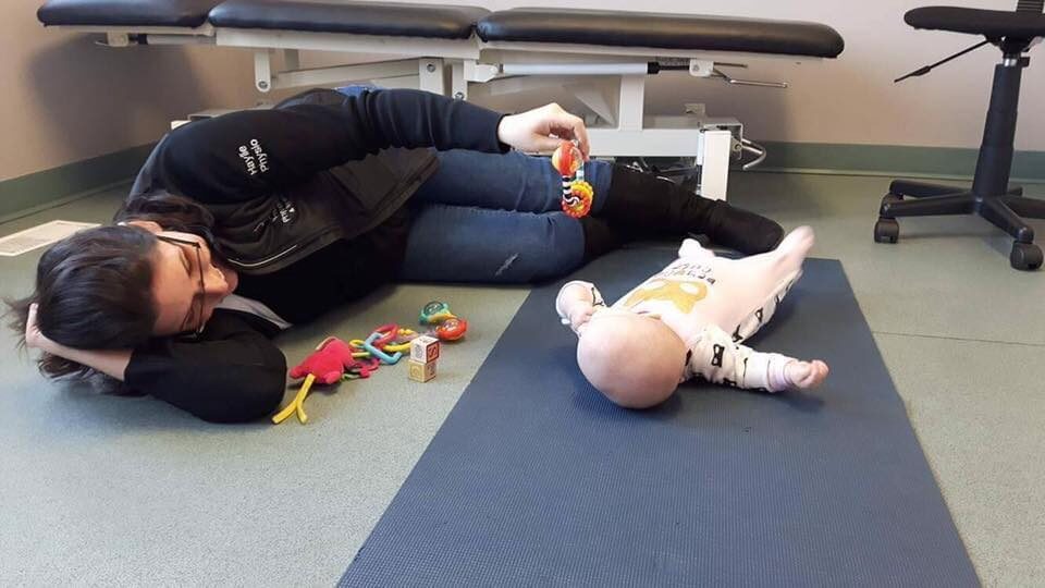
Story
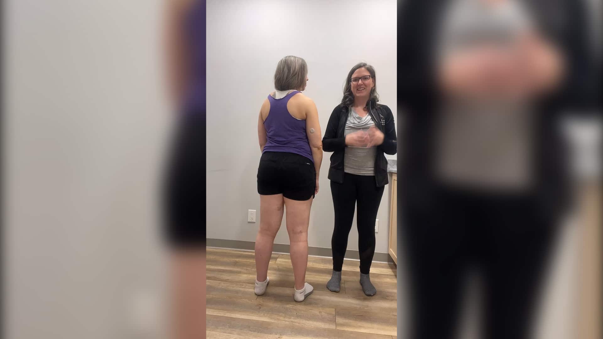
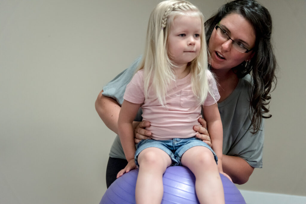
BK is a 65-year-old male that presents to the clinic with the chief complaint of right sided hip pain, ongoing for the past 6 weeks since he ‘twisted funny’ at pickleball. The hip has been more sore in general, with clicking and perceived weakness when he is doing stairs, will “lock” if he brings his hip into too much extension, and is specifically painful in sitting. He reports difficulty sitting normally without experiencing excruciating pain in the right hip. He reports inner thigh muscle tenderness since this recent injury.
- Diane’s question:
In the ISM approach, it is essential to assess the right task otherwise the suboptimal strategy will not be identified, nor the correct driver for this task. Above, there are several tasks reported to be symptomatic. What further questions at this point in the evaluation would you ask to determine if you are going to assess:
- Clicking when in climbing stairs?
- Locking in too much right hip extension?
- Sitting?
- Haylie’s response:
ideally we are selecting a screening task that is the most relevant to the client, when they provide a list of aggravating activities I will ask if there is anything in particular that they would like to work on resolving at the outset so we can focus on that biomechanical assessment. In this instance when discussing with the client of all the symptoms and aggravating factors he was describing, he wished to be able to sit without pain.
He is managing the pain with icing, sitting with the leg straight (demonstrates arching the back/leaning back and to the left while sideflexing the thorax to the right). This client has a history of “Cracking the head of the femur” when he was 50 years of age which he recalls being told he didn’t need to seek physiotherapy for, as well as idiopathic peripheral neuropathy in the bilateral lower extremity. He is a reportedly active individual that walks his dog, plays pickleball, and does pottery. He is currently unable to participate in pickleball and pottery as they are both extremely aggravating and painful; although he has been walking the dog it has been less enjoyable due to pain and discomfort.
The client has had x-rays completed of the right hip that were negative for any arthritic changes or other pathologies.
Other orthopedic complaints include bilateral foot pain for which he did get orthotics in May 2023, and right knee pain. The onset was at the end of May 2023 or early June 2023. He had a break from pickleball from June to September at which time much of his foot/knee pain resolved. When he started back in the fall, he became progressively more sore in the knees until he “moved wrong” in December during pickle ball and the immediate right sided hip/pelvic pain onset. He was able to finish playing the game of pickle ball but had progressive difficulty throughout the rest of the afternoon and evening.
- Diane’s question:
How could a restricted right subtalar joint, with or without a talar tilt during loading, alter the biomechanics of the right knee and impact the right hip during any task that required rotation of the right hip? Give me an example of a direction of subtalar joint restriction (pronation or supination) and follow the impact up the chain.
- Haylie’s response:
We know that the hind foot, which includes the subtalar and talocrural joints are the building blocks of stability and the biomechanical chain of force absorption through closed chain activities. Should the right subtalar joint be in a supinated position (where supination is the position of the talus being positioned medially in the anterior compartment on the calcaneus and posterolateral in the posterior compartment of the talus on the calcaneus).
- Diane’s comment and question:
Let’s revisit the biomechanics of the hindfoot for supination Haylie. When you say in supination the talus is positioned medially in the anterior compartment, are you describing an osteokinematic motion or arthrokinematic? Since you have divided up the motion into the two joints (anterior and posterior), I’m assuming you are talking arthrokinematics. A medial glide of the anterior part of the talus relative to the calcaneus occurs when the talus osteokinematically has rotated medially relative to the calcaneus and this is the motion that occurs in pronation, not supination.
When you say the talus is ‘posterolateral in the posterior compartment’ this is an arthrokinematics description but is it correct? Look at the orientation of the posterior subtalar joint – does it run posterolateral or posteromedial?
- Haylie’s revision/response:
Yes, revisiting the biomechanics of the hind foot. First understanding the language. Pronation and supination for ISM refers specifically to the combined biomechanics of the foot excluding the talocrural and metatarsal phalangeal joints. If we break it down into hind foot and mid foot we refer specifically to the arthrokinematics (the joint surface movements of glides/slides), and osteokinematics (movement of the bones in space).
When we are looking at a squat in general we want to see the following:
- Dorsiflexion of the talocrural joint as we move down into the squat
- Medial rotation of the talus relative to the calcaneous (or a lateral rotation of the calcaneous relative to the talus)
- Fanning of the mid-foot as we move down into the squat
Biomechanically in a squat what this requires is the following:
- Widening of the mortice during dorsiflexion to accommodate the anterior aspect of the talus that is wider than the posterior aspect
- Osteokinematically the talus will plantarflex and adduct in space during pronation (named due to the direction of movement of the anterior portion of the bone). A medial rotation of the talus relative to the calcaneus (or a lateral rotation of the calcaneus relative to the talus) as they are moving simultaneously. This will require anteromedial glide of the anterior compartment, and an anterolateral glide of the posterior compartment of the subtalar joint referring to the talus on calcaneus. The shape of the joint is what will dictate the rotational component
- The fanning of the foot requires the external rotation of the navicular and internal rotation of the cuboid which is relative to the second metatarsal and middle cuneiform. The medial column externally rotates and the lateral column internally rotates which results in lengthening of the foot
Pronation is significantly impacted by ground reactions forces as there is no specific tissues under conscious control that will allow an individual to rotate the talus relative to the calcaneus independently.
So if we then look at normal supination:
- During supination, the talus osteokinematically will dorsiflex and abduct in space. A lateral rotation of the talus relative to the calcaneous (or a medial rotation of the calcaneous relative to the talus – as they are moving simultaneously) occurs. Arthrokinematically, what we see is a posterolateral glide of the talus on the calcaneus in the anterior compartment, and a posteromedial glide of the posterior compartment of the subtalar joint of the talus on the calcaneus
- Supination is related to the shape of the joint but is more dependent on the muscle forces, plantar fascia, windlass mechanism, and the coactivation of the muscles from the leg, and the intrinsic muscles of the foot, yet still requires the ground reaction forces
- Folding of the foot requires internal rotation of the navicular and external rotation of the cuboid relative to the second metatarsal and middle cuneiform. This effectively shortens the foot
Now if we take these normal biomechanical understanding of what is required during a squat (pronation of the foot), we can deduce the following:
- If we limited the subtalar joint into a supinated position, the talus would stay in a relatively dorsiflexed/abducted position with restriction of the ability to move into the plantarflexed/adducted position relative to the calcaneus. This could mean there is restriction in the arthrokinematics of the talus on calcaneus of the anterior compartment into the anteromedial direction and the posterior compartment anterolaterally
- As a result of the restricted ability to pronate at the subtalar joint, there will be compensations to allow for the first ray to remain in contact with the ground during squat (as well as stance, and during ambulation). The impact of this could include an eversion of the foot from the talus tilting in the mortice.
- It is likely that this positioning of the talus and calcaneus will result in changes to the flexor digitorum, flexor hallicus longus, and tibialis posterior, which is likely to add tension to the interosseous membrane between the tibia and fibula, restricting the ability of the mortice to widen
- An external rotation of the tibia relative to the femur follows the supinated hindfoot, and a lateral talar tilt allows the big toe to reach the ground in midstance
- This could conceivably result in internal rotation of the hip, and ultimately result in the ilium being anteriorly rotated with a relative counternutation of the sacrum, if the deep muscles of the pelvis are unable to resist this force
- Widening of the mortice must occur as part of dorsiflexion since the talus is wider at the anterior aspect compared to the posterior aspect. Limitations in dorsiflexion can be related to articular restrictions, or vectors from structures attaching to the interosseous membrane limiting the ability to dorsiflex by function of keeping the mortice from widening
- Arthrokinematics are dependent on ground reaction forces and cannot be consciously controlled by the individual
Cognitive and or Emotional Beliefs
During the discussion on the initial appointment the client revealed that he believes that he never really got better after his femoral fracture and that he has to move carefully in order to avoid injuring himself. BK explained that since the injury he has been trying to move ‘just right’ to ‘ensure I don’t damage the hip further’ and this injury reconfirms for him that he must move in a specific manner to avoid injury. He doesn’t know why exactly he has all these different aches and pains that have been cropping up over the years, but with each passing injury seems to become slightly more vigilant in his quest for ‘moving right’.
- Diane’s question:
How will you frame your assessment to confirm or negate his beliefs?
- Haylie’s response:
I find that this is an ongoing conversation with the client and creating the possibility of change in belief of the client starts with curiosity. Asking questions to clarify such as “has that helped you to feel better to date?” or as we progress through the appointment noticing how simply doing things differently (as taught by Antony Lo) can immediately change the client’s experience coupled with the ability to utilize ISM principles of correction resulting in immediate change for the client can all be very helpful in breaking down beliefs. Helping clients understand the resilience of their body can be immensely helpful in freeing clients from fear of movement.
We were able to make immediate changes to his cognitive beliefs within the ISM evaluation. This was completed by applying corrections through the various areas as will be outlined below resulted in the “Wow that is better” experience for the client; getting this experience and then reflecting with him on his beliefs had him almost immediately recognizing that his vigilance and striving for a stiff ‘just right’ movement was not serving the purpose of improving his pain the way he initially thought. Being able to have the client experience in their body the “yes that is way better” through ISM can be a key factor in breaking down some of these cognitive beliefs.
- Diane’s response:
Yes!! I love this Haylie. You changed the experience in his body and it’s his nervous system reaction to those ‘suggestions of change’, which is what a correction really is – just do something different (I love Antony’s concept on this) that changes his cognitive belief. He figures it out himself with a little help from us.
Meaningful Task
Chief complaint for BK is pain with activity specifically involving squatting or sitting activities including:
- Pickleball (ready position – client is effectively in a squat with racquet forward in his dominant hand (right), with knees and hips flexed)
- Pottery (seated with additional hip flexion to lean forward over the pottery wheel)
- Sitting (in general at home in any chair)
- Diane’s question:
Is his right hip pain always in the same location? Is it nociceptive, neuropathic or nociplastic or a blend and what from his story provides you with evidence to phenotype his pain as you do?
- Haylie’s response:
The pain appears to be located in the same areas, the posterior right pelvis and posterolateral right thigh in addition to the right groin. Since nociceptive pain is pain as a result of noxious stimulus to the tissues that are mechanically or chemically altered, the behaviour of the pain with exacerbation with particular movements and postures his pain would be considered nociceptive in nature as the movement changes and posture exacerbations would be a noxious stimulus that is mechanical in nature. Since the client does not describe any burning/shooting/electric pains it is unlikely to be neuropathic in nature. Given nociplastic pain is arising from the central nervous system, it can arise without the presence of peripheral stimulus and mechanical input and associated with burning/deep throbbing which also is not reported in BK’s case.
- Diane’s further question:
Is his problem going up stairs, and therefore more loading in flexion is relevant, or downstairs, in which more hip extension is relevant? Did he enquire about whether this assessment would help him understand the clicking in extension?
- Haylie’s response:
The client was less concerned about the stairs and more concerned about sitting. I would say that for him going up the stairs would be more concerning since in my notes from assessment indicates going up stairs is more problematic for the client. He was not focused on the clicking sensation specifically.
Meaningful Task for this Session: Sitting
Screening Tasks
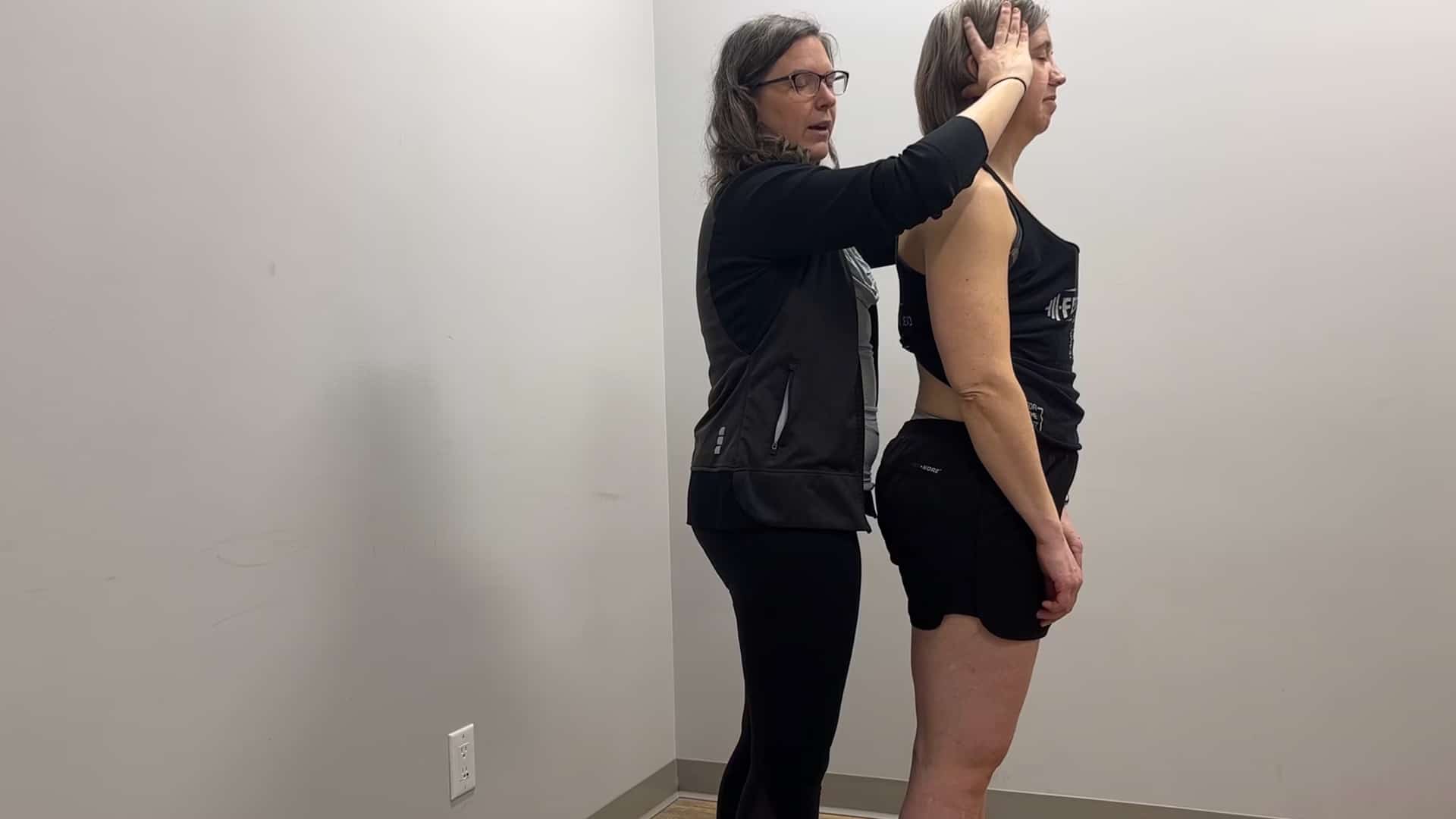
Moving from Standing through Squat to Sitting.
Functional Unit #1 (FU#1)
Standing Start Screen – Findings for FU#1
Since BK’s meaningful tasks (pickleball, pottery, sitting) include movement from standing to squat to sitting, we want to complete a standing start screen. Once we ascertain the asymmetries and postures in standing, we will be able to follow the biomechanically expected motions through the initial screening task – the squat.
Pelvis: Slight left transverse plane rotation (LTPR) LTPR with congruent left intrapelvic torsion (LIPT)
Hips: Right hip is anterior to the left hip as well as to the right acetabulum
- Diane’s question:
Which of the above findings for the right hip (anterior to the left vs anterior to the acetabulum) is relevant to the locking/clicking of this hip and why? Even if the hip doesn’t turn out to be a driver for Mr BK, does this information help you educate him in anyway?
- Haylie’s response:
We know that clicking in and of itself is not necessarily of concern, however it can be disconcerting to clients. It is the biomechanical evaluation and the hip being positioned anteriorly in the acetabulum that is most important for clicking. When we apply the correction of the hip the client no longer experiences the click which for this client was very helpful in assisting him during education. Why this is the more plausible reason for the clicking is a result of the anterior position of the hip in the acetabulum could create a click at the end of range of the femoral head on the anterior limits of the acetabulum, as a result of the change in tension at the hip capsule and with the muscular structures surrounding the hip.
- Diane’s comment:
…or the labrum perhaps?
- Haylie’s response:
Yes of course, and the hip being pulled forward could impact the labrum and could explain the clicking sensation.
Thorax: The client presents with multiple rotations/translations of several thoracic rings as follows:
Thoracic ring 3 (TR3) right translated (→)/left rotated
Thoracic ring 4 (TR4) left translated (←)/right rotated
Thoracic ring 5 (TR5) right translated (→)/left rotated
Thoracic ring 6 (TR6) left translated (←)/right rotated
Thoracic ring 7 (TR7) right translated (→)/left rotated
Thoracic ring 8 (TR8) markedly left translated (←)/right rotated
Thoracic ring 9 (TR9) right translated (→)/left rotated
- Diane’s question:
Every single ring from TR3 – TR9 is rotated opposite to the one above and below. This is a very compressed thorax. In this situation do you expect to find one ring that changes all this? What is the first thing to do to determine where the priority region is?
- Haylie’s response:
I do not expect one ring to correct everything in this instance. The first thing to identify the priority is to determine which thoracic ring is incongruent to the TPR. Anything that is considered congruent to the TPR would be biomechanically appropriate for the TPR and not relevant to the evaluation and identification of drivers. Once we identify this initial incongruent segment (which in our case is TR8), we can follow through task and see if it is worth correcting (as anything that corrects in the task is not pertinent to the task at hand). In this instance TR 6-9 do resolve in the task, as does TR3 which indicates that they are not pertinent to the driver evaluation. TR4 is the first incongruent ring, and when corrected TR5 remained. When attempting to correct TR5 is when it was determined that a complete correction was not able to be achieved and addressing biomechanically was required as outlined below.
At times when clients are dramatically compressed through the thorax it may be helpful to decompress the thorax from gravitational forces through evaluation of the thorax in an alternative position such as downward dog, however was not necessary in this case.
- Diane’s response:
I agree with the last paragraph. I think the first step here is to generally correct this thorax with decompression and see which thoracic rings unwind themselves and which ones require some manual help.
- Haylie’s response:
I see, so when someone comes in and they have this standing start screen posture with the thoracic compression like this, it can be helpful to do some decompression position evaluation to see if there is any immediate improvement and be able to more rapidly identify which may require more manual therapy? Had I done that with this client, it could be that TR5 would have been obvious with less effort/time up front.
- Diane’s comment:
Exactly!
TR 8 is the first thoracic ring that is rotated incongruent to the pelvis
Lumbar Spine: L4-5 Left Rotated (LR), L2-3 neutral, L1-2 Right Rotated (RR)
Squat Task Findings – FU#1
The squat task was chosen for the screening task since it encompasses much of BK’s most problematic daily functions and activities. Should the client biomechanically be disadvantaged during the squat he is likely to be inefficient in his pickleball ready position and a squat is a basic necessity for the lunging motions required. He will also be sitting with poor alignment which may be exacerbating his presenting symptoms with sitting and pottery. During an optimal squat we would expect to see no transverse plane rotation of the pelvis, thorax, shoulder girdle, or cranium, the hips remain centered in the acetabulum, the lumbar vertebra and thorax should remain neutral without rotation/translation, the hips/knees/ankles should move symmetrically into flexion and dorsiflexion, the foot should pronate. The cranial and cervical regions should extend without rotation/translation to keep the eyes level. Any deviation from this would be considered suboptimal biomechanical function (alignment, biomechanics and control) for the task.
Pelvis: RSIJ unlocks at 10% of the task, LTPR exacerbates
- Diane’s question:
Did you happen to notice the timing of the worsening of the TPR of the pelvis and the unlocking of the RSIJ? Which occurred first and what does this suggest?
- Haylie’s response:
The TPR appeared to worsen followed very closely by the unlocking of the RSIJ. This would suggest that whatever is driving the TPR is causing the SIJ unlocking and will be of greater importance to the evaluation.
Hips: Right hip remains anterior to the left hip as well as in the right acetabulum (does not center into the acetabulum) through the task.
Thorax: the thorax as a whole side flexes right during the task with the following changes in specific thoracic ring alignment
TR3 – corrects in task
TR4 – unchanged (remained left translated (←)/right rotated)
TR5 – worsened (increased right translation(→)/left rotation)
TR 6/7/8/9 – correct in task
- Diane’s question:
Am I correct in interpreting your information above to mean that the thoracic rings of interest are now TR4 ← & TR5 →?
- Haylie’s response:
The remaining findings would be as you have stated TR4 ← and TR5 → correct.
- Lumbar: L5 was noted to be left rotated (congruent with the L TPR of the pelvis), with L3 in neutral and L1 and 2 right rotated (incongruent with L5, but congruent with the lower thorax)
Lumbar Spine: L4-5 – correct in task
L2-3 remains neutral, L1-2 rotates to the L
- Diane’s question:
Do you have any notes on where L5-S1 started and what happened in the task? I find it interesting that the rotation of the pelvis worsened (LTPR increased) which would mean the sacrum rotated more to the left, yet you report that L4-5 which was left rotated resolved (became neutral)? I’m curious how L5 tolerated this?
- Haylie’s response:
I do not have any notes on specifically on this unfortunately.
Driver(s) for Functional Unit #1 for the Squat Task
Primary driver: Thorax with the priority ring being TR5
Secondary driver(s): Right hip and pelvis
Clinical reasoning for finding these driver(s): Initially, a complete correction of TR5 was not possible and a correction of TR4 was possible but did not correct TR5. Further assessment for the articular restriction of TR5 will be described later in this report. After restoring articular mobility to all relevant joints of TR5 a full correction was possible and this correction resulted in correction of TR4.
- Haylie’s question:
Is what I wrote above correct? – TR3 resolved in the task but TR4 & TR5 didn’t so your interest here from your task findings are TR4 & 5 – not 3, not 6,7,8,9 – they are all irrelevant regardless of what happens to corrections in standing – he obviously decompresses and lengthens his thorax automatically in a squat for this to happen.
- Diane’s response:
It is perfectly written now!
After release of the articular restrictions in TR5 (see further assessment), correction of TR5 improved L1-2 left rotation and gave the client the best experience in the screening task.
Correction of TR5 did not restore control of the R SIJ or correct the alignment of the right hip. Additionally, correction of the pelvis or hip did not correct TR5; therefore, the right hip and pelvis are secondary drivers for FU#1.
Functional Unit #2 (FU#2)
The decision to move into FU#2 comes from initial observation of the client’s squat screen – first, there is observable right sideflexion and left rotation of the cranium/craniovertebral region. Second, the FU#1 did not completely resolve the presenting cranium/craniovertebral region biomechanical findings. So although the quick screen of FU#1 drivers on head and neck (HN) rotation with and without the driver correction did not appear to negatively impact HN rotation specifically, further analysis of FU#2 is indicated.
Standing Start Screen – Findings for FU#2
Cranial region (Cranium, Occipito-Atlantal – OA, Atlanto-Axial – AA): left intracranial torsion (LICT), sphenoid is RR and incongruent to the ICT. C1 RR
- Diane’s question:
Is the starting position of C1 congruent or incongruent to the left intracranial torsion and is this your expected finding for C1 when the client has a LICT?
- Haylie’s response:
Reviewing this, I know that the biomechanics of the CV region differs from the rest of the cervical spine – given there is no rotation of the C0/C1 (only flexion/extension and side bending with conjunct rotation) the CV region is moving in a biomechanically appropriate way (right side bending and left rotation). However, the position of C1 (RR) is incongruent with the rest of the region. I feel that C1 could reasonably be relatively right rotated in the CV region based on the impact of the alar ligament has between the cranium and C2, and C1 holding the role of ‘washer’ in this area, however overall the CV region should left rotate as a whole. I am not convinced that if it was behaving biomechanically appropriately that I would be able to feel the right rotation compared to the left rotation of the rest of the region.
This being said, if it was moving biomechanically appropriately, we wouldn’t be seeing the side bending right and rotation left during a squat task as the cranium should not need to rotate or sideflex during the squat task.
- Diane’s response:
The common compensatory pattern for a LICT, is the following:
Occiput: left rotated/right sideflexed on the atlas (defines the direction of the ICT)
Atlas: relatively right rotated to the occiput (for a good brain exercise figure out the OA arthrokinematics necessary for these osteokinematics to occur!)
Axis (C2): left rotated (congruent with the occiput) relative to the atlas. Therefore the AA joint is right rotated, the OA joint is left rotated/right sideflexed and this neutralizes the position of the head in space.
In your patient’s case, C1 is incongruent to the ICT which is optimal biomechanics, but we need to know the position of C2 to understand why the head sideflexes right and rotates left in the task – to be continued!
Cervical (C2-7): C2 LR, C3 LR, C4 neutral, C5 and C6 LR, C7 RR
- Diane’s comment:
So C2 is LR therefore the osteokinematic position of the cranium, atlas and C2 are all congruent to what is expected in the task; therefore, it is likely something else is causing the head and neck to RSF/LR.
- Haylie’s response:
This makes so much sense with the CV biomechanics, and I see exactly what you mean by there must be a second vector.
Shoulder Girdle (SG): R SG depressed/protracted, clavicle is parallel to the floor and anteriorly rotated on the R
- Diane’s question:
Is the rotation of the right clavicle congruent or incongruent to its depression?
- Haylie’s response:
The rotation of the right clavicle is incongruent to depression. During depression of the shoulder girdle we would expect to see a posterior rotation of the clavicle.
Thorax (TR1 TR2): TR1 LR, TR2 RR
Squat Task Findings – FU#2
Cranial (Cranium, OA, AA): LICT increases, sphenoid RR increases, C1 RR increases,
Cervical: C2 and C3 LR increases, C4 remains neutral, C5 and C6 LR increases, , C7 RR remains
Shoulder Girdle : R SG depresses further, R clavicle compresses medially and remains anteriorly rotated
Thorax (TR1, TR2): TR1 remains LR, TR2 correct in task
- Diane’s question:
There are many sites of impairment in this unit that don’t resolve in the task. How did you determine where to initiate your corrections?
- Haylie’s response:
I decided to start at the cranium thinking that gravity may be exacerbating issues as a result of compression. Truly it ‘drew my eye’ and thinking back I believe it is since the cranium exacerbated first of all the other findings when observing the task.
Driver(s) for Functional Unit #2 for the Squat Task
Primary Driver: Cranial region (priority cranium)
Clinical reasoning for finding this driver: Correction of the cranial region through co-correction of the LICT and RR sphenoid corrected the RR of C1 and partially corrected C2.
- Diane’s question:
What is it about the position of C2 that makes the finding above predictable?
- Haylie’s response:
C2 is continuing to follow the cranium because of the impact of the alar ligament – our start screen shows the LICT and LR C2, and both are exacerbating into the squat. It would make sense to me that correcting the cranium would improve C2.
The correction of the cranium also corrected C7, the right shoulder girdle and TR1and resulted in the best client experience between both FU#1 and #2 (client exclaimed “WOW! That feels AMAZING! Look at how far I can go!”).
Relationship between FU#1 and FU#2 Drivers
Prior to determining the final task drivers, the articular restrictions within TR5 required treatment.
Further Assessment of the Articular System of TR5
It is important to achieve a complete correction before we can determine if an area is a driver or not. If a complete correction cannot be achieved for any reason, then any articular system impairment pertaining to that body region would need to be addressed prior to finalizing the drivers. During assessment of FU#1 for drivers, a complete correction of TR5 to neutral was not possible and therefore, further analysis of TR5 was required. The underlying articular restriction was identified using two active mobility tasks; breathing mechanics and active range of motion (AROM) testing using thoracic rotation. These two active mobility tests suggested that the right costotransverse joint TR5 was unable to glide inferiorly (restricted active inspiration and right rotation).
- Diane’s question:
What were the specific results of the passive tests for the right costotransverse joint at TR5? Neuromyofascial restrictions can also produce restriction of active inspiration and right rotation. It is the correlation between the active osteokinematic and passive arthrokinematics test findings that confirm an articular system impairment.
- Haylie’s response:
Passive testing of the right costotransverse (CT) revealed a restriction into an inferior glide and posterior roll of the rib on the transverse process for the 5th thoracic vertebra (T5), with appropriate superior glide with anterior roll of the 5th rib on the left transverse process. The end feel was articular in nature into the restricted range (inferior glide with posterior roll). There was no restriction at the z-joint between the right inferior facet of T5 and the superior facet of T6 into either flexion (superior, anterior, lateral) or extension (inferior, posterior, medial)
The therapist restored mobility of the right costotransverse of TR5 using a Grade III sustained passive accessory mobilization (PAM) for inspiration (inferior glide with posterior roll) finding the vector of greatest resistance. There was a cavitation that occurred as the client inhaled, and afterwards full correction to neutral of TR5 was possible.
TR5 continued to left rotate/right translate during the squat screening task; however, when he was instructed to use a connect cue for the 5th rib at the costotransverse “think of letting this spot slide back and down” he was able to maintain the neutral position through squat range available before the hip moved anterior and the TPR of the pelvis worsened resulting in locking of the right SIJ. The client noted a large improvement in the squat task and sitting at the end of the appointment. He was provided with the connect cue for TR5 with breathing and instructed on how to monitor the ring position with his hands until the next appointment.
- Diane’s comment/question:
I’m not sure of the clinical reasoning for moving into motor control training of TR5 BEFORE completing the full ISM assessment for finding drivers? Maybe the cranial region correction would have restored complete control of TR5 without the need to do this? It is easy to slide back into local ‘habits’ isn’t it?!!
- Haylie’s thoughts/comments:
Truthfully the reasoning here was that this was the end of the first appointment and since there wasn’t time to get into the cranium with enough detail, I provided the connect cue as part of home programming. I imagine if I were to see this same sort of client tomorrow, it would not take me as long to run through the driver evaluation and I would likely end up continuing to review the FU #2 and FU #3 drivers to see if those corrections would provide complete control of TR5. It is very tricky to kick the local habits for sure, especially when I do have concern about ensuring that clients feel that they have received enough ‘value’ for their time and money spent!
After this initial appointment, TR5 was not rotated/translated at any subsequent visits. It is believed that TR5 had an initial articular restriction that when addressed during the initial appointment resulted in the ability of the area to complete the align-correct-move (A-C-M). All remaining drivers continued to be present and correlated among each other as outlined above despite the resolution of TR5 at the start of the second visit.
- Diane’s comment:
Therefore, I continue to wonder if that motor control training (connect cue for the right 5th CT joint) was necessary?
- Haylie’s response:
When he returned the second appointment I also felt the same, that likely the TR5 was not as important as I thought at the outset.
The task drivers of FU#1 and 2 at this point were:
- primary cranial region (cranium) and
- secondary pelvis.
After restoring mobility of the right CT joint at TR5, correction of the cranium resulted in complete resolution of alignment of the right hip, and a partial correction of the pelvis. The R SIJ remained stable through 50% of the squat task so remains a secondary driver for the task, therefore the primary driver for the task is the cranial region with the pelvis being a secondary driver, at this point.
At the subsequent visit FU #3 was assessed since the foot is very important for the meaningful tasks outlined above. Although sitting is the client’s primary concern, it is from the ground reaction forces through the foot that will ultimately set us up for the biomechanics that are potentially causing problems in our seated position. This means that evaluation of FU #3 is pertinent in this exam. FU #3 for the lower extremity is important to the squat task and if there is a relationship between the foot and the other identified drivers this relationship would be relevant to our treatment.
Functional Unit #3 (FU#3)
Standing Start Screen – Findings for FU#3
Feet: Significant supination of the right foot.
- Diane’s question:
What were the starting positions of both the left and right tibiofemoral joints?
- Haylie’s response:
The left tibia was externally rotated relative to the femur at 0 degrees knee extension, and the right tibia was internally relative to the femur at 0 degrees knee extension.
Squat Task Findings – FU#3
Feet: Right foot remained supinated and the tibia on the right did not advance over the talus as expected into dorsiflexion. The left foot pronated with appropriate dorsiflexion of the talocrural joint.
Knee: Reverse biomechanics of the right tibiofemoral joint were noted with knee flexion (the tibia externally rotated as the client moved into the squat in the first 5 degrees of knee flexion and remained externally rotated through the squat task).
- Diane’s question:
Are the biomechanics of the right knee congruent, or incongruent, to the biomechanics of the right foot in this task?
- Haylie’s response:
The tibia externally rotating in the squat task would be congruent with the foot being supinated.
Driver(s) for Functional Unit #3 for the Squat Task
A correction of the hind foot is all that is required to determine if there is a foot driver. For BK, a correction of the right hind foot to neutral resulted in an improved subjective experience in the squat task but not as good as the cranium correction. His response was “Well that is better too, but not as good as my head”. Correcting the right hindfoot resolved the biomechanics and alignment of the right knee.
Relationship Between all Unit Drivers
Currently there are three drivers
- Cranial region
- Pelvis
- Right foot
It was difficult to evaluate from the therapist vantage point if correction of the right hindfoot impacted the cranium, however, the therapist utilized client palpation and a mirror to observe the changes and it did appear to improve the overall CV region position through squat, but did not resolve them.
Correction of the cranium improved but did not resolve the right foot nor did correcting the foot completely resolve the cranium or pelvis.
Therefore, it was reasoned that the final squat task drivers were:
- Primary – cranial region (cranium priority)
- Secondary – the foot (created a better response in the pelvis than the cranium)
- Tertiary – the pelvis
Further Assessment & Treatment of Task Drivers
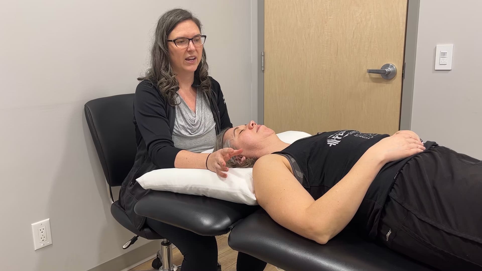
Cranial region (cranium)
Active Mobility
The cranium started in a left ICT with an incongruently rotated sphenoid (RR) and during right rotation of the head and neck zero degrees of active mobility of the posterior cranium (occiput and temporal bones) was noted.
Passive Mobility
A complete correction of the cranium (LICT and RR sphenoid) to neutral was possible; therefore the passive mobility of the cranium was full for the requirements of this task. When the correction was released, the first vector of pull was on the right temporal bone. A short vector of pull into the the sphenosquamous suture was noted. These findings were observed in both the standing and supine positions.
- Diane’s question:
This is a very common vector to find when there is an incongruently rotated sphenoid to the direction of the ICT. It comes from the anterior portion of the cerebellar tentorium. However, it also means that there is a second vector of pull otherwise the ICT would be congruent to the direction of rotation of the sphenoid. Describe for us the anterior attachments of the cerebellar tentorium and how tension here could explain compression of the sphenosquamous (SS) suture. The suture is NOT the impairment here. What finding do you have that negates the suture being the primary impairment?
- Haylie’s response:
The suture would not be the primary impairment as we are able to fully correct the ICT, if the suture was the primary impairment a full correction would not be achievable. The anterior portion of the cerebellar tentorium attaches to the clinoid processes of the sphenoid, and extends back to the transverse ridges on the occipital bone. Therefore if there is tension into the anterior portion the impact could be compression across the sphenosquamous suture. The venous drainage from around the pituitary gland will impact the tension of the anterior portion of the cerebellar tentorium. Whereas the transverse sinus and the straight sinus are more likely to impact the posterior portions of the cerebellar tentorium.
As I write this out I realize that the vector to the SCM on the right is likely the secondary portion that results in the incongruency, otherwise the ICT would be congruent.
Treatment
Given this, the therapist completed the suture technique as learned in ISM to address the sphenosquamous suture – by distracting the sphenoid on the right using the therapists left hand, and holding the temporal bone around the mastoid through placement of the thumb in the clients ear canal, the temporal bone was brought into posterior rotation.
- Diane’s question:
What position is the right temporal bone in when the cranium is in a LICT? The restricted suture techniques I teach in ISM are always into ease, not bind, in other words, into the lesion; so did you rotate the right temporal bone into ease or bind in this technique? Go back to your anatomical description of the anterior attachments of the cerebellar tentorium and hypothesize how this technique may have worked to improve active mobility of the cranium (passive mobility was full) other than by releasing the SS suture.
- Haylie’s response:
Yes my apologies Diane – the LICT would mean that the right temporal bone is anteriorly rotated and I would have started with an anterior rotation going into ease to start and then into posterior rotation which was restricted. It would be more accurate to say that we went into ease (anterior rotation) followed by posterior rotation to achieve full mobility at the right sphenosquamous suture between the temporal and sphenoid bones. The theory here is that the venous system surrounding the pituitary gland would be drained releasing tension from the anterior portion of the cerebellar tentorium allowing for improvement in active mobility of the cranium. Reflecting this will make my next finding predictable, as I have not yet drained the rest of the sinuses.
- Diane’s comment:
But that isn’t what you did right? You posteriorly rotated the temporal bone while fixing the sphenoid. This would ultimately generate tension on the anterior portion of the cerebellar tentorium and squeeze the fluid down the sigmoid sinus to the jugular vein. The treatment technique I teach in Part 3 of the ISM Series for draining to release the anterior cerebellar tentorium doesn’t rotate the temporal bone at all, one stabilizes the greater wing of the sphenoid with one hand while the other distracts (not rotates) the temporal bone thus opening the jugular foramen, allowing the fluid to drain. This is not a suture technique, but the suture technique you chose will also tense the cerebellar tent which is why it works, in my opinion!
- Haylie’s response:
I want to add my ‘ah-ha’ here! It’s funny when things ‘click’ a little more! So since I was treating it as a suture restriction I was doing the rotation and not the distraction technique for the anterior cerebellar tentorium drainage, (which as you state will still tension the tentorium and result in some drainage), then when I felt the remaining pull to the posterior cranium I went into the more specific venous drainage. I know that the suture wasn’t actually restricted because I was able to achieve full passive mobility requirements for the task with the correction, which should have clued me into the anterior cerebellar tentorium and secondary vector for the incongruency from the beginning.
Once the restriction in this area was addressed, the client completed the squat task again with self-reported improvement in the pain of the right hip.
Re-evaluation revealed that the cranium remained in left rotation (LICT) and right side flexion (entire cranium) through the task. Continued treatment is required given the persistence of the suboptimal biomechanical findings.
In standing, additional review of the active mobility of the cranium revealed an improvement in ability of the right temporal bone to posteriorly rotate but it was still an incomplete excursion of expected movement. Another correction of the ICT, coupled with listening for vectors upon release of the correction, was completed and a new intracranial vector in the posterior cranium was identified. When the client was in supine this vector persisted and it was concluded to be coming from the posterior aspect of the cerebellar tentorium.
- Diane’s comment:
I’m not surprised since I believe the first vector was from the anterior aspect of the cerebellar tentorium and that your technique moved fluid into the posterior part of this structure since it is valveless.
During the re-evaluation the client had dramatic improvement in his subjective symptoms moving into the squat – the squat depth and ease of movement was improved when the therapist observed the movement. There was improvement the client’s ability to maintain alignment and control of the pelvis until much later in the task.
- Diane’s comment:
This suggests that whatever you did was able to release the spinal dural tension on the coccyx and thus prevent the increasing LTPR of the pelvis. Maybe the hip wasn’t driving the TPR of the pelvis? Or maybe the right hip was reacting to the right side of the pelvic floor in response to this dural tension? The cerebellar tentorium still doesn’t explain the incongruent rotation of the sphenoid to the LICT though. I suspect there was another vector outside the cranium. Just like the incongruent hip to TPR of the pelvis, this finding suggests two vectors – one on the pelvis, one on the hip. In the cranium, it is usually one compressing the jugular foramen outside the cranium and then the backflow inside the cranium results in tension in the cerebellar tentorium which compresses either the SS suture or the OM suture.
The client was provided with education regarding the cerebellar tentorium and it’s role in the vector analysis in addition to some self-correction of the cranium to be completed with squat movement and prior to sitting.
Right Foot
During the next visit active mobility of the cranium was dramatically improved – not perfect, however ISM is about improvement and not perfection, and given that the right foot has a relationship to the cranium from our previous exam, working the two together seemed like a best use of time.
Active Mobility
Persistent reduction of pronation of the right foot in a squat task was noted.
Passive Mobility
The right hind foot was restricted in two locations, the talocrural joint was limited into dorsiflexion, and the subtalar joint was restricted in medial rotation of the talus relative to the calcaneous and the noted restriction was in the posterior subtalar joint. Arthrokinematically, the talus was restricted in an anterolateral glide relative to the calcaneus in the posterior subtalar joint.
Treatment
The talocrural joint dorsiflexion restriction was addressed first by using a technique where the talocrural joint is held at the end of the available range, and pressure is applied with one hand to the back of the leg to locate the area of greatest impact of anterior movement of the talus in the crus. The muscle that was at fault for this client was two fascicles of the medial head of gastrocnemius which were addressed with release with awareness techniques. The subtalar joint vector was evaluated by bring the posterior subtalar joint to the R1 restriction and then release and listen – a short firm vector to the joint line was identified. Myofascial techniques to the same were applied.
- Diane’s question:
Given our discussion above – which joint line of the subtalar joint was the noted vector at? Was this an articular restriction or myofascial? If myofascial – which muscle?
- Haylie’s response:
This was a joint restriction. I felt the most restriction from the posteromedial aspect of the posterior subtalar joint.
- Diane’s comment:
Therefore, I’m assuming articular mobilization techniques, not myofascial techniques were used to restore mobility.
The client was provided with a home program addition for the foot that included some self-release to the medial head of the gastrocnemius and the medial subtalar joint line on the right.
Once the right hind-foot had been addressed more specifically, a re-evaluation of the drivers was completed which revealed a neutral cranium through 50% of squat task (followed by right sideflexion and left rotation in last 50%), L TPR of the pelvis occurs at 75% of squat task (SIJ unlocking at this point) and the R hip centers during the task.
- Diane’s question:
What is your hypothesis as to what is causing the right sideflexion/left rotation of the head and neck in the last 50% of the squat?
- Haylie’s response:
When I provided the client with connect cues for the cranium as described below in the home programming, he was able to maintain a neutral position throughout the movement. I hypothesized that the neuromuscular habit of contraction of the SCM in the movement pattern practiced over an extended period of time resulted in the continuation of the head and neck right side flexion and left rotation.
Pelvis
Active Mobility of the SIJ
This test must be done in open chain due to the small amplitude of movement at the SIJ.
- Diane’s comment/question:
I’m not sure that the amplitude of motion is the reason this test is done in open chain. What happens at the SIJ the moment the pelvis is under load that is a better reason for why the active mobility test must be done in non-weight bearing, or open chain?
- Haylie’s response:
Yes it makes more sense that weight bearing increases the form closure of the joint and by completing the test in open chain we are removing that factor of consideration.
The amplitude of movement is compared between the left and right sides, symmetry is the expectation. When the right hip is flexed beyond the available motion of the hip, the innominate should posteriorly rotate relative to the sacrum. For this client, there was a different finding each time he lifted the right leg, and the left leg consistently had a posterior rotation of the innominate relative to the sacrum.
- Diane’s question:
Can you explain further what you mean by a ‘different finding’ each time he lifted the right leg and what your interpretation of this inconsistency is?
- Haylie’s response:
Each time he lifted the right leg the motor planning and coordination to complete the task was inconsistent to the last attempt. I found that each time the client completed the active mobility there was a different overall result, and even though at times there was appropriate posterior rotation of the innominate relative to the sacrum, I also found greater posterior rotation of the sacrum relative to the ilium. This a motor planning issue which results in the inconsistency.
- Diane’s comment:
Although, in a healthy individual diversity of motor programming is healthy so this may not be suboptimal for him!
- Haylie’s response:
Interesting! I assumed it would be suboptimal as the opposite side did not show the same variability
Passive Mobility of the SIJ
The client was placed in supine, and the pelvis was positioned symmetrically on the right and left.
- Diane’s question:
Since the pelvis is one ring, what is meant by ‘the pelvis was positioned symmetrically? What was symmetric and why is it important to do this before comparing mobility between the left and right SIJs?
- Haylie’s response:
When I say positioned symmetrically what I am referring to is having the innominate in the same relative position to the sacrum on both sides. For instance, if I have a client in crook lying and feet are equally placed on the table, but the right innominate is anteriorly rotated compared to the left (ie ASIS will be more inferior in comparison), we will not be comparing the right and left SIJ in the same position. If this was the case what I would do is straighten the left leg to the point where the left ASIS is at the same position as the right ASIS and then evaluate the passive mobility on each side. Comparing an anteriorly rotated innominate on the sacrum on the right would not yield the same findings as a posteriorly rotated innominate on the opposite side. I want to compare the two joints in the same position.
The SI joint plane was noted in the anterior/posterior plane and the three parts of the SIJ (top, middle, and inferior) were tested for passive mobility. The greatest restriction was in the middle part on the right, which is often impacted by visceral vectors.
A load, release and listen technique was used to identify the vector compressing the middle part of the right SIJ; the resultant force was noted to be into the abdomen with an inferior inclination. Being that I had taken the Barral Visceral Manipulation, I utilized some VM1 techniques to evaluate and identified the retrocecal ligament as the primary source of this vector.
Treatment
The therapist treated the cecal ligament using Barral release techniques.
Once the restrictive vectors for all unit drivers were released the client was able to achieve a full squat, without pain, he felt strong using the stairs, and he had no pain in sitting.
When the therapist evaluated the drivers in task, the client demonstrated full control of the pelvis, the cranium remained in neutral through task, and the right foot was able to pronate through the task when provided with connect cues. When the cranial connect cue wasn’t utilized at the end of the squat I observed RSF and L rotate of the cranium, and if the foot cue wasn’t used the foot would not move into pronation. As a result the pelvis would not maintain full control, despite the client being pain-free. An important distinction as releasing the vectors alone without the connect cue to maintain alignment, followed by movement progressions would not be effective.
Home Program
The client was given homework throughout treatment to address the motor control of any driver that failed to retain alignment under load. Loads were progressed as treatment continued. One of the best cues for this client was to imagine his head was a hot air balloon, and that he was letting his foot spread out into sand. This cue provided better alignment, biomechanics and control of the pelvis, while maintaining proper alignment of the cranium and the foot through the task. This was part of evaluation of active control which was completed at the end of each appointment in the development of the homework for the client.
Conclusion
In reviewing this case report for ISM certification, I recognize several areas where my own assumptions and habits have affected efficiency. In some cases, my personal biases and treatment preferences led me slightly off track. How quickly we move from the nuanced specifics, to leaping to conclusions without ensuring that we are objectively identifying the driver! For example since moving into the squat and being seated was the primary concern for the client, the evaluation of the hip should have focused on the movement into flexion and not extension. This being said, improvement over perfection, and ultimately the client saw resolution in his presenting complaints.
- Diane’s comment:
The hip was never a driver – so evaluation was never required and was deleted from this report.
Initially I had thought that this issue was a neural issue, however in reflecting there are components of articular, visceral, and myofascial in this case.
This client presented with the same complaint from session to session with steady improvement as we addressed impairments in the three priority areas; the cranium, the foot and the pelvis. The cranium and the foot being more closely related, and the pelvis being the final aspects that completed the plan of care and ultimately allowed the client to return to a pain free meaningful task, as well as his other preferred activities.
Ultimately I would say that the primary impairment was through the dura, which I believe would be considered a myofascial structure, as the dura would connect the cranium to the pelvis and the foot.

Case Study Author


Haylie Lashta
Warman Physiotherapy & Wellness (105 Klassen St West, Warman)
Warman Physio Saskatoon (1260 Baltzan Blvd, Saskatoon)
ISM Series Graduate
105 Klassen Street, Warman, SK, Canada
Clinical Mentorship in the Integrated Systems Model
Join Diane, and her team of highly skilled assistants, on this mentorship journey and immerse yourself in a series of education opportunities that will improve your clinical efficacy for treating the whole person using the updated Integrated Systems Model.
{"first_class":"1","title":"Heading","show_title":"0","post_type":"course","taxonomy":"","term":"","post_ids":"3346","course_style":"recent","featured_style":"custom_block_1","masonry":"0","grid_columns":"clear1 col-md-12","column_width":"200","gutter":"30","grid_number":"1","infinite":"0","pagination":"0","grid_excerpt_length":"100","grid_lightbox":"0","grid_link":"0","css_class":"block-small-thumb","container_css":"","custom_css":""}
We will come together for 3 sessions of 4 (4.5) days over a period of 6-8 months with lots of practical/clinical time to focus on acquiring the skills and clinical reasoning to put the ISM model into practice. Hours of online lecture and reading material and 12 hours of in-person lecture are...
More Info