Story
History of Present Illness:
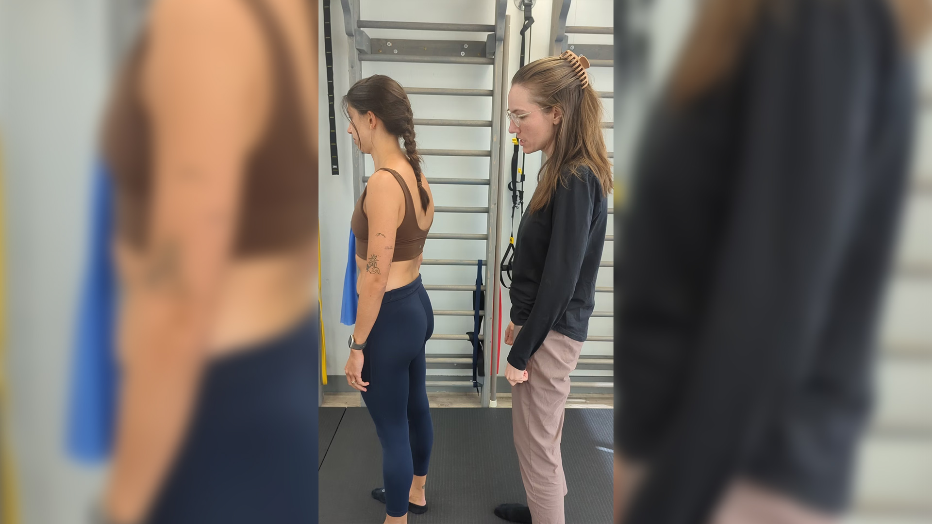
Ms. P is a 27 yo female who was assessed at The ScoliClinic in Dec 2023 for right sided neck tension. She underwent fusion surgery for her right sided thoracic Adolescent Idiopathic Scoliosis. Her thoracic spine was fused from T3-T12 in 2011 and was corrected to a Cobb angle of 22°. She also has a residual left lumbar curve measured at 25°.
- Diane’s question:
I understand that fusion surgery for scoliosis has evolved over the years. Was every thoracic segment from T3-4 to T12-L1 fused in this surgery with the intention of preventing all vertebral mobility? Were the costotransverse joints left free to move?
- Christiane’s response:
The current standard practice for a posterior instrumented spinal fusion surgery for Adolescent Idiopathic Scoliosis at BC Children’s Hospital involves: osteotomies of spinous processes and facet joints, followed by screws implanted into the pedicles, 3 dimensional spine correction, 2 rods connected to the screws on either side of the residual spinous process, and finally the bone that was harvested earlier is laid over the posterior aspect of the spine as a bone graft. The bone graft takes about 3 months to set, but once it has taken, it should be limiting all intervertebral movement rendering the hardware redundant. The costotransverse joints are left intact. Based on Ms. P’s spinal X-rays, it appears that her fusion surgery was done according to these standard procedures, although it is interesting to note that 2 screws are missing at T2 and T3 on the right side. Given the bone graft, the absence of these screws should not have any bearing on her thoracic range of motion.
She started learning more about the postural elements of her scoliosis and being intentional about addressing these about 3-4 years ago. In particular, Ms. P. has been aware of her right scapula’s tendency to tip anteriorly and has been trying to correct it by actively retracting and pulling her scapula back. She feels that it was around the same time as she started working on her posture that she noticed the onset of right sided anterior neck tension. She was previously familiar with upper trap tension bilaterally as a student, but this area of tension felt different: more anterior, along the scalene distribution. The tension is noticeable in sitting and with most exercises involving the right arm. Open kinetic chain exercises tend to feel the worst, such as an overhead shoulder press. The discomfort is overall best described as a near constant feeling of tension that is more or less noticeable based on participation in aggravating activities. There were a few months in the fall where Ms. P noticed sensory changes in her hands bilaterally but no clear pattern of aggravating/easing factors or distribution of the sensory changes emerged. The pain is nociceptive in nature as it has clear mechanical aggravating and easing factors. Ms. P does not appear anxious when she talks about the pain. She believes that if only she could get her right shoulder to remain in a more neutral position, the pain would improve. Ms. P is active and participates in a regular Pilates and yoga practice.
- Diane’s question:
There appear to be two different complaints in this story; a “near constant sensation of tension” in the right anterior aspect of her neck and a positional awareness, or cognitive belief perhaps, of her scapula ‘being wrong’ (tendency to anteriorly tip). Although she experienced some ‘sensory changes in her hands, you did not classify her symptoms as neuropathic, but rather nociceptive. She didn’t appear to report pain, it appears to me that you used this term, not the patient, – is tension classified as pain?
- Christiane’s response:
Good points Diane, I agree that the neck tension Ms. P complains of could be classified as neuropathic as opposed to nociceptive. The positional awareness of her scapula did not come across as a primary complaint in her subjective, more so a cognitive belief as to why she was experiencing such longstanding neck tension.
Past Medical History:
Ms. P was deaf for the first year of her life until amniotic fluid was drained from her ears. She has always had joint hypermobility but has never been diagnosed with a connective tissue disorder. She suffers from urinary incontinence with heavy laughter and occasionally with running since ~8 years of age. She has pursued some pelvic floor PT for it recently with no real changes to symptoms. She had a tear of her right MCL, ACL and meniscus in 2013 playing basketball which was surgically repaired. She gets grabbing pain in the upper lumbar spine with occasionally feelings of instability, felt centrally to the spine, with transitional movements such as getting out of bed or coming off the floor. This lumbar pain has been ongoing for the past 4-5 years, worse in past 1-2 years.
- Diane’s question:
So to clarify, none of these significant complaints are her meaningful complaint for her sessions with you at this time? Do you feel there is any relationship between the tension in her neck (which appears to be her meaningful complaint from the story reported above), her urinary incontinence (which doesn’t appear related to pregnancy or delivery), the low back pain or the injury to her right knee?
- Christiane’s response:
That’s right. She has a number of significant complaints, but felt that the neck tension was impeding her quality of life at the time the most and therefore asked for us to focus our assessment on the neck. At first, I wasn’t sure whether we would find any links between these various complaints given that the meaningful tasks for each one are so different. But having completed a full assessment and treatment with the lumbar spine and the right foot being co-drivers, I feel differently. Ms. P’s inability to control L2 had a direct impact on her neck tension, as described below; and likely resulted in more pointed lumbar pain with the transitional movements noted here when the loads on the spine are higher. The pelvic floor symptoms would warrant their own assessment, but urinary incontinence is often related to lack of pelvic floor motor control (especially when pelvic floor weakness, prolapse and any other pelvic specific issues have been ruled out by a pelvic floor physiotherapist). The pelvic floor is part of the deep core unit along with multifidus and transverse abdominus. Our assessment revealed some atrophy of multifidus as well as some diaphragmatic dysfunction. In addition, Ms. P’s residual thoracic ring rotations could be impacting the neural drive to her transverse abdominus. So it doesn’t seem surprising that the pelvic floor may be left overloaded and not always able to prevent urinary leakage in situations of high intra-abdominal pressure such as heavy laughter. Finally, sub-optimal foot biomechanics will have a direct impact on knee biomechanics via the connection of the hindfoot to the tibia. Whether her right knee injury pre-disposed her to current suboptimal right foot biomechanics or vice versa, we will not know!
Meaningful Task
The meaningful task is a right sided shoulder overhead press which we will also use as a screening task. Symptoms in the neck are felt without any weights, so we will perform our assessment using an unweighted overhead shoulder press. The starting position will be standing.
Screening Task
Functional Unit #1 (FU#1)
Start screen findings (standing):
Pelvis: left rotated in the transverse plane (L TPR) with congruent left intrapelvic torsion (L IPT). Slight lateral sway to the right
Hips: right hip is anterior relative to the left hip (congruent with pelvis) and slightly anterior relative to acetabulum
Thorax: general thoracic right rotation residual from right thoracic curve
- TR 4 & 5 are rotated right, left translated (←)
- TR 6 is rotated left, right translated (→)
- TR 7 & 8 are rotated right, left translated (←)
- TR 10 are rotated right, left translated (←)
- Diane’s comment and question:
You have described the osteokinematics of this structural, fused, scoliosis as having the same pattern as a non-structural scoliotic thorax. From what I have read about the intrathoracic positioning of a thoracic scoliosis (Brink et al 2019), the axis of rotation for axial rotation is posterior to the spinous process (as opposed to within the vertebral body). This impacts the direction of translation that accompanies rotation such that translation and rotation occur to the same side, contrary to the direction of translation that occurs in a non-structural scoliotic thoracic ring, which is contralateral as you describe above. How can you be sure that when you palpate a laterally translated thoracic ring (i.e. TR 4 & 5), it is rotated right and not left as per the described biomechanics in Brink et al (2019)? I also understand that the rotated vertebra is towards the anteriorly rotated rib in a structural scoliosis, opposite that of a non-structural scoliotic thorax. When we meet Jan 31,2024 with Ms P – let’s update and review your opinion on this. It doesn’t change how individual rings are corrected because we don’t force translation ever, we let the ring unwind itself, but from my experience it does feel very different.
- Christiane’s response (update after co-consultation with Diane present):
Careful examination and clinical reasoning have led us to different findings and conclusions than the ones listed above:
- Active left rotation of the trunk revealed further “apparent” left translation of TR4 but right translation of TR5. Which ring is limiting left thoracic rotation? In a non-structural scoliotic spine, we might assume that TR4, a ring which translates left and is right rotated would impede left rotation. However, when we corrected TR4 <- in Ms. P (by inducing posterior rotation to the left rib and palpating anterior rotation at the right rib) and prevented it from further translating left, her left rotation worsens. Correction of TR5 -> (by inducing posterior rotation on the right rib and palpating anterior rotation on the left rib) improved left rotation. Ms. P’s thorax therefore appears to be behaving in a similar fashion to a typical structural scoliotic thorax whereby left rotation of a ring has opposing osteokinamatics to a non-structural scoliotic thorax: left thoracic ring rotation is coupled with anterior rotation of the left rib and posterior rotation of the right rib and right translation of the entire thoracic ring. Therefore, TR 4 left translation actually facilitates left rotation, and TR 5 right translation potentially impedes left thoracic rotation.
- Diane’s comment:
However, Ms. P’s thoracic vertebra were surgically fused and no inter-ring translation should be possible; so what were we feeling and interpreting as inter-ring translation? Christiane explains below how we determined that the rings were NOT translating at all!
- Christiane’s response:
- TR 4, 5, 7, 8 and 10 appeared left translated in the standing start screen when palpating Ms. P’s thorax, but in fact cannot be given that a fused thorax should not permit inter-ring translation. NO inter-ring translation at any level of the thorax was confirmed using the lateral translation mobility test (Lee 2018) whereby one hand stabilized and prevented motion at TR5, and the other hand induced a lateral translation force at TR4 on the contralateral side. An abrupt bony end-feel confirmed that lateral translation of a whole ring is not possible in a thorax that has undergone a spinal fusion surgery. The costotransverse joints; however, are left intact during a posterior spinal fusion explaining why motion is palpated in the ribs and some thoracic rotation range of motion is preserved. How can one reconcile these 2 findings: lateral translation of rings palpated at rest and accentuated during thorax rotation, and the inability of a posteriorly spinal fused thoracic ring to translate? The best hypothesis we have come up with is that in a posteriorly spinal fused thorax, the lateral movement one palpates in the mid-axillary line is only apparent, not true lateral translation of a thoracic ring. The axis of rotation of the rib at the costovertebral and costotransverse joints does not lie in the pure coronal plane (Beyer et al. 2014); therefore, the same singular point on a rib will travel laterally on a transverse plane when the rib anteriorly rotates giving the impression of right ring translation with right rotation.
- In summary, our clinical interpretation of the thoracic ring start screen position in standing in Ms. P are:
- TR 4 & 5 are rotated left, the left ribs are anteriorly rotated, the right ribs are rotated posteriorly and there is NO inter-ring translation,
- TR 6 is rotated right, the right rib is anteriorly rotated, the left rib is posteriorly rotated and there is NO inter-ring translation
- TR 7 & 8 are rotated left, the left ribs are anteriorly rotated, the right ribs are rotated posteriorly and there is NO inter-ring translation
- TR 10 is rotated left, the left rib is anteriorly rotated, the right rib is rotated posteriorly and there is NO inter-ring translation
- Diane’s comment:
To be clear for the reader, these are NOT the osteokinematics of structural scoliotic thoracic rings, these are the osteokinematics of structural scoliotic thoracic rings that have undergone a posterior spinal fusion.
- Lumbar spine: residual left lumbar curve with apex at L3. Left rotation throughout lumbar spine with increasing rotation from L5 to L2-3.
The thorax is overall in right rotation and therefore is incongruent to the pelvis, hips, and lumbar spine which are all in left rotation. However, given the structural nature of the thorax right rotation from her residual curve, it is likely that her nervous system and body are well adjusted to the overall right rotation of her thorax. However, thoracic rings 4, 5, 7, 8, and 10 will be of particular interest during the task as these are left rotated and incongruent with the rest of the thorax, as well as TR 6 as this ring is further right rotated relative to the other rings within the thorax
Screening task: right shoulder overhead press, unweighted:
Pelvis: left transverse plane rotation and intrapelvic torsion increases
Hips: slight increase of right hip anterior translation relative to left hip and acetabulum
Thorax:
- TR 5, 8, 10 all worsen, increasing in left rotation
- other rings do not improve nor worsen
Lumbar spine: general curve correction
- L4-5: right rotate out of the start screen position of left rotation to close to full correction
- L1-3: also right rotate out of the start screen position of left rotation but only partially
- Diane’s question:
Below, you tell us that the FU#1 driver was the lumbar spine and the priority segment was L2; however, above you just note that L1-3 only partially corrected in the task. Did something in particular draw your attention to L2?
- Christiane’s response:
L2 was the most left rotated segment noted during the start screen and the segment that stayed in the most left rotation during the screening task. When I corrected the lumbar spine, I noted that correcting L2 corrected L1 and L3. L2 is therefore our segment of interest within the lumbar spine.
- Diane’s comment & question:
So the left TPR/IPT of the pelvis increased in the task; however, L4-5 derotated (from a left rotated position noted in the standing screen to ‘close to full correction’). This would result in an incredible amount of strain on the left bands of the iliolumbar ligament – especially the horizontally oriented bands. What is your hypothesis from your findings so far as to what could be causing the pelvis to rotate left more, yet the lumbar spine to unwind in the transverse plane? If we were pursuing her low back pain, I would be concerned about this finding.
- Christiane’s response:
Interesting observation Diane! I am not sure I have a full answer, but here are some thoughts. The majority of people with a dominant lumbar curve have the pelvis rotated in an incongruent direction to the lumbar spine and congruent to the thoracic spine. Typically that looks like a right rotated thorax and pelvis, and a left rotated lumbar spine (Wang et al. 2014). I assume that in those cases the iliolumbar ligaments would adapt accordingly during the years of pubertal growth during which Adolescent Idiopathic Scoliosis typically develops. Here, Ms. P’s pelvis is left rotated in congruence with the lumbar spine in standing which would be atypical for her curve type. An X-ray of Ms. P’s spine dating back from 2016 indicates that she was standing with a pelvis in right rotation at the time, following the more typical structural scoliotic lumbo-pelvic relationship. The iliolumbar ligaments would therefore already be accustomed to incongruence between the pelvis and lumbar spine, making the incongruence noted during the meaningful task less unlikely. Ms. P’s tendency to stand with a pelvis in left rotation may have developed post 2016 following her knee injury and dysfunction originating in her right foot or knee. During the task the pelvis left rotates further following the medial tilt of the right hindfoot whereas the lumbar spine is not constrained to follow the pelvis due to previously developed increased laxity in the iliolumbar ligament.
- Diane’s comment and question:
Great hypothesis! One thing I love about ISM is this: every time we have a clinical hypothesis we try to develop a test to confirm or negate that hypothesis. To your point and evidence cited above, you would expect Ms. P to have a congruent TPR of her pelvis and thorax and the lumbar spine rotated incongruent to both regions. In this screening task of a unilateral overhead shoulder press, she doesn’t present with this pattern; she presents with her pelvis rotating further in an incongruent direction to the thorax and hypothesize that this is due to (or driven by) an increasing medial tilt of her right hindfoot. What assessment test could you do to confirm, or negate, your hypothesis that the foot is what is driving the increasing LTPR/IPT of the pelvis in this task?
- Christiane’s response:
I could check whether or not the pelvis still rotates further left during the task when I am correcting the foot.
Driver for FU#1
Lumbar spine with L2 being the vertebra of interest within the lumbar spine. Correction of L2 allowed the rest of the lumbar spine to come out of left rotation, and corrected the right hip, pelvis, and most of the thorax (some TR8 left rotation remaining). As the thorax did not fully correct with correction of the lumbar spine, the thorax is considered a secondary driver within FU#1.
- Diane’s question:
For my clarity since I don’t correct lumbar vertebrae often, you did a manual correction of L2-3 which was rotated left (greatest degree of left rotation of all lumbar segments), and this caused the TPR/IPT of the pelvis to unwind as well as all noted TRs except TR8)?
- Christiane’s response:
I used my left digits 1 and 3 to create mild traction at the spinous processes of L1 and L3 and then used my second digit on the left transverse process of L2 to induce right rotation. As her nervous system acclimated to the correction, I found I was even able to get a good correction by applying very light pressure on the left transverse process of L2, traction no longer being needed.
- Diane’s further question:
The second part of my first question remains to be answered. Did all of the rib positions of rings TR4, 5, 6, 7, & 10 fully corrected/stacked with this L2 correction? Further question, if this is true what does this tell us about your hypothesis of what was driving the LTPR (incongruent to the thorax) of the pelvis?
- Christiane’s response:
L2 correction fully corrected the thorax (all except TR8 LR) and the pelvis. This could indicate that the L TPR of the pelvis is driven by the lumbar spine versus the lower limb. Or at the very least, it indicates that the rotation of the lumbar spine is in relationship to the rotation of the pelvis; meaning that we can expect a foot driven pelvis to also have an impact on the lumbar spine.
- Diane’s comment:
A quick look at the right foot with the lumbar spine corrected would have answered your question. If the lumbar spine was driving the TPR of the pelvis, which in turn was driving the right foot, then the lumbar spine correction would not only correct the TPR of the pelvic but also the right hindfoot lateral tilt.
The shoulder press overall feels better with a L2 correction, but some neck tension remains at end range shoulder flexion. In contrast, correction of the thorax, pelvis and right hip felt either worse or unchanged to Ms. P. A thorax correction did not improve pelvis nor hip alignment, a pelvis correction did not improve hip nor thorax alignment. A right hip correction corrected the pelvis but only partially corrected the thorax. The remaining feeling of right shoulder tension with end range flexion warrants further investigation of FU2.
- Diane’s question:
Christiane, you obviously have way more experience than I do when working with structural scoliosis. Can you describe for me, and the readers, how you correct an individual thoracic ring in an unfused structural scoliosis, for e.g. when TR8 is rotated left/translated left, the left rib is anteriorly rotated and the right rib is posteriorly rotated? Tell us where your hands are and what you do and what you feel for during this correction.
- Christiane’s response:
Similar to a ring correction in a non-structural scoliotic thorax, I start by palpating the translated ring on the left and right side. I stand behind the patient, starting the correction by gently lifting both ribs, giving some time for the nervous system to adjust, and then posteriorly rotate the left rib (which for TR8 in this case was anteriorly rotated). The technique used for correcting a structural scoliotic ring translation is no different than a non scoliotic thoracic ring translation, only the rotation is happening in the opposite direction (eg: left translation is coupled with left rotation), and the correction may feel different to the therapist depending on how severe the curve is and how impacted the articulating surfaces of the costotransverse joints are. In a fused thorax, a ring correction will feel very similar again to the clinician, only it is good to know that only rib rotation is being corrected as ring translations are no longer possible due to fused vertebral bodies. A rotated ring still appears translated relative to the rings above and below due to the change in position of the rib as it rotates (see explanation in earlier response).
- Diane’s comment:
In both the structural scoliotic thoracic ring and the non-structural thoracic ring, the correction is applied through the rib that is anteriorly rotated and no translation is part of this correction. The clinician allows the ring to derotate as it needs to, regardless of the biomechanics. It is not uncommon to feel the ring translate as part of the automatic response to posteriorly rotating the anteriorly rotated rib in both cases; however, the direction of translation and rotation differs once one’s attention is set to feel this specifically.
Functional Unit #2 (FU#2)
Start standing screen:
Cranial: left intracranial torsion (L ICT), sphenoid in right rotation. There is incongruence of rotation between the ICT and the sphenoid
Cervical spine: C2 and C3 are both right rotated, left translated (←), C5, C6, and C7 are left rotated right translated (→)
Shoulder girdle: overall left rotated
Right scapula: depressed, anteriorly tipped, protracted, slight upward rotation
Right head of the humerus: slightly anterior relative to glenoid fossa
Left scapula: elevated, retracted, winging, downward rotation
Clavicles: in left rotation
TR1 and TR2: both are right rotated
There is incongruence between the cranium and C2 (Diane’s comment: Well spotted!) as well as between TR1 & 2 and the shoulder girdle, and between the upper and lower cervical segments.
- Diane’s question:
Clavicles: Left rotation is a common terminology for shoulder girdle position but not clavicles in ISM; however, it is not wrong. Can you clarify which clavicle was anteriorly rotated and which was posteriorly rotated when the clavicles are in ‘left rotation’. Also, I’m not sure how much of her thoracic structural scoliosis (convex right) above TR3 (highest point of fusion) required unwinding above and whether you consider the position of TR 1 & 2 (RR) to be ‘normal’ for Ms. P. or abnormal? What about C7?
- Christiane’s response:
- Clavicles: when describing the clavicles in left rotation, I mean that the right clavicle is anteriorly rotated and the left clavicle is posteriorly rotated. This left rotated positional finding describes the osteokinematics of the clavicles that one expects with a left head rotation task.
- Upper thoracic ring findings: given Ms. P’s X-ray from 2016, her right thoracic curve actually ends at T7, and she has a mild left upper thoracic curve from C7-T5. Therefore TR1 & 2 being in right rotation would be incongruent to her left upper thoracic curve. C7 being in left rotation would be more congruent to the general position of the upper thorax, but would still be considered incongruent given the amount of left rotation it was in and how it differed from TR1 & 2. Apologies, I should have mentioned the left upper thoracic curve in Ms. P’s history above.
Screening task: right shoulder overhead press, unweighted:
Cranium: worsens, increase in left intracranial torsion and sphenoid right rotation
Cervical spine: overall worsens
C7 left rotation increases
C2 right rotation increases
Shoulder girdle:
Right clavicle draws medially, right scapula corrects
The right humeral head stays slightly anterior to the glenoid fossa and the scapula corrects itself, coming into upward rotation during the task.
TR1 & 2: unwind to full correction
- Diane’s question:
During a right shoulder press (arm elevation) what should the right clavicle do? Did it? During full right arm elevation the lower cervical and upper thoracic segments in a non-structural scoliosis should rotate to the right. You mention that her TR 1 & TR2 ‘unwound’ which I assume you mean they come out of right rotation? What is full correction for Ms. P’s TR1 & TR2 in a shoulder press considering her structural scoliosis?
- Christiane’s response:
During a right shoulder press, the right clavicle should posteriorly rotate, and should not translate medially. In this case, the clavicle only partially rotated posteriorly and drew medially. TR1 & 2 came out of right rotation, but likely stayed in enough right rotation to facilitate the shoulder press task. A full correction of TR1 & 2 in Ms. P would mean coming into mild left rotation in standing to match the rest of the upper thorax, but would allow for some right rotation during the right shoulder press task.
- Diane’s further question:
There are a lot of incongruencies to one another in this screening task; however, which segments are also incongruent to the requirements of the right shoulder press task? These are the ones that should be first corrected. I think there are 3.
- Christiane’s response:
The cranium, C7, and the right clavicle are all incongruent to the right shoulder press task. One would expect a right ICT, C7 right rotation, and posterior rotation without medial translation of the clavicle with a right shoulder press task.
- Diane’s response:
I’m not sure if the cranium should rotate into a right ICT if the head remains in neutral (i.e. not looking at the right hand); however, the right humeral head should center, and not remain anterior to the glenoid fossa.
Driver for FU2:
Right shoulder girdle, specifically the right clavicle within the right shoulder girdle. Correction of the right clavicle corrects the rest of the shoulder girdle and significantly improves the task experience. It also corrects the cervical spine and the cranium (ICT and sphenoid rotation) and the head of the humerus. In contrast, correction of the cranium felt no different and although it corrected the cervical spine, it only partially corrected the right clavicle. Correction of C7 (which corrects the rest of the cervical spine) only partially corrected the shoulder girdle and fully corrects the cranium.
FU#2 driver: Primary driver right Shoulder Girdle (clavicle)
FU#1 driver: Primary driver Lumbar Spine (L2), secondary driver Thorax (TR8)
Relationship of FU#2 driver to FU#1 driver:
The right shoulder girdle only partially corrects L2 and L2 only partially corrects the clavicle. Co-correction of the right clavicle and L2 feels even better than with individual corrections, but a slight amount of tension in the neck is felt at end range shoulder flexion, warranting assessment of FU3. The shoulder girdle (clavicle) and the lumbar spine (L2) are co-drivers.
- Christiane’s response:
The humeral head sits anteriorly relative to the glenoid fossa in standing and remains slightly anterior throughout the screening task. However, correction of the clavicle fully corrects the humeral head in standing and throughout the screening task. If an impairment within the upper extremity (hand, wrist or elbow) was impacting the task, the impairment would be affecting the task via the glenohumeral joint. As correcting the glenohumeral joint did not improve the experience of the task nor the biomechanics of the task, one can assume that any impairments at the right hand, wrist or elbow were not impacting this particular task.
- Diane’s response:
It may depend on the elbow, hand and wrist position as to whether this is true or merely an assumption.
Functional Unit #3 (FU#3)
Start standing screen:
Feet:
- Right foot:
- Hindfoot: calcaneus and talus close to neutral, slight medial talar tilt
- Forefoot: 1st metatarsal internally rotated relative to the medial cuneiform, hallux valgus
- Left foot:
- Hindfoot: in mild lateral tilt (calcaneus and talus)
- Forefoot: 1st metatarsal internally rotated relative to the medial cuneiform, hallux valgus
Right knee: tibia in external rotation relative to femur. Appropriate screw home mechanism observed when Ms. P comes in and out of knee flexion in standing
Screening task: right shoulder overhead press, unweighted:
Feet:
- Right foot: overall worsens. Slight external rotation of talus relative to calcaneus resulting in a medial tilt of hindfoot. 1st metatarsal brought into further internal rotation
- Diane’s question:
During a right shoulder press (unweighted) would you expect the feet to supinate, pronate or remain in neutral? Is external rotation of the talus relative to the calcaneus part of pronation or supination of the foot and can you hypothesize why this may be a good foot response for this task?
- Christiane’s response:
I would expect supination of the right foot with a right shoulder press task. External rotation of the talus relative to the calcaneus is part of supination, meaning it may actually be a good foot response for this task. However a medial tilt of the hindfoot is not expected as the calcaneus should medially rotate relative to the talus rather than follow the talus into external rotation leading to a medial tilt of the whole hindfoot.
Right knee:
No changes observed at the knee
Driver for FU#3:
right foot with co-correction of hindfoot and forefoot improves the experience of the task
- Diane’s question:
This question relates to the previous one. Tell me what you specifically did to the right foot that improved the experience of the task? This information will be important for a question that comes up in your further assessment of this foot.
- Christiane’s response:
I created some distraction at the subtalar joint, anchoring the calcaneus in a neutral position rather than a medial tilt. I held the subtalar joint with one hand while also providing some distraction and external rotation at the forefoot through the navicular, medial cuneiform and first metatarsal. In hindsight, I probably should have provided light facilitation of supination at end range shoulder flexion rather than hold the foot in a neutral position throughout the task.
- Diane’s response and question:
If the calcaneus tilted medially relative to the talus you would be ‘spraining’ the lateral side of the talocalcaneal joint. At which hindfoot joint (subtalar or talocrural) does the medial tilt occur at? When that joint is corrected, what do you consider next in the hindfoot – check the hindfoot correction in your support or the ISM Flow to review a proper correction of the hindfoot please.
- Christiane’s response:
Right, a full hindfoot correction involves neutralizing both the talocrural and subtalar joints. Because my correction technique was more intuitive than it was biomechanical, I am not 100% certain of what I was truly doing at both joints to improve Ms. P’s experience. If I could go back in time, I would be more methodical, assessing and correcting the talocrural joint and then the subtalar joint, while also facilitating some mild supination during the task.
- Diane’s response:
Reflection is one of the best ways we learn! My question; however, remains…. At which joint of the hindfoot does the medial tilt occur at? It occurs between the talus and the mortice, not the subtalar joint
Summary of functional unit drivers
FU1: lumbar spine (L2), thorax (TR8) secondary
FU2: shoulder girdle (right clavicle)
FU3: feet (right hindfoot and forefoot)
Relationship of all 3 drivers: Correcting the right foot only partially corrects L2 and the right clavicle. L2 and clavicle co-correction only partially correct the right foot. L2 and right foot correction only partially correct the right clavicle.
The best correction is obtained with a co-correction of the right foot, L2 and clavicle meaning they are co-drivers. The single best correction was obtained by correction of L2 resulting in close to full correction of the clavicle and the right foot, therefore the lumbar spine was considered the first of the co-drivers to further assess.
Co-drivers: lumbar spine (L2), shoulder girdle (right clavicle), feet (right hindfoot and forefoot)
Secondary driver: thorax (TR8)
- Diane’s comment:
And the so called ‘fused’, or any ring within her structural scoliosis, was not a player at all in this task!
Further Assessment of the Drivers
Further assessment of the driver for FU#1 (primary driver – Lumbar spine (L2)):
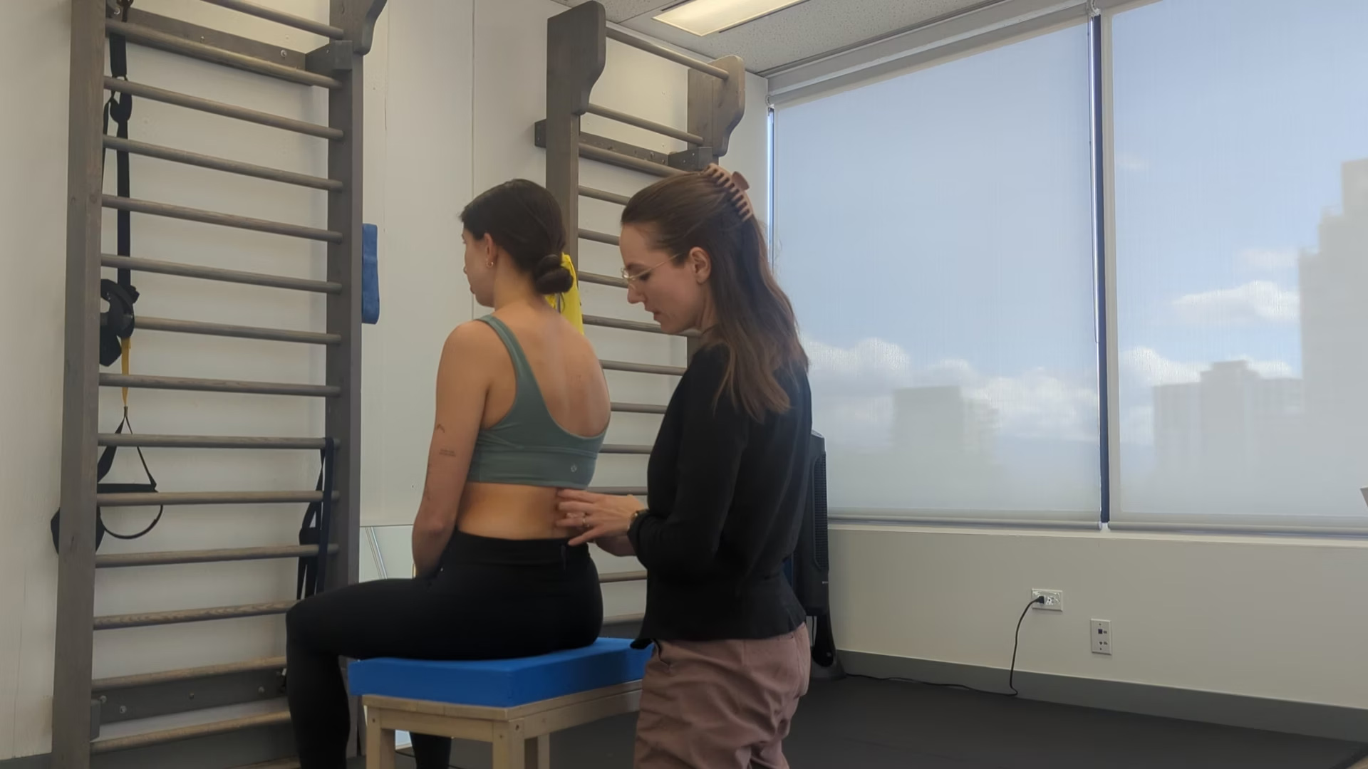
Active mobility testing of the lumbar spine:
During the right shoulder press task, L2 was unable to fully come out of left rotation. Active mobility of the L2 vertebra is assessed during active mobility of the lumbar spine:
- Flexion: decreased flexion through L2-3 compared to L1-L2 and L3-L4-5. Overall left rotation of lumbar spine with highest amount at L2 and L3
- Right side flexion: some discomfort, ~1/2 total range of motion. Decreased range of motion appears concentrated at L2 and L3, also increased left rotation at these segments is noted
- Left side flexion: full range of motion, no discomfort, de-rotation of lumbar spine
- Extension: full range of motion, equal contribution from all segments
Based on these findings, we can hypothesize that there will be a decreased superior glide at the facet joint of L2 on L3
- Diane’s question:
This supposes the restriction is articular. If so, which zygapophyseal joint do you hypothesize is restricted, left or right? How would these findings differ IF the system impairment was a myofascial restriction of say superficial fibres of multifidus or lumbar longissimus for example?
- Christiane’s response:
In the case of an articular restriction, I would hypothesize the superior glide of the left zygapophyseal joint to be restricted. An articular restriction is distinguished from a myofascial restriction by assessing for ease of correction of the joint in question. A purely myofascial restriction allows for a full correction applied with minimal force whereas a purely articular restriction cannot be corrected without targeted manual therapy to the joint. In this case, we ended up having both a myofascial and articular restriction as a partial correction of L2 and L3 was easily obtained and vector analysis indicated tension within a fibre of quadratus lumborum affecting the joint. Mild articular restriction in the superior direction in the left facet joint of L2 on L3 was also found.
- Diane’s response:
Excellent reasoning!
Passive mobility testing of the lumbar spine:
As hypothesized, some articular stiffness was noted at the left facet joint of L2 on L3 in the superior direction. Following mobilization, flexion range of motion was fully restored, however some limitation of right side flexion range of motion remained at L2-3 accompanied by left rotation of the same segment leading to further investigation of any neuromuscular, fascial, or visceral vectors which may be affecting the L2-3 segments in right side flexion. An articular impairment is no longer suspected given the restoration of flexion range of motion. A correct, release and listen technique at L2 revealed an inferior and lateral, relatively a superficial and long vector, which was likely a hypertonic fibre of the quadratus lumborum. Following soft tissue release of the hypertonic fibre, right side flexion range of motion was almost fully restored and the L2-3 segments no longer rotated in the functional task despite a short vector remaining at L2 the writer was unable to release (likely of visceral or diaphragmatic origin).
- Diane’s question:
Can you describe for non-ISM readers what you mean when you use the term ‘listen’. Where is term derived from? Take a look at the anatomy of the lumbar portion of longissimus, superficial fibres of multifidus, deep segmental fibres of psoas and tell me how you can be sure that it was quadratus lumborum you were treating? How did you release this hypertonic fibre?
- Christiane’s response:
By “listen” I mean: passively moving the segment of interest to a neutral position, and then slowly releasing the correction. While I release the pressure I was applying, I am paying attention to what may be pulling the segment of interest out of its neutral alignment. I am noticing the speed, intensity, length, and direction of the pull. This is a term borrowed from the world of osteopathy, first coined by Dr. Rollin Becker (osteopath), and further developed and applied by J.P Barral and Gail Wetzler. In this context, I was using active listening skills (listening for the movement of L2 during active spine range of motion), passive listening #1 skill (listening to the passive movement of L2) and finally a passive listening #2 skill as part of vector analysis (listening to the vectors acting on L2 when releasing my correction).
I hypothesized that I was treating quadratus lumborum (QL) based on the direction and length of the vector I listened to upon release of L2. I found the specific vector I was interested in releasing by stabilizing L2 with one hand and pulling various fibres of surrounding muscles away from L2 until I felt the strongest pull into left rotation acting on L2. The vector was going in a lateral and inferior direction towards the iliac crest and was palpated superficiality. A fibre of lumbar longissimus or a superficial fibre of multifidus would have been palpated more medially to the spine. A fibre of psoas would not have been palpable so superficially. I released the hypertonic fibre of QL by shortening the fibre of interest, waiting for a palpable decrease in tone, and stretching the same fibre.
Active control of the lumbar spine:
Active control of the lumbar spine was tested with Ms. P in sitting, arms outstretched at 90° shoulder flexion, with the writer providing resistance to isometric shoulder flexion, extension, and trunk rotation while palpating the spinous processes of L1, 2, 3, 4. Pre-release of the lumbar spine, segments L2 and L3 went into left rotation with resisted shoulder flexion and extension and resisted trunk rotation bilaterally. Post-release of the lumbar spine, Ms. P was able to control segments L2 and L3 with all resisted movements except shoulder flexion, without any cuing of lumbar multifidus. Post release of the right shoulder girdle (discussed below), Ms. P was able to control L2-3 with resisted shoulder flexion without cuing of lumbar multifidus.
- Diane’s question:
What is your hypothesis as to how the vectors impacting the right clavicle could inhibit the segmental fibres of multifidus at L2-3 and thus segmental lumbar control of L2 and L3?
- Christiane’s response:
The link between the right clavicle and L2 and L3 multifidus activation may be the phrenic nerve as phrenic nerve dysfunction has been linked to excessive subclavius activity (impacting clavicular biomechanics) and altered activity of the diaphragm (Bordoni et al. 2013). The position of L2 and L3 may be impacted directly by diaphragm activity or indirectly via the quadratus lumborum which is connected to the diaphragm by the lateral raphe. The lumbar multifidi are innervated by the posterior rami of the associated nerve roots and one can hypothesize that neural drive to the multifidi of L2 and L3 may be affected negatively by the positional finding of these vertebra. Therefore, one hypothesis is that correction of right clavicular biomechanics led to normalization of phrenic drive, positively impacting the position and motor control of L2 and L3 via improved subclavian, diaphragmatic, QL, and multifidus activity.
- Diane’s response:
Cool! I hadn’t heard of the link between the phrenic nerve and excessive subclavius activity. I will check out the reference citation below!
Passive control of the lumbar spine:
The tests for anterior and posterior stability were negative at L2 on L3.
The writer chose to perform this test due to Ms. P’s history of joint hypermobility, reports of feeling unstable in her lumbar spine with transitional movements, and significant right sided multifidus atrophy noted at L2-3. Ms. P could not recall any specific traumatic event which could have led to injury of the passive structures of the lumbar spine, but has a history of playing high level basketball as a teenager and wondered if there could have been a forgotten fall or collision.
- Diane’s question:
I’m not aware at this point if you go into this right segmental multifidus at L2-3 further below and if this question is too early, just tell me and I’ll delete it here. Given that Ms. P can’t recall a recent ‘event’ or injury to her low back it is likely a long time ago as you suggest. What does the evidence tell us about the structural changes in multifidus over time? You noted ‘significant atrophy’ on the right side of multifidus at L2-3 yet releasing the right clavicle (shoulder girdle) restored full control at L2-3. When there is significant atrophy of the deep fibres of multifidus, given what we know from the timeline of changes, how is this possible?
- Christiane’s response:
Good question! Release of a neuromuscular vector at the clavicle cannot lead to instant hypertrophy of lumbar multifidus. My guess is that release of subclavius and pec major facilitated improved neural drive to L2 and L3 multifidus which was sufficient to recover motor control of these segments under low loads. However strengthening of this muscle will likely be needed to restore and maintain lumbar motor control under higher loads.
- Diane’s response:
Absolutely agree!
Further assessment of the driver for FU#2 (secondary driver – Right Shoulder girdle (clavicle)):
Active mobility testing of the right shoulder girdle:
Prior to testing active mobility of the right shoulder girdle, the driver within the right shoulder girdle was confirmed to be the clavicle. Correcting the clavicle corrects both the scapula and the humeral head, allowing us to focus solely on the clavicle with the active and passive mobility testing.
Posterior rotation of the right clavicle was assessed with right head and neck rotation, the range of motion was decreased compared to the left clavicle during left head and neck rotation but did not translate medially. Anterior rotation of the right clavicle was also assessed during left head and neck rotation, the range of motion was also decreased compared to the left clavicle during right head and neck rotation. An inferior glide and an anterior roll were both noted at the sternoclavicular joint on the right with shoulder elevation but were decreased in amplitude compared to the left side.
- Diane’s question & comment:
During right arm elevation, the clavicle should posteriorly rotate osteokinematically and yet you are correct about the sternoclavicular joint (SCJ) arthrokinematics – the medial end of the clavicle glides inferiorly and rolls anteriorly. There are two joint compartments of the SCJ. At which joint does the clavicle glide inferiorly and at which does it roll anteriorly? What is the impact of rotation of the upper two thoracic rings (think about the manubrium) on these arthrokinematics and then hypothesize where this clavicular roll is coming from. When the clavicle is medially compressed one of these movements is restricted – which one? Since both the inferior glide and anterior roll were noted it suggests that in the position you tested this in, the clavicle was not medially compressed. Why?
- Christiane’s response:
The SCJ is a complex joint as it contains an intra-articular disc allowing the larger convex surface of the medial end of the clavicle to articulate with the smaller concave surface of the manubrium. The inferior portion of the joint also articulates with the first costal cartilage. The disc separates the joint into a medial and lateral compartment where the medial compartment is inferior to the lateral compartment. With a right arm elevation task, osteokinematically we expect right rotation of the shoulder girdle complex and of thoracic rings 1 and 2. The first 2 thoracic rings are joined anteriorly by the manubrium, meaning the manubrium also rotates right in a right arm elevation task. The more medial and inferior portion of the right sternoclavicular joint is intimately linked to the first rib via a costoclavicular and sternoclavicular ligament and is therefore the part of the joint where the anterior roll occurs. The more lateral and superior portion of the sternoclavicular joint is where the inferior glide occurs. When the clavicle is medially compressed, the roll of the clavicle is restricted. Given that I was able to palpate a roll occurring here, the clavicle could not have been stuck in medial compression at the time of assessment.
- Diane’s response:
Well reasoned!
Passive mobility of the shoulder girdle:
A correct, release and listen technique was used to identify the dominant vectors acting on the right clavicle. The right clavicle was passively distracted from the sternum. On release of this distraction, the listening indicated a relatively short medial vector which corresponded to subclavius. After release of subclavius, the medial vector listening mostly resolved but some anterior rotation of the clavicle was still felt upon release leading to release of the clavicular fibres of pectoralis major.
- Diane’s question:
How did you differentiate that the medial vector of pull was not from the clavicular fibres of pectoralis major from the start? I’m also a bit confused now because here you note that the clavicle was medially compressed and yet it defies the biomechanics of the active mobility test findings? It was medially compressed only in arm elevation and not during head and neck rotation and not in sitting but it was in sidelying?? Something seems off here?
- Christiane’s response:
Good point. I did note that the amplitude of rotation palpated on the right during active mobility testing was reduced compared to the left. Could it make sense that there was elevated tone in certain fibres of pectoralis major and/or subclavius that were causing full medial translation of the clavicle in certain ranges only, but still allowing some rotation to occur at the SCJ during active mobility testing? Given how short the medial vector felt, my guess would be that it was a subclavius vector vs. pectoralis major.
- Diane’s response:
Strategies are task and position specific and what we find in one task (head and neck rotation could be quite different than in arm elevation.
Active control of the shoulder girdle:
Ms. P held her right shoulder in close to full flexion and was instructed to hold it there while the writer progressively loaded it. As the load placed on the shoulder increased, her clavicle had increased difficulty stabilizing and started drawing medially. A cue to maintain clavicular elongation increased her tolerance to load.
- Diane’s question:
I’m confused again. In your CRF and in your Screening Task report above you report that the ‘Right clavicle draws medially’ during the task. Here you state that it ‘started drawing medially’ when the shoulder was loaded. Can you clarify this finding for me please.
- Christiane’s response:
Ah sorry, I should have specified that this active control testing was performed after release of the lumbar spine and the neuromuscular vectors holding the clavicle in medial translated. These findings indicate that addressing these two drivers increased Ms. P’s ability to flex the shoulder and withstand load, but that either another driver needs to be addressed and/or some motor control training is needed to train optimal shoulder biomechanics under load.
Passive control of the shoulder girdle:
Passive control of the right shoulder girdle was not assessed as no traumatic injury to the shoulder girdle was recounted in the patient’s story.
Further assessment of the driver for FU#3 (secondary driver – Right Foot):
Active mobility testing of the feet:
We are most interested in assessing active mobility of the right foot during a supination task as that is the motion required during a right shoulder press. This was assessed using a standing body twist to the right. The hindfoot was in a slight medial tilt during the starting position and ended in an increased medial tilt position by the end of the right standing twist task. The forefoot followed the hindfoot by tilting right as a block into more external rotation, the distal end of the first metatarsal struggling to stay on the ground.
Passive mobility testing of the feet:
- Right talocrural joint: dorsiflexion was limited to ~75%. The end feel corresponded to a soft tissue stretch indicating a neuromuscular vector. A correct, release and listen dorsiflexion technique revealed a long superior and medial vector up the lower leg. Palpation with monitoring of the positioning of the talus revealed hypertonicity in the distal tibialis posterior.
- Diane’s question:
In an ISM further assessment of a driver, active and passive mobility testing is made meaningful when confined to the requirements of the task. Does Ms. P have sufficient dorsiflexion at the right talocrural joint for the shoulder press task?
- Christiane’s response:
Great point! Ms. P had sufficient dorsiflexion for a shoulder press task. The fibres of tibialis posterior released via this passive listening technique may be relevant in the task nonetheless if those same fibres contributed to the medial tilt of the hindfoot observed during the task.
- Diane’s further question:
Ms. P stands in a medial hindfoot tilt and I’ve questioned above which joint this tilt comes from. Further assessment here of passive mobility shouldn’t be so much about the talocrural joint but which joint?
- Christiane’s response:
a medial tilt is more likely to come from the subtalar joint, therefore assessment of passive mobility should mostly be focused at this joint. Especially given a shoulder press task which requires minimal dorsiflexion.
- Diane’s response:
Actually the tilt of the hindfoot comes from the talocrural joint and here it suggests her center of mass is lateral to the base of support of this foot and that could be secondary to her structural scoliosis.
- Right subtalar joint: a correct, release and listen technique revealed a long superior and medial vector up the lower leg indicative of a neuromuscular vector. Palpation with monitoring of the positioning of the calcaneus revealed hypertonicity in tibialis posterior, about half way up the calf
- Diane’s question:
I have the same question here. Does the shoulder press task require the right foot to supinate? If not, then this restriction is not relevant. Her ST joint did supinate slightly in the screening task (slight external rotation of the talus was noted), yet her experience was better when her foot was ‘corrected’ although I’m not sure what exactly was done in this correction that improved her experience.
- Christiane’s response:
The suboptimal biomechanics noted at Ms. P’s right foot during the shoulder press task is her hindfoot going into a medial tilt. Her talus starts going into external rotation, in congruence with supination biomechanics, but her calcaneus tilts medially rather than rotating medially relative to the talus. Her experience improved with correction of the hindfoot, preventing a medial tilt (described in detail above).
- Diane’s further question:
Hindfoot tilts are most often secondary to restricted/compressed subtalar joints and the tilt actually occurs at the talocrural joint. Could you passively move the calcaneus into medial and lateral rotation relative to the talus and if not (as I suspect), which arthrokinematics glide of the anterior and/or posterior subtalar joint(s) were restricted?
- Christiane’s response:
I believe I was able to passively move the calcaneus into medial and lateral rotation relative to the talus. If the joint was restricted, I would suspect it to be restricted in lateral rotation which is associated with pronation. This type of restriction would be caused by a restriction of a lateral glide in the anterior portion of the joint and/or a medial anterior glide in the posterior aspect of the joint.
- Midfoot:
- The navicular needs to be able to internally rotate relative to the second metatarsal for optimal supination biomechanics to occur. On passive mobility testing, the navicular was able to internally rotate but was slightly limited with a tissue stretch end feel. A correct, release and listen technique at the talonavicular joint revealed a long superior vector into the lower leg. Palpation of the tibialis posterior while monitoring the navicular revealed some additional trigger points in the distal portion of the muscle which were pulling the navicular into an externally rotated position.
- The cuboid needs to be able to externally rotate for optimal supination biomechanics to occur. On assessment, the cuboid was in a dorsal position relative to the 5th metatarsal and calcaneus. On passive mobility testing the cuboid was able to externally rotate but was slightly limited with a tissue stretch end feel. A correct, release and listen technique at the calcaneocuboid joint revealed a short plantar vector pulling the cuboid towards the calcaneus indicating a neuromuscular vector within the abductor digiti minimi. An articular vector was not suspected in this case given the writer’s ability to correct the cuboid easily.
- Forefoot:
- The medial cuneiform and 1st metatarsal were assessed together. Both of these bones should be able to internally rotate for optimal supination biomechanics to occur. On passive mobility testing, both bones were able to internally rotate. A correct, release and listen technique at the 1st metatarsal and medial cuneiform joint indicated first a long vector leading to the calcaneus followed by a short internal rotation vector leading to a neuromuscular vector in the flexor and adductor hallucis brevis muscles.
- In hindsight, I should have also assessed the lateral cuneiform along with the 4th and 5th metatarsals for a fuller picture of the lateral compartment of the foot.
Active control of the feet:
Ms. P had difficulty controlling her hindfoot during a double heel raise, an increase in medial tilt was noted. After release of her foot, she was better able to control her hindfoot with cuing to lengthen through her medial longitudinal arch.
- Diane’s question:
Does she do a double heel rise in a shoulder press task? Is this meaningful to assess for this task? Consider my questions above for all this section OK?
- Christiane’s response:
Ms. P does not do a double heel rise in a shoulder press task, but given that some supination does facilitate optimal biomechanics in a shoulder press task, a heel rise task could be used to train supination in later stages of motor control training. I agree that for simply assessing active control we could have stuck to assessing whether good supination biomechanics were restored after the releases discussed above during the shoulder press task rather than adding in a new and more complex task for the right foot.
- Diane’s response:
I agree with the last sentence above. As training progresses, active control of the right foot could be assessed by adding more load to the right shoulder press task.
Passive control of the feet:
Passive control of the right foot was not assessed as no traumatic injuries to the foot or ankle were recounted in the patient’s story.
Hypothesis
Ms. P’s right sided neck tension in sitting, and with movement of the right arm, is likely stemming from a combination of neuromuscular tension and lack of motor control in the lumbar spine all the way up to the right clavicle and down to the right foot as co-correction of all three regions resulted in the best overhead shoulder press experience.
The link between the right clavicle, L2, and the right foot is not obvious at first sight, but further reading into the neural and fascial connections between the thoracic outlet, the diaphragm, and the thoracolumbar sling helps shed some light. The diaphragm and the quadratus lumborum are connected together by the lateral raphe, and play an important role in stabilizing the lumbar spine (Bordoni, Zanier, 2013).
Further the clavicle is connected to the pleura, pericardium and diaphragm via the endothoracic fascia and pretracheal fascia. The pretracheal fascia wraps around the clavicle and further connects it to the cervical spine.
Another possible link between the lumbar spine/diaphragm and the right clavicle is via the phrenic nerve as it anastomoses with the subclavian nerve, innervating subclavius. Phrenic disorders have been associated with excessive subclavius activity in the literature1. The neuromuscular link between the diaphragm and the right clavicle seems more likely in this case as no fascial vector leading to the pericardium or pleura was identified.
Foot and pelvis positioning have a documented effect on each other, mainly mediated by the shank (Khamis et al. 2015). We can hypothesize that if a lateral tilt of the hindfoot tends to be associated with anterior pelvic tilt, a medial hindfoot tilt may be associated with posterior pelvic tilt. If we consider just the right lower extremity examined in our case above, we can extrapolate that the medial hindfoot tilt noted on assessment may be contributing to posterior rotation of the right innominate and to overall left intrapelvic torsion.
- Diane’s comment and question:
A posterior pelvic tilt is a movement of the entire pelvic ring on the hips and lumbar spine. Posterior rotation of the right innominate is not a posterior pelvic tilt, it is a relative movement between the innominate and sacrum. When the right innominate rotates posterior, is this part of a left or right intrapelvic torsion? Something is off with this hypothesis and the findings as I think you will quickly see!
- Christiane’s response:
Ah yes, of course. Left intrapelvic torsion is associated with right sided ANTERIOR innominate rotation, not posterior rotation as I described above, meaning my hypothesis does not hold up. And yet I clearly observed unwinding of the pelvis with a foot correction. Another hypothesis could stipulate that the positioning of the hindfoot is in relationship to the pelvis via the right hip, which is slightly anterior to the acetabulum. A medial tilt of the hindfoot would presumably bring the tibia into external rotation, which may pull the right femur into external rotation, and thus contribute to the right femoral head being anterior relative to the acetabulum, pulling the pelvis into left transverse plane rotation. The converse could likely also be true where left TPR of the pelvis could lead to the right femoral head being in an anterior position relative to the acetabulum, the femur and the tibia in an externally rotated position, pulling the hindfoot into a medial tilt.
- Diane’s response:
These are both better hypotheses. Sometimes, the mechanisms that cause the relationship between two impairments (foot and pelvis) are difficult to discern. Yet in ISM, we don’t need to know the ‘how’, just the fact they are related allows us to treatment plan.
The pelvis and the lumbar spine are in close relationship via the thoracolumbar fascia meaning that left rotation of the lumbar spine could be contributing to left rotation of the pelvis and thus contributing to right foot positioning.
- Diane’s question:
In standing, when the pelvis is in a left TPR, what is the congruent position of the left and right feet? Is Ms P’s right foot congruent, or incongruent, with her pelvis TPR?
- Christiane’s response:
When the pelvis is in left TPR, the congruent position of the left and right feet would be mild pronation of the right foot and mild supination of the left foot. Therefore Ms. P’s right foot is incongruent to her pelvis TPR.
- Diane’s response:
Correct, so the left rotation of the pelvis is unlikely causing the right medial hindfoot tilt.
We also cannot forget the role the scoliosis is likely playing in amplifying these links. A right thoracic curve is associated with global right translation and rotation of the thorax which clinically is often correlated to left rotation of the shoulder girdle.
We could hypothesize that Ms. P’s shoulder girdle is predisposed to left rotation; and that her lumbar vertebra already being in some left rotation structurally, are predisposed to vectors acting on them amplifying the left rotation.
To summarize, we can hypothesize that the L2 co-driver is pointing to dysfunction in the diaphragm, affecting phrenic neural drive and potentially leading to overuse of subclavius and medial compression of the right clavicle. L2 may also be contributing to the medial tilt noted in Ms. Ps’ right hindfoot via its relationship to the pelvis, femur, and shank. The client’s scoliotic spine is only amplifying these findings via the stress placed on her tissues given the structural torsion through her thoracic and lumbar spines.
Ms. P will benefit from release of the vectors affecting the alignment of L2, her right clavicle, and her right foot, followed by motor control training of these areas and further strengthening integrated into an overhead shoulder press and other open kinetic chain shoulder exercises, to achieve long term relief of her right sided neck tension.
Initial Treatment Session
Release:
Lumbar spine: A left sided articular vector at L2-3 was first released with Ms. P lying in right sidelying. The superior glide of the facet joint at L2-3 was restored using about 10 reps of a grade 4 mobilization. Following the articular mobilization, a correct, release and listen technique was used (the L2 vertebra was taken out of left rotation as the writer applied gentle traction at the spinous processes of L1 and L3, and gently guided the transverse process of L2 into right rotation) to identify the remaining neuromuscular vector: superficial fibres of the quadratus lumborum. The specific fibres involved were identified by stabilizing L2 and palpating the soft tissue until a left rotation vector is felt at L2. The fibres of interest were released by shortening the fibres, waiting for tone to dissipate, and followed by gentle massage.
- Diane’s comment:
Nicely described!!
Right shoulder girdle: The right subclavius muscle was released with Ms. P in left sidelying. The writer gently made her way into the space posterior to the clavicle, progressively applying pressure to the posterior aspect of the clavicle. The subclavius release was followed by massage of the clavicular fibres of pectoralis major.
- Diane’s comment:
An interesting way to access subclavius. I usually go inferior to the clavicle especially one that is held in anterior rotation since the superficial lamina of the deep cervical fascia is very taut in this position and limits access to the posterior clavicle.
Right foot: The tibialis posterior was released with Ms. P sitting on the edge of a plinth, foot dangling off the floor. The trigger points of interest were identified with palpation of the muscle, and monitoring for movement at the talocrural joint, subtalar joint, and the talonavicular joints. The writer applied deep pressure to the calf with her fingers, shortening the vector of interest first for tone to subside before following up with gentle massage and cues to think of the medial longitudinal arch being able to lengthen. Further release was provided along the sole of the foot, particularly on the lateral aspect of the foot to release abductor digiti minimi and in the plantar aspect of the first metatarsal/interosseous space to release flexor hallucis brevis and adductor hallucis brevis.
Align:
Lumbar spine: The left rotation through her L2 vertebrae was significantly improved in standing (a full return to neutral is not expected given Ms. Ps’ left lumbar structural curve). No further left rotation was noted during her meaningful task indicating that a connect cue is not necessary to control the lumbar spine.
- Diane’s question:
Do you think the ‘significant atrophy of right multifidus noted at L2-3’ is in relationship to her lumbar scoliosis and persistent left rotation? When would you consider addressing this segmental deficit with motor control training?
- Christiane’s response:
Yes, the multifidi on the concavity of scoliotic curves have been documented to consistently be weaker and have higher fat infiltration compared to the convex side, especially near the apex of the curve (Wang et al. 2022). I would consider training multifidus segmentally as Ms. P introduces higher loads to her motor control retraining, especially if I was observing loss of control of her lumbar spine at these higher loads or if Ms. P was noticing recurrence of symptoms.
Right shoulder girdle: Ms. P’s residual right thoracic curve predisposes her to an anteriorly tipped and protracted right shoulder girdle. A cue to expand through her right upper and anterior thorax using her breath to allow the clavicle to lengthen away from the sternum and the scapula to slide posteriorly was given; however, Ms. P’s ability to expand through her thorax was limited, due to a number of rings remaining held in rotation. There are likely one or more vectors that remain to be released around the thorax impacting Ms. P’s breathing mechanics.”
Further assessment and release of the thorax would likely be warranted in a subsequent session. Ms. P. was nonetheless able to control her right clavicle and shoulder girdle effectively during her meaningful task.
- Diane’s question:
When she was able to do this, did she note any change in her neck tension during the right shoulder press?
- Christiane’s response:
She did! The neck tension was no longer present during the task.
Right foot: Post release, Ms. P’s standing alignment revealed a habitual tendency to sway her pelvis forward and have her weight go through her heels. A cue was given to shift her pelvis posteriorly bringing her centre of mass over her midfoot, allowing her forefoot to splay and lengthen through her medial longitudinal arch. Some attention was required to maintain this alignment during her meaningful task.
Connect:
No segmental multifidus cuing was provided given Ms. P’s ability to control her lumbar spine during the meaningful task. She was also able to control her right shoulder girdle without further cuing of the inner core unit. Strengthening of the right foot intrinsics should be considered in subsequent treatment sessions once Ms. P has integrated how to let go of tibialis posterior and lengthen through her medial longitudinal arch in standing.
Move:
An unweighted right sided shoulder press with particular attention placed on right shoulder girdle and right foot positioning was used as a home exercise program in the early stages of retraining. Later stages of retraining will involve a bilateral shoulder press, with added weight.
Follow up treatment
Ms. P reported a significant improvement in her right sided neck tension as well as her lower back pain post treatment. Her right foot needed some further release and motor control training, but the lumbar spine and right shoulder girdle releases were relatively well maintained. Some minor right sided neck tension was still identified by the client with a right sided shoulder press.
Next steps would include:
- Further assessment and treatment of the thorax (secondary driver)
- Further motor control training with incremental increase in load of the right shoulder girdle and foot and monitoring of L2
- Potential referral to a therapist trained in visceral assessment and manipulation to treat the remaining vector(s) at L2
References
Beyer et al. In vivo thorax 3D modelling from costovertebral joint complex kinematics. Clinical Biomechanics, 2014
Bordoni B, Zanier E. Anatomic connections of the diaphragm: influence of respiration on the body system. Journal of Multidisciplinary Healthcare. 2013
Khamis S, Dar G, Peretz C, Yizhar Z. The relationship between foot and pelvic alignment while standing. Journal of Human Kinetics. 2015
Wang et al. BMC Musculoskeletal Disorders (2022) 23:891 https://doi.org/10.1186/s12891-022-05841-5
Wang et al. Characteristics of the pelvic axial rotation in adolescent idiopathic scoliosis: a comparison between major thoracic curve and major thoracolumbar/lumbar curve. The Spine Journal, 2014
Case Study Author

Christiane Junod
The ScoliClinic
ISM Certified Practitioner
1665 West Broadway #670, Vancouver, BC V6J 1X1, Canada
Clinical Mentorship in the Integrated Systems Model
Join Diane, and her team of highly skilled assistants, on this mentorship journey and immerse yourself in a series of education opportunities that will improve your clinical efficacy for treating the whole person using the updated Integrated Systems Model.
{"first_class":"1","title":"Heading","show_title":"0","post_type":"course","taxonomy":"","term":"","post_ids":"3346","course_style":"recent","featured_style":"custom_block_1","masonry":"0","grid_columns":"clear1 col-md-12","column_width":"200","gutter":"30","grid_number":"1","infinite":"0","pagination":"0","grid_excerpt_length":"100","grid_lightbox":"0","grid_link":"0","css_class":"block-small-thumb","container_css":"","custom_css":""}
We will come together for 3 sessions of 4 (4.5) days over a period of 6-8 months with lots of practical/clinical time to focus on acquiring the skills and clinical reasoning to put the ISM model into practice. Hours of online lecture and reading material and 12 hours of in-person lecture are...
More Info