Story
Ms. O was assessed January 28th 2023 at Habitat Health Clinic. Ms. O is a 31 year old female who is moderately active, eats well and sleeps well. She has moderate-severe stress levels at work and in personal life. Subjectively Ms. O reports headaches and neck tension which is worsened by working with arms outstretched. She also has a burning sensation in the right scapular region which is more frequent than the headaches. Often the symptoms occur at the same time. She experiences pain after she has worked more than 4 hours or if she completes weighted upper body activities after not doing them for a while. Running also aggravates her symptoms.
Her occupation is physical therapist which requires arms to be outstretched for 70% of her work day. She also has a long commute 1.5 hours per day where her arms are outstretched and this aggravates her symptoms.
When her headache begins it starts in the right suboccipital region and makes her neck feel stiff to rotate right. She feels her headaches are linked to her neck tension and possibly the thorax pain. Her headaches developed 8 years ago when in school and the neck and thorax pain started 5 years ago. Initially she thought her scapular pain was a shoulder injury from yoga. The quality of headache, neck and thorax pain is dull and throbbing. Her headaches make it difficult for her to concentrate when at work and make it hard for her to take part in high intensity activities like running which she loves.
Past treatment has included massage which can worsen symptoms, acupuncture improves symptoms, chiropractic improves symptoms, and strengthening programs for the upper body which can aggravate symptoms. Other practitioners have told Ms. O that her pain is due to weakness but she doesn’t fully buy in because the strength exercises make things worse.
Her pain would be classified as Nociplastic as it originally started over time with no threat or damage to tissue and has gone on for over 8 years. It is in a local area in her head and neck but cannot be pinpointed to a certain event which would have caused damage to the area.
- Diane’s comment:
I’m not sure I totally agree with this phenotyping of her pain. Aside from the timeline, what are the key characteristics of the nociceptive, neuropathic and nociplastic phenotypes? Then consider words used in your story above such as burning or tension/stiffness and the fact that she can identify mechanical factors that aggravate and therapies that can relieve her pain and see if you still think this is only nociplastic situation.
- Allison’s response:
Nociceptive pain is pain that has an immediate threat to tissue. It creates inflammatory markers and causes the nociceptors to transmit pain signals to the brain. This type of pain comes with signs of inflammation which takes 6 weeks to heal and can be relieved with anti-inflammatories.
Neuropathic pain is associated with damage to the nerve tissue and is often accompanied with burning, prickling, radiating, and sensitivity to pressure or touch.
Nociplastic pain is associated with changes to the immuno-endocrine system. It affects widespread areas of the body and is often not treatable with anti-inflammatory medications. This type of pain is associated with conditions such as fibromyalgia and can affect the body systemically.
On reflection, I now think her pain would be classified as Nociceptive pain. This is based on the definition of local nociceptors being affected due to an immediate tissue threat or tissue damage. Ms. O reports that the mechanical load of her arms as well as certain neck and head movements affected the local areas of pain she describes. Often her pain is relieved with anti-inflammatories or holding her neck, which indicates local tissue inflammation. It is hypothesized that poor mechanics are the cause of tissue re-injury when she sustains load on the area. It takes a few days for the pain to subside and then re-injury occurs because her body position and biomechanics have not changed.
Her goals are to get back to working without having headaches and neck tension. She would also like to get to the root of the scapular problem so that she can prevent the ongoing pain. Ms. O also enjoys long distance running, working out with weights and getting back to these activities are a priority.
Past Medical History
Ms. O sustained right elbow fractures and dislocation 2005. She had pin in and out surgeries six months apart. She has had chronic headaches for many years which were vision related and corrected after she was prescribed glasses. The current headaches began 8 years ago and feel more like tension headaches. Ms. O sustained two whiplash injuries, one in 2007, she was tubing off the back of a boat and was thrown. After this she had trouble lifting her head off the pillow. She did not have treatment after this. In 2019, Ms. O slipped down stairs and hit her tailbone. She reported that she felt the impact go right to her head. After that she had concussion symptoms and neck pain.
- Diane’s question:
There are many traumas in Ms. O’s past here to consider when planning your assessment. Which two functional units require investigation based on this story so far?
- Allison’s response:
Given the whiplash and her inability to lift her neck off the pillow I would want to look into functional unit # 2. I would also want to have a look at the pelvis in functional unit # 1 given her fall and the sensation of the impact that traveled from her tailbone to her head.
- Diane’s comment:
Agreed, and you would likely consider the entire FU#1 and not just the pelvis since the pelvis could be a victim of another impairment within the unit.
Meaningful Complaint
Ms. O has pain in the head, neck and right scapular region after working with her arms outstretched in a seated position, running, and/or doing upper body strengthening exercises. Her head and neck pain get worse as the day goes on and is relieved by Tylenol, massaging the back of her head, squeezing her neck as well as deep breathing and relaxation techniques. Her symptoms affect her ability to think clearly when at work and do the physical activities that she loves. When Ms. O performs this task she feels as though her arms are heavy and uneven, oftentimes this feeling is accompanied by right thorax pain. She also feels as though her right arm is longer than her left.
- Diane’s question:
There are some biomechanical findings in her meaningful complaint report, but also some suggestion that her sympathetic nervous system (SNS) may be upregulated. What leads me to say this? If her primary driver is the SNS, how will this impact your biomechanical ISM assessment?
- Allison’s response:
Ms. O reports that massaging her neck and doing deep breathing helps to relieve pain. She also reported a moderate amount of stress in her life. This may indicate that her sympathetic nervous system is working overtime which could cause heightened pain signaling. If her primary driver is the SNS then it would be necessary to help down regulate this in order to have an accurate biomechanical assessment. If her SNS is heightened we will not be able to make body corrections without a sympathetic response.
- Diane’s comment:
If the dural/neural tension caused by an upregulated SNS is causing the musculoskeletal system to adapt, the fact that correction of any body region makes something else worse reinforces the fact that the SNS is the driver of the MSK.
- Allison’s reply:
I hadn’t thought of it this way. I usually just do the dural/neural tests and if they are negative I assume it’s not a dural/neural driver. Certainly I see this pattern often and I do tend to still give flossing or tensioners while they correct the segments. But what would I use as my objective measure? Also would that mean there are no MSK co drivers?
- Diane’s reply:
Listen to this video in the ISM flow under Release Techniques for the Cranium https://learnwithdianelee.com/course/ism-flow/release-techniques-for-the-cranium/
The dural/neural system is NEVER a driver since it isn’t a skeletal body region. You can have intra-cranial or intra-spinal dural vectors that are impairing mobility of the cranium and/or spine; however, in this instance correcting the cranium/spinal segment/thoracic ring improves the experience of the neurodynamic task, improves other sites of impairment and Passive Listening #2 on release of the correction produces a pull into the impaired dura. This is totally different from a dural/system impairment that is underpinned by trauma and an upregulated SNS. This condition causes congestion in the venous sinuses around/between the dural layers (cranium and/or spinal column) and it is this congestion that takes out the ‘crimp’ of the dura and reduces its mobility. The MSK system then adapts by shortening to accommodate this tension. Any attempt to correct the alignment of the MSK results in further impairment elsewhere and worsening of the neurodynamics of the neural/dural system. This was done on day 3 of part 3 and you’ll find more information in the thorax book as well for a reminder.
Your objective measure is the impact of any MSK correction on the positive neurodynamic test. If correcting the skeleton improves ND mobility, then you find the vector that is impeding the ABC’s of that region. If correcting the skeleton worsens ND mobility, then you have a lack of mobility in the nerve/dura itself that is usually part of a wider body problem and underpinned by an upregulated SNS. Unless the trauma causing this upregulation is addressed, the dural/neural relief from your techniques will only be temporary. This has nothing to do with MSK co-drivers.
Cognitive Beliefs
Ms. O feels that her elbow injury may have caused some stickiness in the dural/neural system, that along with tension in the neck she feels activates her headaches. She also feels like the aching in the shoulder girdle seems to contribute to headaches. Other clinicians in the past have given her arm and shoulder strengthening exercises; however, these exercises seem to aggravate her symptoms so she stops her rehabilitation. This may be limiting because it could be that these strengthening exercises are just not quite the right exercises for correcting her particular muscle imbalances.
- Diane’s question:
Your patient is a physical therapist so she may have some understanding that dural/neural tension can occur. However, what did she mean by the term ‘stickiness in the dural/neural system’? Were these her words or your interpretation? How does she hypothesize the relationship between aching in the shoulder girdle (which you previously said she described as burning) and her headaches? Did you clarify whether she meant ‘aching’ vs ‘burning’ in the region of the scapula? Why would this be an important question to ask?
- Allison’s response:
I believe she feels that there is scar tissue tacking down the neural sleeve in her elbow which is pulling on the nerve root in her neck. She said she thinks the nerve is pulling from her elbow to her neck so I interpreted the dural/neural portion.
She didn’t elaborate on what she thinks the relationship is between shoulder and headaches; she just knows they happen together. She also described her neck tension happening at the same time and she thinks the tension in her neck and thorax is creating headache pain.
She used the term burning and aching interchangeably during the interview. It would be important as a burning could indicate an overactive muscle or neuropathic sensation whereas an ache could indicate injury to the joint or local inflammation to tissue
- Diane’s comment:
Burning is also a neuropathic sensation.
- Diane’s further question:
What do we know about dural system impairments and another hypothesis for why upper extremity exercises may exacerbate her symptoms other than ‘incorrect exercises for muscle imbalances’?
- Allison’s response:
The dural system may be in a state of fluid congestion due to cranial dysfunction or a twist in FU# 2 which would interfere with drainage of the head. Thus putting more load on the upper body would exacerbate poor cranial alignment and create more pressure in the dural system and the head. exacerbating her symptoms.
- Diane’s comment:
You mentioned above that Ms. O has moderate stress in her life, and we discussed the possibility that her SNS may be upregulated. Another hypothesis could be that the SNS is compromising arterial blood flow to the upper extremities creating an O2 deprivation when exercising her upper limbs.
- Diane’s question:
There are several ‘yellow flags’ in this story that suggest a biomechanical ISM assessment may not go well here and that a dural mobility assessment may be better to proceed with. I know from your prior clinical reflection form of this individual that you were able to find unit drivers relative to her meaningful task; however, I’m curious if you thought of doing a dural mobility assessment prior to a possible provocative ISM biomechanical assessment? What was your clinical reasoning at this point for choosing which approach?
- Allison’s response:
I did think about the dural system as being impaired so I assessed her spinal dural mobility using the slump test as well as upper limb neural mobility tests 1, 2, and 3. The dural/neural mobility tests were all negative. I believe there may be a dural/neural component but I don’t believe the dura system is the only cause of current complaints. As far as the unregulated SNS I made sure to ask if she had ever experienced any light headedness, dizzy spells or over sensitivity during her previous physical assessments. She did not report any of those symptoms so I proceeded with a biomechanical assessment with caution making sure to ask if any uncomfortable sensations came up locally or elsewhere in the body.
Meaningful Task
Based on the information gathered in the patient’s history the meaningful task chosen was:
- Working, or driving, with arms outstretched in sitting
Screening Tasks
The screening task chosen was a double arm lift in the seated position, with the start screen being sitting.
Functional Unit #1 (FU#1)
Start Screen: Sitting
The sitting start screen was first evaluated as the patient lowered into a sitting position in order to determine if she was sitting on a locked pelvis. If she was not sitting on a locked pelvis we would have to correct the pelvis to make sure the findings in FU#2 were not being influenced by drivers in FU#1. That is the reason why I chose to start at FU#1 even though the symptoms were in FU#2. The sitting start screen allows us to determine what position the patient starts in. This picture is unique to each client based on their postural habits, previous injuries and even their cognitive state. Noting where the body position starts allows the practitioner to see the changes in alignment and biomechanics during the task. This gives us more insight into the areas of interest.
Findings
Pelvis: Rotated to the right in the transverse plane (TPR) with a congruent right intrapelvic torsion (IPT).
Hips: Left hip anterior to right hip and congruent with the right rotation of the pelvis
Thorax:
TR3 was translated left/rotated right which is incongruent to the rest of thorax
TR4-TR10 translated right/rotated left
TR3 is rotated right and TR4-TR10 are rotated left, thus it will be important to investigate TR3 and TR4 as the position of rotation is incongruent. Incongruence can lead to poor mechanics, positioning and control of the local area and surrounding body segments. In order to determine whether this area is one that needs to be corrected, we will first observe how the two segments behave in the task. Do the biomechanics improve in the task or get worse? If the mechanics get worse, or stay the same, it will be important to correct the alignment of both and see what happens to the task mechanics and patient experience.
- Diane’s question:
In ISM, we use the word ‘congruent’ or ‘incongruent’ in two ways. The first is how you have used the word congruent above (the pelvis TPR and IPT, the left hip position congruent with the TPR of the pelvis, and TR3 being incongruent to the rest of the thoracic rings); when a body region or segment/ring is rotated in the opposite direction to another region/segment/ring. What is the second way we use this term?
- Allison’s response:
The first way would be positionally as listed above. The second way would be referring to the biomechanics of a movement and whether or not the body segment moves in a way that is congruent for that. For example when turning to the right all of the rings of the thorax should translate left and rotate right in order for the movement to have even load transfer, but if one ring is translating right and rotating left it would be incongruent for the movement of right transverse plane rotation for the thorax.
Lumbar spine: lumbar spine follows the sacrum into right rotation in the transverse plane and is congruent with the sacrum
- Diane’s question:
How do we know which way the sacrum is rotating from any information you have provided so far?
- Allison’s response:
When the pelvis was assessed in standing, the position of the innominate bones and sacrum were noted. The left innominate was anteriorly rotated and the right was relatively posteriorly rotated. The sacrum was in a right rotated position and unlocking of either SIJ was not detected in standing or in the sitting assessment. This indicates that the sacrum was rotated right in the transverse plane.
Screening Task: Seated Double Arm lift
As noted, the seated double arm lift task is the position where Ms. O feels most of her discomfort. This task will be used to help determine where she is using suboptimal biomechanics, alignment or control. After noting areas of incongruency we can then determine how those segments behave while in the seated double arm lift task. This allows us to group segments of interest so that we can eliminate non drivers. We can then go forth and correct these segments to determine the primary driver for each functional unit.
Findings
Pelvis: rotates further into right TPR/IPT
Hips: Both move congruently with the pelvis. Left hip moves more forward compared to the right
Thorax: TR3 translates left rotates right more, TR4 translates right rotates left more
Lumbar spine: Follows pelvis into further right rotation
Driver for FU#1
Thorax: priority rings are TR3 and TR4 (glued).
This is supported by the following findings:
TR4-10 did not change in the task, whereas the pelvis, hips and TR3 and TR4 got worse. Correcting TR3 and TR4, as well as, correcting the positioning of the pelvis and left hip also made the arm lift task feel lighter and arms more even, but did not fully correct the patient’s sensation. The arms were still slightly heavy and felt uneven. There was also a visible lag in the time it took to lift the right arm. The right scapula did not upwardly rotate as quickly as the left, which indicated that the biomechanics did not correct thus more information from FU#2 needs to be gathered.
I believe there may be another driver but most likely not in FU#1 as body segments that got worse in the task were accounted for in the corrections.
Functional Unit #2 (FU#2)
Start Screen : Sitting
Findings
Cranial region: Temporal bones, occipital and sphenoid are rotating right, thus the intercranial torsion is net right and congruent between the cranial bones.
- Diane’s question:
The temporal bones don’t rotate to the right – what do they do when the occiput rotates to the right? What are the bones and the osteokinematics of a right ICT?
- Allison’s response:
The left temporal bone is rotated anteriorly and the right temporal bone is rotating posteriorly. The occiput is rotated right. The sphenoid is rotated right and congruent with the ICT.
Cervical region: C2 is translated right and rotated left which is incongruent to the ICT and to C3 and C4
C3 and C4 are translated left and rotated right
C5 was translated right rotated left which was incongruent with C3, C4, and C7
C6 neutral
C7 translated left rotated right incongruent with C5 and C2
Thorax: TR1 rotated right in the transverse plane, TR2 translated left and rotated right
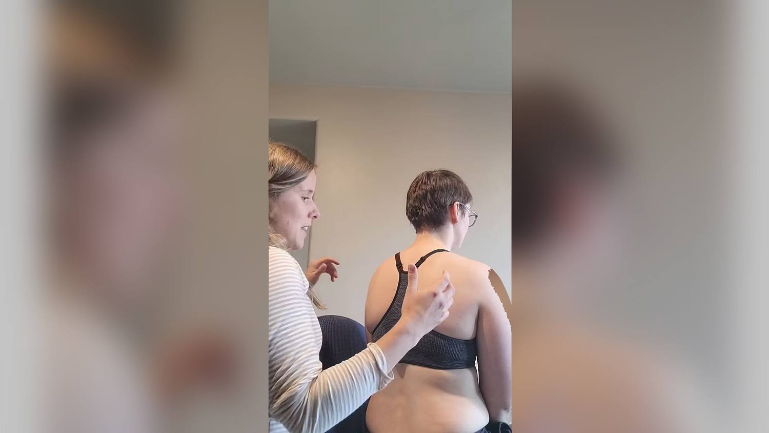
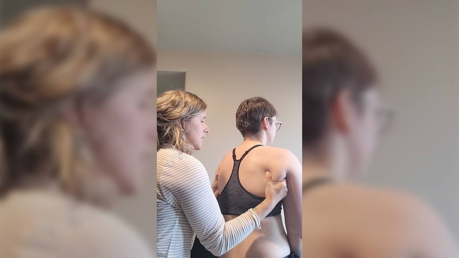
Shoulder Girdle: The TPR of the shoulder girdle is net right. Left scapula elevated, Right scapula depressed (both shoulders scapula and glenohumeral joints are congruent to clavicles).
- Diane’s question:
This sounds like a very compressed cervical region with multiple alternately rotated segments. The left scapula is elevated and the right one depressed, and you state that the clavicles are congruent, thus I assume you mean the left clavicle is more elevated than the right. What is the congruent rotation of each clavicle (anterior/posterior) for this to be true?
- Allison’s response:
The left clavicle is anteriorly rotated and the right is posteriorly rotated
Seated Double Arm Lift
Findings
Cranial region: Right ICT/ right rotated sphenoid remain
Bervical Spine: C2 translates right rotates left further
C3-C4 remain the same
C5 corrects in task
C7 corrects in task
Shoulder girdle
- Right TPR corrects
- Right scapula protracts more than it should for the task (arm lift to 90 degrees). The right scapula did not upwardly rotate as quickly as the left.
- Left scapula has appropriate biomechanics
Thorax: TR1 corrects in task, TR2 corrects in task
Driver for FU#2
The cervical region is the driver for FU#2 with C2 as priority for the following reasons:
C2 goes further into right translation and left rotation in the task and the timing of this is immediate. A correction of C2 corrects C3 and C4, improves right scapular mechanics, T1 and T2 as well as the cranium. With C2 corrected, the patient’s experience improved with the sensation of lighter arms and more even length; however, this correction created a twinge in the right shoulder blade/thorax area. This suggests that there is another driver or a co-driver.
- Diane’s comment:
TR1 and TR2 corrected in the task so I’m not sure what you mean when you say that correcting C2 corrects T1 and T2 (which should be written as TR1 and TR2). These corrections should consider only sites of impairment in the task itself. TR1 and TR2 were noted above to have corrected in the task – so I’m a bit confused here. Also, there is no mention as to what the correction of C2 did to the right scapula which did not upwardly rotate quickly enough, by your judgment. Did the right scapular biomechanics change with the correction of C2? Your missing driver here could be the right shoulder girdle.
- Allison's response:
C2 did correct the right scapular mechanics and correcting the right shoulder girdle did not improve the mechanics of C2.
Relationship of Drivers from FU#1 and FU#2
Correcting the thorax (TR3 & 4) in FU#1 partially corrects the task in that the experience for the patient was the best out of all the corrections in FU#1; however, the right scapular mechanics are not improved and the driver for FU#2 (C2) goes further into right translation and left rotation. When correcting the driver for FU#2 (C2) TR3 goes further into left translation, right rotation and T4 goes further into right translation and right rotation. When both unit drivers are corrected simultaneously the task and experience are fully corrected. Thus the neck and thorax are co-drivers with priority segments being C2, TR3 and TR4 (which are glued).
- Diane’s comment:
My remaining comment would be about the impact of this co-correction on the right scapula – did it improve? Yes it did.
FU#3 Lower was not assessed as the body segments were observed to not change during the task.
FU#3 Upper was only minimally investigated in order to help refute the patient’s belief of her elbow being involved in her symptoms.
Arm and hand
- Right elbow appeared slightly hyperextended
Right arm anatomically shorter no change in task when elbow is corrected
No driver was found for functional unit #3
Summary of Functional Unit Drivers:
FU#1: Thorax: priority segments TR3 and TR4
FU#2: Cervical: priority C2
FU#3: no drivers found
Summary: Neck and Thorax are co-drivers with priority segments being C2, TR 3 & 4 (which are glued).
Further Analysis of the Drivers
Further analysis of the driver(s) involves analyzing the following to determine the underlying system impairments:
- Active mobility
- Passive mobility
- Active control
- Passive control
Further Assessment of the Driver for FU#1 (Thorax: TR3 & 4)
Active Mobility Testing for TR3
During an arm lift task the thoracic ring should maintain neutral alignment without rotation, translation or compression. Active mobility of TR3 was monitored during thoracic rotation. As the TR3 was found to be in left translation and right rotation, it is necessary to determine whether this ring can return to neutral from that position. Active listening revealed that during right rotation the ring was able to move with appropriate biomechanics in a smaller range. During left rotation the ring did not rotate but translated slightly more to the left. Flexion and extension were also tested and the ring could extend bilaterally but flexion on both sides was slightly limited. Because the TR3 was able to move in other planes of motion it is hypothesized that the impairment lies in either the neural, myofascial or visceral vector. To assess further we will correct the ring, release and listen to determine where the vectors arise from.
Active Mobility Testing for TR4
Active mobility was monitored for TR4 during thoracic rotation to determine if the ring could move back to neutral. Active listening revealed that during rotation to the left the ring translated further right and rotated left. During right rotation the ring did not move thus flexion and extension were tested. The ring was limited in flexion and extension on the right side, thus more information from the correct release and listen needs to be gathered in order to determine which system holds the impairment.
Passive Mobility Correct Release and Listen of TR3 and TR4
Thoracic rings 3 and 4 were assessed together as it was determined that they were glued, which means there is a vector between the two restricting independent mobility. When assessing thoracic ring 3 and 4 a gentle contact on the bottom part of the ring was made and held. During passive listening number one there was a vector in between TR3 and TR4 that was compressing them together. The vector was short and felt like intercostal muscles.
A full correction was achieved of TR3 and TR4 with moderate difficulty using more posterior rotation of the rib and medial translation of the rings.
- Diane’s clarification:
For clarity, the ring isn’t posteriorly rotated during a correction, the rib of the ring is posteriorly rotated.
The end feel did not register as a stiff capsule, but it required more effort than a gentle hold. If a small amount of pressure had corrected the positioning it would have been a visceral or neurological vector. If it was too stiff to fully correct, then the articular system would have been the culprit. When the correction was slowly released, passive listening #2 revealed first the compression of the ribs together (intercostals), then TR3 was pulled to the left, anterior and superior, which I determined was the serratus anterior. The first vector of pull on TR4 was a long, inferior pull from the back which felt like iliocostalis. The second vector on TR4 felt like a short, medial and inferior pull from the front TR4 which I determined was the pectoralis minor.
Active Control of TR3 and TR4
In order to determine whether the patient could maintain appropriate biomechanics during the task, the vectors of pull must first be released. The vectors of the intercostals, left serratus, and pectoralis minor were released with a release with awareness technique. A muscle manipulation technique was also applied to the iliocostalis on the right. Dry needling was used to release the superior, medial, and inferior border of the scapula thus affecting the 9 fascicles of serratus anterior.
After this, I assessed the position of the TR3 and TR4 as the client lifted the arms. This increased the load in order to determine how well the client could control the position of these rings. This is the basis of active control. After vectors were released her Thoracic rings 3 and 4 tended to translate/rotate opposite to each other and become compressed.
- Diane’s comment:
Thoracic rings 3 & 4 were rotating opposite to each other before the vectors were released so this isn’t a different or new finding. What is new is found in your comment ‘and become compressed’. This statement implies that the vectors preventing independent movement of these rings were released; however, those that compress the two rings together were still active, or that there was insufficient motor control of the deep system of these rings to prevent compression via the superficial system.
In order to activate the segmental muscles which control alignment of two thoracic rings, a cue was given “create space in the thorax using the back pressure from the lungs” to help Ms. O regain alignment and control of her thoracic rings. When applying additional load to the thorax she was unable to maintain optimal alignment of TR3 & 4 so the load was reduced by using only the arm lift with a cue to retain alignment. This will be her homework.
Passive Control of TR3 and TR4
Passive control was completed in order to determine the integrity of the ligamentous structures. There were no passive control issues evident.
- Diane’s question:
What findings were in either her story, or objective examination that suggested this was indicated at this time?
- Allison’s response:
Ms O had reported that she fell in her history. If there is trauma to an area it is important to test the passive system.
- Diane’s comment:
This was not reported in the story of this report.
- Allison’s response:
Ms. O had reported a slip on the stairs where she hit her tailbone. Because it was on stairs at an angle, I wondered if she also hit her thorax on a step.
Further Assessment of the Driver for FU#2 (Cervical: C2)
Active Mobility Testing of C2
During an arm lift task, the cervical spine should maintain a neutral stable position without compression, rotation or translation. The position of C2 was assessed during active cervical rotation in order to determine whether or not the C2 could rotate back to neutral. The start position of the C2 was right translated and left rotated. During right rotation of the head and neck, active listening revealed that C2 failed to right rotate even to the neutral position. C2 was able to rotate left with appropriate biomechanics.
Passive Mobility & Listening – C2
Passive mobility of C2-3 was performed in supine, by placing hands on the anterior portion of the tubercle of the transverse process of C2. It was then corrected using a small amount of traction and then right rotating the vertebra back to a neutral position. The vertebra corrected quite easily and the passive listening 1 revealed a vector on a superior angle on the right side, which was determined to be the rectus capitis posterior major. The fact that it was easy to correct suggests the impairment lay in the neuro-muscular system. When C2 was let go the right side moved first translating right with a short strong vector followed by the left which felt like a longer pull coming from the side and down into the thorax which was determined to be a middle scalene and the investing fascia.
- Diane’s question:
So there appears to be two vectors attaching to C2 resulting in right translation/left rotation. Which vector is dominant and why?
- Allison’s response:
The rectus capitis posterior major vector pulling the C2 into right translation/left rotation is more dominant because it was the first vector detected during passive listening. It was also strong and caused a greater amplitude of movement.
Active Control of C2
Prior to testing active control of C2, the vectors of the right rectus capitis posterior major and the left middle scalene were released using the release with awareness technique. After those vectors were released there was still a vector compressing the C2 which seemed to connect the C2 into the thorax, this was determined to be the semispinalis cervicis . The vector of semispinalis cervicis was released by correcting C2, TR3 and TR4 then applying the stack and breathe technique in order to release the vector between the two segments. Once the vectors were released, the C2 was monitored during a double arm lift task to determine if segmental motor control was restored subsequent to this release. The C2 did translate to the right slightly so to prevent this a cue of ‘imagine a guy wire attaching the sternum to the back of the head, pulling tight and lifting up toward the ceiling’ was used which helped to restore control of C2 during the double arm lift task.
Passive control of C2
Passive control tests were completed in order to determine the integrity of the ligamentous structures. The test was completed with the patient in supine and the therapist stabilizing the inferior vertebrae of the neck and performing anterior and posterior shear as well as lateral shear tests.
- Diane’s question:
What from the story, or your objective findings, suggested that these tests were indicated?
- Allison’s response:
Ms O had reported that she fell in her history. If there is trauma to the body it is important to test the passive structures.
- Diane’s comment:
There is no report whether these tests were positive or negative, I can only assume they were negative.
- Allison’s response:
Correct. All passive control tests were negative.
Neural mobility tests were performed in order to help refute the patient’s cognitive belief of neural vectors in her elbow causing headaches and neck tension. Correcting both of the functional unit codrivers during the task helped to create the ‘wow’ moment, thus the patient was able to accept the co-driver hypothesis.
- Diane’s question:
What was the ‘wow’ moment? What changed in the elbow, or her experience that gave your patient buy-in for your hypothesis?
- Allison’s response:
The ‘wow’ moment was created when both of the drivers for functional unit #1 and FU#2 were corrected. Her arms felt lighter, more even and she did not have any twinges in the right thoracic region. When we completed the neural mobility testing it was negative which indicated that it was not actually her neural system causing the headaches. We also corrected the elbow and had her complete the meaningful task and it did not make a noticeable difference.
Hypothesis
It is hypothesized that Ms. O’s symptoms when lifting the arms are the result of poor biomechanics in the cervical and thoracic spine as well as impaired control of the thoracic rings 3 & 4 when completing the double arm lift task. There was a common vector between both the cervical spine and the thoracic rings 3 & 4 that was causing the thorax and c-spine to be co-drivers. Correcting both the cervical vertebra and the TR3 and TR4 was necessary to improve the feeling of the task and restore the right scapular mechanics.
Initial Treatment Session
Release
TR3 and TR4 were both co-corrected, then the client was told to direct their breath into my hands. This technique is also known as the ‘ring stack and breathe’ technique. Once a softening was felt, the rings were gently compressed and then let go as the client took in a deep breath, in a proprioceptive neuromuscular facilitation technique. To create length in the intercostals, the client was guided into side flexion to the right and left as they directed their breath to either side with the intention of creating space in between the rings.
Dry needling was used on the left serratus vector. Needles were used at the superior, medial, and inferior border of the left scapula thus affecting the 9 fascicles of serratus anterior.
The right iliocostalis was released in prone using a muscle manipulation technique. This was released in prone.
The release with awareness technique was also used to release the left middle scalene, right pec minor attaching to ring 4 on the right, and right rectus capitis posterior major. The vector of right semispinalis cervicis was released by correcting both C2, TR3 and TR4 then applying the stack and breathe method in order to release the vector between the two segments.
The patient was also encouraged to roll the back of the skull with a ball to release the suboccipitals and the right iliocostalis as part of her home program.
Align
In order to bring awareness to TR3 and TR4 they were corrected and an alignment cue of “creating space in the thorax using the back pressure from the lungs into this area” was given for the thorax in order to create space between the rings 3 and 4. The correction by the therapist was then released and the patient was told to try and maintain that same correction feeling with the cue. This was to be completed once per hour daily for the duration of 5 deep breaths and when doing the arm lift task. The alignment cue of “widening the back of the head” was prescribed to keep the C2 in alignment. This was to be completed once per hour, daily, for 5 deep breaths and when doing the arm lift task.
Connect
A connect cue – ‘imagine a guy wire attaching the sternum to the back of the head, pulling tight and lifting up toward the ceiling’ – was used to connect into the deep neck flexors when doing any tasks that require the arms to be outstretched. A connect cue – ‘a fish hook lifting and lengthening the space between the ribs at the back’ – was used to activate the segmental multifidus.
Move
The double arm lift was used as the movement task doing 3 sets of 10 repetitions daily (slowly) and using the align and connect cues. The patient will use feedback from the sensation of the task. If it is feeling light and arms even without pain in the right thorax then she is cueing correctly.
Follow Up Treatment
The patient reported feeling really good after the initial treatment and was not having any headaches. Subsequently, she attempted the upper body strengthening exercises previously prescribed by a different therapist (mainly bicep curls and shoulder press).
This resulted in a reactivation of the pattern of over-activation of the right iliocostalis, the intercostals between TR3 and TR4 and serratus anterior on the left. The arm lift task was reassessed and improvement in the C2 positioning was noted, the TR3 and TR4 were feeling slightly glued again but the translation/rotation of each was not as evident as the initial assessment.
Given the findings and issues with lifting weights, more education on how to slowly add load, along with proper alignment and cueing, was given in order to maintain the active control of the neck and thorax. The same vectors as noted in the first treatment session were released. The patient was more adept at using the connect and align cues so the load on the double arm was increased to hold arms out for 5 seconds. Ms. O was also encouraged to continue with her cues during her workday.
Next steps would be:
- to complete specific strengthening for scapular control given the poor mechanics.
- Integrate into everyday tasks like cleaning walls
- Increase load in arm lift with 1Lbs or 2Lbs weights
Summary
The patient did end up transitioning to 2 Lbs weights 4 weeks after initial treatment once she regained control of her rings 3 and 4. Her right thorax pain has been diminishing as she strengthens her ‘guy wire cue’ however she still feels it from time to time while treating clients. Ms. O’s headaches have decreased in frequency and the sensation has changed from a muscle tension feeling to the occasional ‘full feeling’ headache. Further assessment will be necessary to determine the root cause of these sensations.

Case Study Author

Clinical Mentorship in the Integrated Systems Model
Join Diane, and her team of highly skilled assistants, on this mentorship journey and immerse yourself in a series of education opportunities that will improve your clinical efficacy for treating the whole person using the updated Integrated Systems Model.
{"first_class":"1","title":"Heading","show_title":"0","post_type":"course","taxonomy":"","term":"","post_ids":"3346","course_style":"recent","featured_style":"custom_block_1","masonry":"0","grid_columns":"clear1 col-md-12","column_width":"200","gutter":"30","grid_number":"1","infinite":"0","pagination":"0","grid_excerpt_length":"100","grid_lightbox":"0","grid_link":"0","css_class":"block-small-thumb","container_css":"","custom_css":""}
We will come together for 3 sessions of 4 (4.5) days over a period of 6-8 months with lots of practical/clinical time to focus on acquiring the skills and clinical reasoning to put the ISM model into practice. Hours of online lecture and reading material and 12 hours of in-person lecture are...
More Info